

Translate this page into:
Application of organometallic chemistry in the formulation of a modern therapeutic drug by Ag nanoparticles green-mediated by Allium to treat the breast cancer
⁎Corresponding authors. zjkblk@sina.com (Jinku Zhang), 15833867775a@sina.com (Jirui Sun)
-
Received: ,
Accepted: ,
This article was originally published by Elsevier and was migrated to Scientific Scholar after the change of Publisher.
Peer review under responsibility of King Saud University.
Abstract
In the recent study, we decided to survey the capacities of metallic nanoparticles formulated by Allium monanthum (AgNPs) as a novel chemotherapeutic drug in the treatment of several types of breast cancers. Characterization of AgNPs was done by UV–Visible Spectroscopy (UV–Vis), Fourier Transformed Infrared Spectroscopy (FT‐IR), Transmission Electron Microscopy (TEM), and Field Emission Scanning Electron Microscopy (FE‐SEM). For investigating the antioxidant properties of AgNO3, Allium monanthum, and AgNPs, the DPPH test was used in the presence of butylated hydroxytoluene as the positive control. To survey the cytotoxicity and anti-breast cancer effects of AgNO3, Allium monanthum, and AgNPs, MTT assay was used on the breast adenocarcinoma (MCF7), breast carcinoma (Hs 578Bst), infiltrating ductal cell carcinoma (Hs 319.T), infiltrating lobular carcinoma of breast (UACC-3133), inflammatory carcinoma of the breast (UACC-732), and metastatic carcinoma (MDA-MB-453) cell lines. DPPH test revealed similar antioxidant potentials for Allium monanthum, AgNPs, and butylated hydroxytoluene. Silver nanoparticles had very low cell viability and anti-breast cancer properties dose-dependently against MCF7, Hs 578Bst, Hs 319.T, UACC-3133, UACC-732, and MDA-MB-453 cell lines without any cytotoxicity on the normal cell line. The best result of anti-breast cancer properties of AgNPs against the above cell lines was seen in the case of the UACC-3133 cell line. According to the above findings, the silver nanoparticles containing Allium monanthum aqueous extract can be administrated in humans for the treatment of several types of breast cancer especially breast adenocarcinoma, breast carcinoma, infiltrating ductal cell carcinoma, infiltrating lobular carcinoma of breast, inflammatory carcinoma of the breast, and metastatic carcinoma.
Keywords
Silver nanoparticles
Chemotherapeutic drug
Breast cancer

1 Introduction
Cancer is a genetic disease that includes 277 types of diseases. There are also more than 100,000 types of chemicals in our environment, of which only 35,000 have been analyzed and about 300 of them produce cancer. The remaining 65,000 chemicals in nature have not yet been tested (Stahl et al., 2013; Pennathur et al., 2013; Enzinger and Mayer, 2003). Cancer occurs due to uncontrolled cell division, which is the result of environmental factors and genetic disorders. The four key genes involved in cancer cell conduction include DNA repair genes, tumor suppressor genes, oncogenes, and programmed death genes (Pennathur et al., 2013; Akhtar, 2013). If a genetic mutation is produced in a cell, normal cells go out of their way and are affected by new commands that progress to cancer cells. In addition to chemicals, sunlight, shortwave, viruses and bacteria also have a special role in causing cancer (Zhang et al., 2012). Cancers have existed since the beginning of mankind. In recent decades, advances in computer molecular medicine have been able to not only study the causes and mechanisms of this deadly disease but also to perform better in its early diagnosis and treatment (Akhtar, 2013; Zhang et al., 2012). More than 50% of cancers are currently being treated, especially if diagnosed early. Cancer can be treated in several ways: surgery, chemotherapy, radiation therapy, immunotherapy, gene therapy, or a combination of these (Enzinger and Mayer, 2003; Akhtar, 2013; Zhang et al., 2012).
According to the high side effects of chemotherapeutic drugs such as mouth sores, weight loss, diarrhea, vomiting, hair loss, fatigue, and nausea, the formulation of modern chemotherapeutic drugs is necessary (Zhang et al., 2012). Recently, scientists have understood that metallic nanoparticles have excellent anticancer properties (Front, 2018; Sintubin et al., 2009).
In recent centuries, the application of nanotechnology has played an important role in the development of science. Nanoscience has shown that if we reduce the size to nanometers, unique properties such as optical properties, electrical conductivity, hardness, and chemical reaction will be obtained (Arunachalam et al., 2003; Warma, 2010; Raut et al., 2010). Nanoparticles are widely used because of their high surface-to-volume ratio, small size, and excellent reactivity. One of the most important advances in nanotechnology is the production and application of nanoparticles in the biological sciences (Raut et al., 2010; Hemmati et al., 2020). Nanoparticles are generally effective in a wide variety of sectors that if their production is based on green chemistry, they have great applications in the fields of food, medicine, cosmetics and health. Nanoparticles centered on inorganic materials such as magnetic metals, their oxides and alloys, and semiconductors have the most studies and potential in biomedicine from diagnosis to treatment of diseases (El-Aassar et al., 2021). The effects of nanoparticles should be predictable, controllable and get the desired results with minimal toxicity. Among all nanoparticles, metallic nanoparticles made fastidious attention because of its wide applications in chemical, electrical, optical, bioremediation, sensor, and biological fields (Arunachalam et al., 2003; Warma, 2010; Raut et al., 2010; Hemmati et al., 2020). Typically, metallic nanoparticles of controlled size and shape have been synthesized by various physical (microwave and ultraviolet (UV) irradiation, laser ablation), chemical, and biological ways. Chemical synthesis generally utilizes chemicals and solvents, which are associated to environmental and human health impacts. In addition, it demands extreme conditions (e.g., pH, temperature) which are not optimal (Zangeneh et al., 2019; Shahriari et al., 2019; Singh et al., 2018). On the other hand, biological nanoparticle synthesis (plants and microorganisms mediated) is a relatively new, eco-friendly, and promising area of research with a considerable potential for expansion (Singh et al., 2018; Hemmati et al., 2020; Ahmeda et al., 2020). Numerous medicinal plants have shown potential to produce stable metallic nanoparticles within a few seconds (Zangeneh et al., 2019; Shahriari et al., 2019; Singh et al., 2018; Hemmati et al., 2020; Ahmeda et al., 2020). Microorganisms are also equally capable of adsorbing metallic atoms and accumulating metallic nanoparticles by secreting large amounts of enzymes, which are involved in the enzymatic reduction of metallic ions (Singh et al., 2018; Hemmati et al., 2020; Ahmeda et al., 2020). These biologically synthesized metallic nanoparticles have become an attractive and potential option to explore as a tool for biosensors, immunoassays, targeted drug delivery, photoimaging, photothermal therapy (PTT), and photodynamic therapy (PDT) (Singh et al., 2018; Hemmati et al., 2020). Metallic nanoparticles have been formulated due to the antibacterial, anti-parasitic, antifungal, antiviral, antioxidant, wound healing, anti-inflammatory, and anti-cancer properties (Zangeneh et al., 2019; Shahriari et al., 2019; Singh et al., 2018; Hemmati et al., 2020; Ahmeda et al., 2020). Among the above therapeutic potentials, anticancer properties are important. Interestingly, in human cancer and cell biology, various types of metallic nanoparticles, such as metallic nanorods, nanocages, nanostars, nanocubes, and nanospheres, have become effective tools. Their application in cancer diagnostics and therapeutic development is due to their favorable optical and physical properties that provide a potential platform for developing cancer theranostics (Hemmati et al., 2020; Ahmeda et al., 2020). Hemmati et al. (2020) introduced a chemotherapeutic drug formulated by metallic nanoparticles containing Thymus vulgaris leaf aqueous extract for the treatment of several types of blood cancer (Ahmeda et al., 2020). In addition, another study was showed that metallic nanoparticles synthesized using Camellia sinensis leaf aqueous extract had excellent anti-acute myeloid leukemia in the in vivo and in vitro conditions. In the previous study, metallic nanoparticles significantly removed the acute myeloid leukemia cell lines i.e., Murine C1498, 32D-FLT3-ITD, and Human HL-60/vcr and regulated the histopathological, immunological, biochemical, and hematological parameters in the animals (Hemmati et al., 2020).
Following the above explanations, we decided to survey the capacities of metallic nanoparticles formulated by Allium monanthum (AgNPs) as a novel chemotherapeutic drug in the treatment of several types of breast cancers.
2 Materials and method
2.1 Material
Dimethyl sulfoxide (DMSO), Antimycotic antibiotic solution, hydrolysate, decamplmaneh fetal bovine serum, Ehrlich solution, 4-(Dimethylamino)benzaldehyde, 2,2-diphenyl-1-picrylhydrazyl (DPPH), carbazole reagent, borax-sulphuric acid mixture, Dulbecco's Modified Eagle Medium (DMED), and phosphate buffer solution (PBS) all were achieved from Sigma-Aldrich company of USA.
2.2 Synthesis of AgNPs
At the beginning of the aqueous extracting, the fresh and healthy parts of Allium monanthum leaves were collected. After shade drying in a mixer, 50 g of powdered plant sample was extracted with distilled water with increase of polarity at a ratio of 1:15 (v/v). At the end, for concentrating, rotary evaporator was used (El-Aassar et al., 2021; Zangeneh et al., 2019; Shahriari et al., 2019).
A reported procedure (with some modifications) was used to green-synthesis of AgNPs (El-Aassar et al., 2021; Zangeneh et al., 2019; Shahriari et al., 2019). First, 25 mL of the plant extract (0.2 g in 25 mL of water) was added to 50 mL of 0.1 M AgNO3. Then, the mixture was stirred for 24 h at 30 °C. After the time, the silver nanoparticle was formed. The obtained AgNPs was washed three times with water:ethanol and centrifuged at 10000 rpm for 15 min. Finally, the precipitate was dried at room temperature. The synthesized nanoparticles as a dark brown powder were kept in a vial for chemical characterization and biological activity evaluation.
2.3 Measurement of cell toxicity of AgNPs
In this experiment, the following cell lines have been used for investing the cytotoxicity and anti-human lung cancer effects of the AgNO3, Allium monanthum, and AgNPs using an MTT assay:
-
Normal cell line: HUVEC.
-
Breast cancer cell lines: Breast adenocarcinoma (MCF7), breast carcinoma (Hs 578Bst), infiltrating ductal cell carcinoma (Hs 319.T), infiltrating lobular carcinoma of breast (UACC-3133), inflammatory carcinoma of the breast (UACC-732), and metastatic carcinoma (MDA-MB-453)
These cells were maintained in a DMEM medium with 10% bovine embryos and 1% penicillin/streptomycin antibiotic (to prevent fungal growth). Prerequisites for cell growth at 37 °C are 5% CO2 with 95% moisture, which was provided by the NÜVE incubator (EC160 model). For MTT assay, when the cells reached at least 70% cell growth, they were separated from the bottom of the flask by trypsin-ethyldiamine tetraacetic acid and centrifuged at 1700 rpm for 6–1 min. Cell precipitate was prepared in suspension in 1 mL of culture medium. The viability of cells in cell suspension was determined by mixing it with an equal proportion of trypan blue, and counting them with a neobar slide under a light microscope. After confirming that the cells were not infected, cells with a viability of more than 90% were used for testing (Arulmozhi et al., 2013). To investigate the effect of nanoparticles on cancer cell proliferation, tetrazolium (MTT) salt colorimetric method was used. For this test, 104 cells were added to each 96-well plate well. After 24 h of incubation, concentrations of 1–1000 μg/ml were treated on cancer and normal cells for 24, 48 and 72 h. After these times, 20 μl of MTT solution and 200 μl of base culture medium were added to each well. The plate was placed in a dark CO2 incubator at 37 °C for 4 h in the dark. After this time, 100 µl of DMSO was added to each well. 492 and 630 nm optical readings were placed in the ELISA reader (DANA model DA3200). The cell viability was computed by the following formula (Arulmozhi et al., 2013):
2.4 Assessment of the antioxidant potential of AgNPs by DPPH
The ability of hydrogen atoms or electrons to give off different compounds and nanoparticles in this test is measured by the degree of decolorization of the 2 and 2-diphenyl-1-picryl-hydrazyl purple solution in methanol. In this method, DPPH (Sigma-Aldrich) was used as a stable radical compound. Thus, 100 μl of various dilutions of nanoparticles in methanol was added to 10 mL of 0.005% DPPH solution in methanol. After 1 h of incubation at the absorption room temperature, the samples were read against Blank at 518 nm. The DPPH inhibition percentage was computed by the following formula (Hosseinimehr et al., 2011):
In this formula, “Control A” shows the negative control of light absorption that lacks nanoparticles, and “Sample A” expresses the amount of light absorption of different concentrations of nanoparticles (Hosseinimehr et al., 2011).
2.5 Statistical analysis
To compare the results, in addition to the formula mentioned above, which was calculated as an average of 3 repetitions of experiments. The results were analyzed using SPSS software version 22 and the statistical differences between the treatments were examined by t-test.
3 Results and discussion
3.1 Morphology analysis of silver nanoparticles synthesized using Allium monanthum
FE-SEM analysis is one of the common chemistry tests for determining the morphology and size of several materials such as metallic nanoparticles. In the present study, the FE-SEM image of silver nanoparticles synthesized using Allium monanthum aqueous extract is shown in Fig. 1. The nanoparticles appeared as an agglomerated structure. The hydroxyl groups present in Allium monanthum could be responsible for agglomeration. In addition, FE-SEM images indicated the range size of 10–35 nm and the shape of spherical for silver nanoparticles.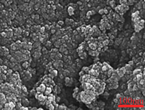
FE-SEM image of silver nanoparticles.
3.2 FT-IR analysis of silver nanoparticles synthesized using Allium monanthum
The FT-IR spectrum of silver nanoparticles is shown in Fig. 2. The formation of AgNPs is approved by the presence of the peaks at wavenumbers of 469, 511 and 572 cm−1. Similar peaks with some differences in the wavenumber have been reported for green-synthetic AgNPs by other research groups (El-Aassar et al., 2021). The other peaks in the spectrum are attributed to the functional groups of different organic compounds in extract, which are linked to the surface of AgNPs. The presence of secondary metabolites such as phenolic, flavonoid, saponins, Quinones, Terpenoids in extract has been reported previously (El-Aassar et al., 2021; Zangeneh et al., 2019; Shahriari et al., 2019; Singh et al., 2018; Hemmati et al., 2020; Ahmeda et al., 2020). The peaks in 3348 and 2952 cm−1 are related to O—H and aliphatic C—H stretching; the peaks from 1502 to 1714 cm−1 are corresponded to C⚌C and C⚌O stretching, and the peaks at 976 cm−1 could be ascribed to —C—O and C—O—C stretching.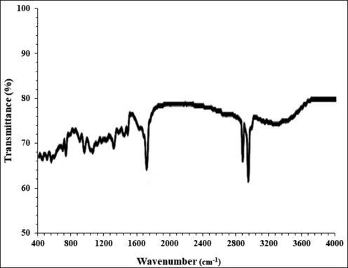
FT-IR of the silver nanoparticles.
3.3 UV–visible spectroscopy of silver nanoparticles synthesized using Allium monanthum
UV–Vis spectroscopic analysis showed the presence of an absorption peak at 428 nm which confirmed the formation of the silver nanoparticles (Fig. 3).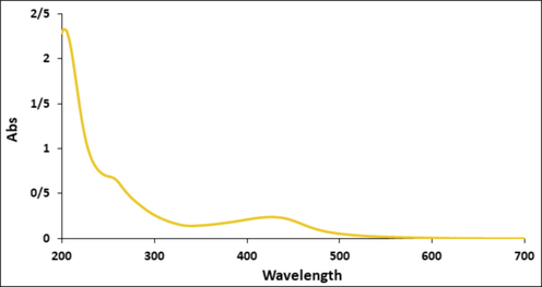
UV–Vis of the silver nanoparticles.
3.4 Cytotoxicity potential of silver nanoparticles synthesized using Allium monanthum
Nanotechnology is a new branch of science with a wide range of applications and nanoparticles with different compositions and sizes, shapes and surface chemical properties can have different biological and biomedical applications. Reducing the size of materials at the nanoscale can often cause electrical, magnetic, structural, morphological, and chemical changes. Nanoparticles typically have a higher percentage of atoms on their surface, which increases surface reactions (El-Aassar et al., 2021; Zangeneh et al., 2019; Shahriari et al., 2019; Singh et al., 2018). Proper design of nanomaterials can be used to target specific cancer cells. Nanoparticles have antibacterial and magnetic properties by penetrating microorganisms due to their high surface-to-volume ratio and small size (Hemmati et al., 2020; Ahmeda et al., 2020). In addition, due to their photocatalytic, catalytic and ionic properties, they are widely used in the fight against human pathogenic microbes, bacteria, fungi and viruses. A study has shown that concomitant use of metallic oxide and doxorubicin reduces the reproductive toxicity of doxorubicin (Ahmeda et al., 2020). By producing active bases such as oxygen ions and hydroxides, silver nanoparticles disrupt the metabolism, proliferation and respiration of microorganisms by destroying organic structures and strongly interacting with enzymes and proteins in the electron transfer system, and it can kill more than 650 types of gram-negative and gram-positive bacteria resistant to common antibiotics in vitro up to 99.9% (El-Aassar et al., 2021; Zangeneh et al., 2019; Shahriari et al., 2019; Singh et al., 2018). Metallic nanoparticles in cell cultures and human tissues yield toxins that raise inflammatory products such as cytokines and oxidative stress, ultimately leading to cell death (El-Aassar et al., 2021; Zangeneh et al., 2019; Shahriari et al., 2019; Singh et al., 2018). Larger nanoparticles are seen by the nuclei and mitochondria, causing mutations in DNA, destruction of the mitochondrial structure, and even cell death. Solubility and density, surface baroelectricity, surface structure, shape, chemical composition, and size and dimensions are the key factors in determining the toxicity of nanoparticles. The exact effect of silver nanoparticles on cancer cells is not fully understood, but increasing ROS production is one of the possible mechanisms (Zangeneh et al., 2019; Shahriari et al., 2019). When nanoparticles are in contact with cancer cells, the cellular defense mechanism is activated to minimize damage. However, if the ROS production stimulation inside the cell by nanoparticles exceeds the cell antioxidant defense capacity, the cells are destroyed during the process of apoptotic cell death (Singh et al., 2018; Hemmati et al., 2020; Ahmeda et al., 2020). The electrostatic interaction of nanoparticles causes them to be absorbed into target cells. Positively charged nanoparticles are attracted to cancer cells with a high percentage of anionic phospholipids and certain groups of charged proteins and carbohydrates on their outer surface (Singh et al., 2018; Ahmeda et al., 2020).
The morphological parameters of silver nanoparticles which affect anticancer properties of these nanoparticles against several cancer cell lines are size, form, and surface coating. Among the above parameters, the role of size of silver nanoparticles is the most (Namvar et al., 2014). Previously, it was showed whatever the size of silver nanoparticles reduced, the ability of these nanoparticles for transferring to the cancer cell lines and killing them increased (Namvar et al., 2014). As can be observed in Fig. 1 of our study, silver nanoparticles had uniform spherical morphology in the range size of 10–35 nm. The size of silver nanoparticles at lower than 50 nm is very suitable for the killing of tumor cell lines in vivo and in vitro (Namvar et al., 2014). About the anticancer properties of silver nanoparticles, they have used for the treatment of several cancers including human lung cancer, mammary carcinoma, uterus cancer, lung epithelial cancer, Lewis lung carcinoma, colon cancer, and human glioma (Singh et al., 2018).
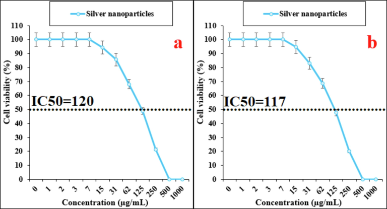
In the present experiment, the treated cells with several concentrations of the present AgNO3, Allium monanthum, and AgNPs were examined by MTT test for 48 h regarding the cytotoxicity properties on normal (HUVEC) and breast adenocarcinoma (MCF7), breast carcinoma (Hs 578Bst), infiltrating ductal cell carcinoma (Hs 319.T), infiltrating lobular carcinoma of breast (UACC-3133), inflammatory carcinoma of the breast (UACC-732), and metastatic carcinoma (MDA-MB-453) cell lines (Figs. 4-7).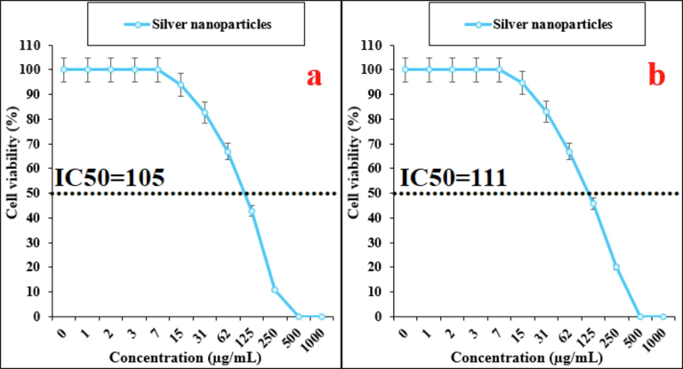
The anti-breast cancer activities of silver nanoparticles against breast adenocarcinoma (MCF7 (a)) and breast carcinoma (Hs 578Bst (b)) cell lines.
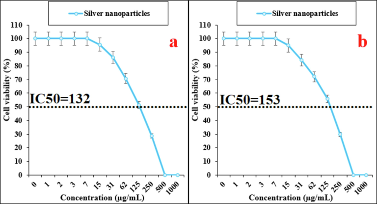
The anti-breast cancer activities of silver nanoparticles against infiltrating ductal cell carcinoma (Hs 319.T (a)) and infiltrating lobular carcinoma of breast (UACC-3133 (b)) cell lines.
The anti-breast cancer activities of silver nanoparticles against inflammatory carcinoma of the breast (UACC-732 (a)) and metastatic carcinoma (MDA-MB-453 (b)) cell lines.
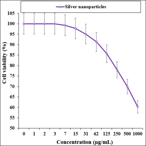
The cytotoxicity activities of silver nanoparticles against HUVEC (normal) cell line.
The absorbance rate was determined at 570 nm, which indicated extraordinary viability on normal cell line (HUVEC) even up to 1000 μg/mL for AgNO3, Allium monanthum, and AgNPs.
In the case of breast cancer cell lines, the viability of them reduced dose-dependently in the presence of AgNO3, Allium monanthum, and AgNPs (Figs. 4-6).
3.5 Antioxidant properties of silver nanoparticles synthesized using Allium monanthum
Probably the anti-breast cancer properties of silver nonparties against breast adenocarcinoma (MCF7), breast carcinoma (Hs 578Bst), infiltrating ductal cell carcinoma (Hs 319.T), infiltrating lobular carcinoma of breast (UACC-3133), inflammatory carcinoma of the breast (UACC-732), and metastatic carcinoma (MDA-MB-453) are related to their antioxidant activities. The previous researches have revealed that antioxidant compounds such as medicinal plants and silver nanoparticles as single electron donors can stabilize and scavenge the free radicals, which in conditions of oxidative stress may begin angiogenesis or carcinogenesis (Rehana et al., 2017; Del Mar et al., 2016; Jeong et al., 2012; Oganesvan et al., 1991). High levels of free radicals are observed in various cancerous cells and several accumulating evidence suggests that free radicals function as key signaling molecules stimulate different growth-related responses that finally begin tumorigenesis and angiogenesis (Radini et al., 2018; Beheshtkhoo et al., 2018; Sangami and Manu, 2017). In detail, Free radical-induced development of cancer involves malignant transformation due to DNA mutations and changed gene expression through epigenetic mechanisms which in turn causes the uncontrolled proliferation of cancerous cells (Beheshtkhoo et al., 2018; Sangami and Manu, 2017). Many researchers reported a remarkable role of antioxidant compounds such as medicinal plants and silver nanoparticles in growth inhibition of prostate, ovary, breast, endometrial and lung, and colon cancer cells with removing free radicals (Katata-Seru et al., 2018; Sankar et al., 2014).
In our study, the antioxidant effects of the silver nanoparticles synthesized using Allium monanthum aqueous extract were evaluated by DPPH assay revealed concentration-dependent effects i.e., an increase in the concentration of the silver nanoparticles leads to an increase in antioxidant activities. The DPPH radical (Hi-media) is stable due to the delocalization of a spare electron over the molecule, thus preventing dimer formation. This radical is used in the DPPH radical scavenging capacity assay to quantify the ability of antioxidants to quench the DPPH radical. The dark purple color of DPPH will be lost when it is reduced to its nonradical form stable organic nitrogen centered free radical with a dark purple color which when reduced to its nonradical form by antioxidants becomes colorless. DPPH radicals are widely used in the model system to investigate the scavenging activities of several natural compounds. When the DPPH radical is scavenged, the color of the reaction mixture changes from purple to yellow with decreasing of absorbance at wavelength 517 nm (Gultekin et al., 2016; Rehana et al., 2017).
In the concentrations of studied, the best result was seen in the high concentration or 1000 µg/mL (Fig. 8). Comparative analysis of the individual antioxidant assays showed significant variations in the exertion of radical scavenging effects. Among all materials tested (AgNO3, Allium monanthum, and AgNPs), the silver nanoparticles indicated more excellent inhibition effects against DPPH. In contrast, standard (butylated hydroxytoluene) demonstrated lower antioxidant effects compared to the silver nanoparticles.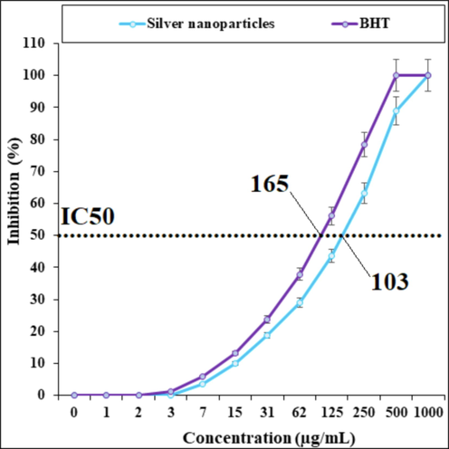
The antioxidant properties of silver nanoparticles and BHT against DPPH.
Usually, many free radicals such as Reactive oxygen species (ROS) are produced in the procedure of mitochondrial respiration which is responsible for oxidative stress in the human body. This oxidative stress damages human body DNA and develops several oxidative diseases like nephrotoxicity, hepatotoxicity, and hematotoxicity. The oxidative stress can be efficiently reduced with the help of antioxidant materials such as metallic nanoparticles (Gultekin et al., 2016). The synthesized silver nanoparticles exhibit higher antioxidant activity for the formation of free radicals into the living system (Gultekin et al., 2016). The silver nanoparticles have redox properties and play a significant role in deactivating free radicals in the living system (Rehana et al., 2017). In recent years, researchers evaluated plants and bio mediated synthesized nanoparticles for antioxidant activity. The reason behind the antioxidant activity of green or biosynthesized nanoparticles could be due to the presence of metabolites compounds such as phenolic compounds, flavonoids, carbohydrates, and other sugar substances (Del Mar et al., 2016; Jeong et al., 2012; Oganesvan et al., 1991). Also, many researchers reported phenolic and flavonoids attached to the nanoparticles exhibited the antioxidant activity. Previously has been indicated that Allium monanthum is rich in antioxidant compounds (Noda and Kawano, 1988). Several studies were carried out in the nanotechnology field using various medicinal plants, but still, no report is available on silver nanoparticles synthesized using Allium monanthum aqueous extract.
4 Conclusions
In our research, the silver nanoparticles have been appropriately characterized and confirmed using FE-SEM, TEM, FT-IR, and UV–Vis. In the FT-IR test, the presence of many antioxidant compounds with related bonds caused the excellent condition for reducing of silver in the silver nanoparticles, so that the antioxidant properties of silver nanoparticles was the better than the butylated hydroxytoluene as the positive control. The silver nanoparticles indicated suitable antioxidant and anti-breast cancer activities against breast adenocarcinoma (MCF7), breast carcinoma (Hs 578Bst), infiltrating ductal cell carcinoma (Hs 319.T), infiltrating lobular carcinoma of breast (UACC-3133), inflammatory carcinoma of the breast (UACC-732), and metastatic carcinoma (MDA-MB-453) cell lines without any cytotoxicity effect on the normal cell line. Seemingly, the silver nanoparticles synthesized using Allium monanthum leaves aqueous extract can be used for the treatment of several types of breast cancers in human after confirming in in vivo and clinical trial experiments.
Funding
Beijing-Tianjin-Hebei Basic Research Cooperation Special Project (2019): “Visualized Stem Cell Targeted Tumor Therapy Techniques for Precise Diagnosis and Treatment of Tumors” Project Number: H2019104018.
Declaration of Competing Interest
The authors declare that they have no known competing financial interests or personal relationships that could have appeared to influence the work reported in this paper.
References
- Green synthesis and chemical characterization of gold nanoparticle synthesized using Camellia sinensis leaf aqueous extract for the treatment of acute myeloid leukemia in comparison to daunorubicin in a leukemic mouse model. Appl. Organometal. Chem.. 2020;34:e5290
- [CrossRef] [Google Scholar]
- Areca nut chewing and esophageal squamous-cell carcinoma risk in Asians: A meta-analysis of case–control studies. Cancer Cause Control. 2013;24:257-265.
- [Google Scholar]
- Ellagic acid encapsulated chitosan nanoparticles for drug delivery system in human oral cancer cell line (KB) Colloids Surf. B Biointerfaces. 2013;110:313-320.
- [Google Scholar]
- One-step green synthesis and characterization of leaf extract-mediated biocompatible silver and gold nanoparticles from Memecylon umbellatum. Int. J. Nanomed.. 2003;8:1307-1315.
- [Google Scholar]
- Green synthesis of iron oxide nanoparticles by aqueous leaf extract of Daphne mezereum as a novel dye removing material. Appl. Phys. A. 2018;124:363-369.
- [Google Scholar]
- Tentative identification of the composition of Agaricus bisporus aqueous enzymatic extracts with antiviral activity against HCV: A study by liquid chromatography–tandem mass spectrometry in high resolution mode. J. Function Foods. 2016;24:403-419.
- [Google Scholar]
- (a) Wound dressing of chitosan-based-crosslinked gelatin/ polyvinyl pyrrolidone embedded silver nanoparticles, for targeting multidrug resistance microbes, Carbohydrate Polym.; 2021, 255, 117484. (b) Microencapsulation of lectin anti-cancer agent and controlled release by alginate beads, biosafety approach, Int. J. Biol. Macromol. 2014, 69, 88–94. (c) Zhaleh, M., Zangeneh, A., Goorani, S., et al., 2019. In vitro and in vivo evaluation of cytotoxicity, antioxidant, antibacterial, antifungal, and cutaneous wound healing properties of gold nanoparticles produced via a green chemistry synthesis using Gundelia tournefortii L. as a capping and reducing agent. Appl. Organometal. Chem. 33, e5015.
- Polydopamine nanomaterials: Recent advances in synthesis methods and applications. Bioeng. Biotechnol.. 2018;6:109.
- [Google Scholar]
- J. Turk. Chem. Soc. A: Chem.. 2016;3:623-636.
- (a) Hemmati, Joshani, Z., Zangeneh, A., et al., 2020. Biosynthesis and chemical characterization of polydopamine-capped silver nanoparticles for the treatment of acute myeloid leukemia in comparison to doxorubicin in a leukemic mouse model. Appl. Organometal. Chem. 34, e5277. doi: 10.1002/aoc.5277. (b) Zangeneh, M.M., 2020. Green synthesis and formulation a modern chemotherapeutic drug of Spinacia oleracea L. leaf aqueous extract conjugated silver nanoparticles; Chemical characterization and analysis of their cytotoxicity, antioxidant, and anti-acute myeloid leukemia properties in comparison to doxorubicin in a leukemic mouse model. Appl. Organometal. Chem. 34, e5295. doi: 10.1002/aoc.5295. (c) Mohammadi, G., Zangeneh, M.M., Zangeneh, A., et al., 2020. Chemical characterization and anti-breast cancer effects of silver nanoparticles using Phoenix dactylifera seed ethanolic extract on 7,12-Dimethylbenz[a] anthracene-induced mammary gland carcinogenesis in Sprague Dawley male rats. Appl. Organometal. Chem. 34, e5136. doi: 10.1002/aoc.5136. (d) Hamelian, M., Zangeneh, M.M., Shahmohammadi, A., et al., 2020. Pistacia atlantica leaf extract mediated synthesis of silver nanoparticles and their antioxidant, cytotoxicity, and antibacterial effects under in vitro condition. Appl. Organometal. Chem. 34, e5278. doi: 10.1002/aoc.5278. (e) Hemmati, S., Rashtiani, A., Zangeneh, M.M., et al., 2019. Green synthesis and characterization of silver nanoparticles using Fritillaria flower extract and their antibacterial activity against some human pathogens. Polyhedron 158, 8–14. (f) Mahdavi, B., Paydarfard, S., Zangeneh, M.M., et al., 2019. Assessment of antioxidant, cytotoxicity, antibacterial, antifungal, and cutaneous wound healing activities of green synthesized manganese nanoparticles using Ziziphora clinopodioides Lam leaves under in vitro and in vivo condition. Appl. Organometal. Chem. 33, e5248. doi: 10.1002/aoc.5248. (g) Jalalvand, A.R., Zhaleh, M., Goorani, S., et al., 2019. Chemical characterization and antioxidant, cytotoxic, antibacterial, and antifungal properties of ethanolic extract of Allium Saralicum R.M. Fritsch leaves rich in linolenic acid, methyl ester. J. Photochem. Photobiol. B 192, 103–112.
- Green synthesis and chemical characterization of Thymus vulgaris leaf aqueous extract conjugated gold nanoparticles for the treatment of acute myeloid leukemia in comparison to doxorubicin in a leukemic mouse model. Appl. Organometal. Chem.. 2020;34:e5267
- [CrossRef] [Google Scholar]
- The radioprotective effect of Zataria multiflora against genotoxicity induced by γ irradiation in human blood lymphocytes. Cancer Biother. Radiopharm.. 2011;26:325-329.
- [Google Scholar]
- Macrophage immunomodulating and antitumor activities of polysaccharides isolated from Agaricus bisporus white button mushrooms. J. Med. Food. 2012;1:58-65.
- [Google Scholar]
- Green synthesis of iron nanoparticles using Moringa oleifera extracts and their applications: Removal of nitrate from water and antibacterial activity against Escherichia coli. J. Mol. Liq.. 2018;256:296-304.
- [Google Scholar]
- Cytotoxic effect of magnetic iron oxide nanoparticles synthesized via seaweed aqueous extract. Int. J. Nanomed.. 2014;19:2479-2488.
- [Google Scholar]
- Noda, Shozo; Kawano, Shoichi, 1988-06-01. The Biology of Allium monanthum (Liliaceae) I. Polyploid Complex and Variations in Karyotype. Plant Species Biol. 3(1), 13–26.
- Phenolic and flavonoid compounds of Ziziphora clinopodioides. Chem. Nat.. 1991;27:247.
- [Google Scholar]
- Biosynthesis of iron nanoparticles using Trigonella foenum-graecum seed extract for photocatalytic methyl orange dye degradation and antibacterial applications. J. Photochem. Photobiol., B. 2018;183:154-163.
- [Google Scholar]
- Extracellular synthesis of silver nanoparticles using dried leaves of pongamia pinnata (L) pierre. Nano-Micro Lett.. 2010;2:106.
- [Google Scholar]
- Evaluation of antioxidant and anticancer activity of copper oxide nanoparticles synthesized using medicinally important plant extracts. Biomed. Pharmacother.. 2017;89:1067-1077.
- [Google Scholar]
- Environ. Technol. Innov.. 2017;8:150-163.
- Anticancer activity of Ficus religiosa engineered copper oxide nanoparticles. Mat. Sci. Eng. C. 2014;44:234-239.
- [Google Scholar]
- Biosynthesis of gold nanoparticles using Allium noeanum Reut. ex Regel leaves aqueous extract; characterization and analysis of their cytotoxicity, antioxidant, and antibacterial properties. Appl. Organometal. Chem.. 2019;33:e5189
- [CrossRef] [Google Scholar]
- Gold Nanoparticles in Diagnostics and Therapeutics for Human Cancer. Int. J. Mol. Sci.. 2018;19:1979.
- [Google Scholar]
- Lactic acid bacteria as reducing and capping agent for the fast and efficient production of silver nanoparticles. Appl. Microbiol. Biotechnol.. 2009;6:741-749.
- [Google Scholar]
- Oesophageal cancer: ESMO Clinical Practice Guidelines for diagnosis, treatment and follow-up. Annal. Oncol.. 2013;24(Suppl):51-56.
- [Google Scholar]
- Extracellular synthesis of silver nanoparticles using dried leaves of pongamia pinnata (L) pierre. Nano-Micro Lett.. 2010;2:106.
- [Google Scholar]
- Preparation, characterization, and evaluation of cytotoxicity, antioxidant, cutaneous wound healing, antibacterial, and antifungal effects of gold nanoparticles using the aqueous extract of Falcaria vulgaris leaves. Appl. Organometal. Chem.. 2019;33:e5216
- [CrossRef] [Google Scholar]
- Epidemiologic differences in esophageal cancer between Asian and Western populations. Chinese J. Cancer. 2012;31:281-286.
- [Google Scholar]
Further reading
- Silver nanoparticles as an effective disinfectant: A review. Mat. Sci. Eng. C. 2019;97:954-965.
- [Google Scholar]
- Designed inorganic nanomaterials for intrinsic peroxidase mimics: A review. Sen. Act. B Chem.. 2019;283:18-34.
- [Google Scholar]
- Antimicrobial properties of ZnO nanomaterials: A review. Ceram. Int.. 2017;43:3940-3961.
- [Google Scholar]
- In vitro antileishmanial and antioxidant potential, cytotoxicity evaluation and phytochemical analysis of extracts from selected medicinally important plants. Biocat. Agri. Biotech.. 2019;19:101117
- [Google Scholar]
- Superparamagnetic MFe2O4 (M = Fe Co, Mn) nanoparticles: Tuning the particle size and magnetic properties through a novel one-step coprecipitation route. Chem. Mater.. 2012;24:1496-1504.
- [Google Scholar]
- Synthesis and antimicrobial activity of copper nanoparticles. Mater. Lett.. 2012;71:114-116.
- [Google Scholar]
- Biological degradation of plastics: a comprehensive review. Biotech. Adv.. 2008;26:246-265.
- [Google Scholar]
- Developments and challenges in the manufacturing, characterization and scale-up of energetic nanomaterials – A review. Chem. Eng. J.. 2018;350:939-948.
- [Google Scholar]


