

Translate this page into:
Chitosan coated molybdenum sulphide nanosheet incorporated with tantalum oxide nanomaterials for improving cancer photothermal therapy
⁎Corresponding author. sekarbioline@gmail.com (Srinivasan Rajasekar)
-
Received: ,
Accepted: ,
This article was originally published by Elsevier and was migrated to Scientific Scholar after the change of Publisher.
Peer review under responsibility of King Saud University.
Abstract
In recent years, two-dimensional nanomaterials (2D) prominent for site specific photothermal treatment (PTT), which are one of the most interesting strategy due to their maximizing cancer cell killing efficiency without the normal cells. Several robust methods are established for 2D material synthesis and improving the photothermal conversion efficiency (PCE), biocompatibility, and photostability in cancer PTT. Such preferred mechanism like nanomaterial decoration on to their surface would enable access to tunable 2D nanomaterial properties to improve cancer PTT. Here, we first time report a robust route for deposition of tantalum (TaO2) on to chitosan (CS) coated molybdenum sulphite (MoS2) nanosheet surface via electrostatic interaction, which assists to improve cancer PTT efficiency. Detailed studies prove that prepared TaO2-CS-MoS2 nanomaterial shows lack of toxicity, photostability and PCE was calculated from 26 °C to 47.2 °C under the 808 nm irradiation/5 min. Therefore, the TaO2 deposition particularly interest to promote the photostability, biocompatibility and PCE of bare MoS2 nanosheets. Therefore, the possible mechanism is highly expected to improve biological features in cancer PTT.
Keywords
MoS2 nanosheet
Tantalum oxide
Chitosan
Cancer photothermal therapy

1 Introduction
Photothermal therapy (PTT) has been realized as a talented route to destroy cancer cells with high efficiency (Nam et al., 2018). This attracted strategy exhibits unique features including low systemic effects, low cost, minimal invasiveness, high selectivity and stability (Khafaji et al., 2019). On the other words, well-established PTT agent may enable advances in a precise site-specific heat generator particularly inside the cancer cells under exposure of NIR light, ultrasound, radiofrequency and magnetic field (Cheng et al., 2014). Tremendous number of noble metal isotropic and anisotropic nanoparticles (ANPs) (Alkilany et al., 2012), as well as some 2D materials such as transition metal dichalcogenides (TMDCs), black phosphorous, (Geng et al.,2018) and graphene (Orecchioni et al., 2015) were extensively explored as photothermal conversion agents for cancer PTT. Among them, TMDCs such as MoS2, WS2, Bi2Se3, TiS2 and MoSe2 have been widely demonstrated in the cancer PTT. This is because of their unique properties such as owing to their high PCE, good biocompatibility and photothermal stability (Zhang et al., 2017; Li et al., 2018; Shu et al., 2018; Xu et al., 2018). Recently, Molybdenum disulfide (MoS2) has great attention for PTT due to its remarkable photothermal conversion efficiency (PCE) in the near-infrared (NIR) range and better biocompatibility (Zhang et al., 2018; Liu et al., 2018). Although, most of previous reports has been demonstrated that the modification in the outer or inter-layer sheets of MoS2 exhibit enhanced PCE with excellent biocompatibility, stability and photothermal conversion efficiency (PCE) (Murugan et al., 2019a,b; Chen et al., 2016). For instance, the surface region of 2D nanomaterial deposition by small sized nanoparticles (NPs) dramatically influenced kupffer cell engulf of 2D materials in vital organs, which in turn helps rapid excretion, increased tumor accumulation and prolonged circulation time in blood (Cheng et al., 2016). Hence, the metal nanostructure decoration on the exterior of MoS2 suggests a strategy to access new form of 2D materials for cancer PTT. In addition, tantalum oxide (TaO2) nanoparticles is an ideal material for imaging due to their strong X-ray attenuation as well as combine to PTT agent it can influence the therapeutic ability (Freedman et al., 2014). For example, amalgamation of tantalum oxide (TaO2) nanoparticles (NPs) into polypyrrole (PPy) NPs improved imaging and photothermal ablation of tumor was estimated to be 66.5% at intravenously injection and 100% for intra-tumoral injection, respectively (Jin et al., 2014). Another report demonstrated that the prepared core/shell nanoparticles using tantalum oxide showed multimodal imaging features including computed tomography (CT), photoacoustic and fluorescence imaging, pH-and thermal-sensitive drug release in cancer therapy (Jin et al., 2017).
Herein, we report the hydrothermal synthesized MoS2 nanosheet decorated with TaO2 for cancer PTT. It is a two-step process, wherein MoS2 nanosheet coated with chitosan (CS) were obtained in the first step and then selectively deposition with TaO2 in the second step. CS has been widely used as a coated material due to their enhanced biocompatibility enables to get the superior nanomedicine. CS had hydrophilic (—OH) and hydrophobic (—NH2) groups that put forth better degradability, cytocompatibility, and mucoadhesive ability (Fathi et al., 2018). The inherent properties of CS give it an upper hand in terms of promotion of cancer PTT, as well as an effective biopharmaceutical material in cancer applications. The findings revealed that the TaO2 decorated MoS2 nanosheets exhibited much better biocompatibility and high PCE. Hence, the surface phase interaction of TaO2-CS-MoS2 nanosheet as novel functional material provides a new strategy for cancer PTT.
2 Materials and methods
Sodium molybdate dehydrate (Na2MoO4·2H2O), thiourea (CH4N2S), L-Cysteine, chitosan (CS), tantalum pentoxide (Ta2O5), sodium hydroxide (NaOH), hydrochloric acid (HCl). All reagents used as received without any further purification.
2.1 Synthesis of molybdenum disulphide (MoS2) nanosheet
Hydrothermal route used to fabricate MoS2 nanosheet at 200 °C for 24 h. In brief, the precursor solution of 50 mg Na2MoO4.2H20 mixing with 35 mg of thiourea in 50 mL of deionized water. Then, it was sonicated for 15 min at room temperature and subsequently adjusted to pH 3.5 by adding 0.1 M HCl. Afterward, the reactant was transferred to a teflon-lined autoclave and heated to 200 °C for 18 h in a hot air oven. After completion of the reaction, the black color precipitate was separated from the solution by centrifugation at 8000 rpm for 10 min and washed several times with milliQ water and ethanol to remove any impurities. The black color MoS2 powder was obtained by drying the precipitate under vacuum at 80 °C overnight (Saada and Bissessur, 2012).
2.2 Synthesis of chitosan coated MoS2 (CS-MoS2) nanosheet
In a round bottom flask, 0.3 g of MoS2nanosheet and 30 mg of L-cysteine was dispersed in 25 mL of milli-Q water under continuous stirring of solution. After 5 h of stirring, 1% CS solution prepared in acetic acid (1%) was added in a drop wise manner at room temperature (RT) to form CS coated MoS2 nanosheet. After 5 h stirring, the final product was separated by centrifugation and washed with distilled water and ethanol for several times. The resultant nanostructures (CS-MoS2) were dried overnight under vacuum (Rayappan et al., 2017).
2.3 Synthesis of tantalum oxide materials
For synthesis of TaO2, 0.05 M of Ta2O5.6H20, and 200 µL of 0.5 M NaOH was dissolved in 25 mL of Milli-Q water and the solution was kept overnight under mild stirring. The product obtained was collected by centrifugation and washed several times with milli-Q water and ethanol. The final product was dried at 40 °C for 6 h. After cooling the sample to RT, the powder sample was washed with water for 15 min under ultrasonication and dried under vacuum.
2.4 Synthesis of CS-MoS2 nanosheet incorporated with TaO2
Briefly, the concentration of 0.5 mg/mL of as-synthesized TaO2 and 100 mg of CS-MoS2 was added in 25 mL of water and the mixture was stirred overnight at RT. Aliquots were collected and purified by repeated centrifugation and washing steps with water and ethanol. The final product (TaO2-CS-MoS2) was dried under vacuum and stored for further use.
2.5 Nanomaterials and their characterization
The morphological features and composition of synthesized TaO2-CS-MoS2 nanomaterials were observed using transmission electron microscopy (TEM, JEOL JEM-2100). For X-ray diffraction (XRD) investigations, XRD pattern was recorded on a D/max-2550 PC X-ray diffractometer (XRD; Rigaku, Japan) from 20 to 100°. The structure of TaO2-CS-MoS2 nanomaterials was also investigated by Fourier transform infrared (FTIR, Nicolet 6700 Thermo Fisher, USA) spectrometer in the scanning range of 4000–400 cm−1.
2.6 Cell line and cell culture condition
The MCF-7 breast cancer cell line was obtained from National Centre for Cell Sciences (NCCS), Pune, India. Then, the cell line was maintained in DMEM media with addition of proper supplements such as 10% (v/v) FBS, 1% (v/v), 100 μg/mL streptomycin and 100 U/mL penicillin. Then, the cells were grown in a humidified incubator at 37 °C under atmosphere supplemented with 95% air and 5% CO2. The cell culture medium was changed every day, and cells were trypsinized and harvested before reaching confluence.
2.7 Cell viability assay
The HBL-1000 and MCF-7 breast cells were placed in a 96-well plate at a density of 2 × 10⁵ cells/well and grown for 24 h. The grown cells were treated with various concentrations (6, 12, 25, 50, 100 μg/mL) of bare MoS2, and CS-MoS2-TaO2 nanosheets at 37 °C for 24 h. After washing the cells with PBS to remove unbound sheets, 20 μL MTT solution (3-(4, 5-dimethylthiazol-2-yl)-3 2, 5-diphenyltetrazolium bromide) at 0.5 mg mL−1 were dropped into the 96-well plate for MTT assay. The cell viability was calculated as a percentage of viable cells after treated with nanomaterials compared with the untreated cells (Murugan et al., 2017).
2.8 Photothermal performance measurement of bare MoS2 and CS-MoS2-TaO2 nanosheets
Photothermal performance experiments of bare MoS2 nanosheet, and CS-MoS2-TaO2 nanosheets were carried out to examine the photothermal conversion efficiency at a laser light source at 808 nm (continuous-wave NIR laser device with power of 0.5 W/cm2). 0.4 mL of aqueous suspensions containing 100 μg/mL of bare MoS2 or TaO2-CS-MoS2 nanosheets were placed in cuvette and irradiated with 808-nm NIR laser at a power of 0.5 W/cm2 for 5 min. The temperature increase was monitored for every 30 s using a thermocouple thermometer to determine the PCE of bare MoS2 and TaO2-CS-MoS2 nanosheets.
2.9 Measurement of intracellular ROS generation
To obtain quantitative information about the intracellular free radicals such as peroxide and superoxide free radicals, 5 μg/mL of 2′,7′-Dichlorofluorescein diacetate (DCFH-DA) was added to each well in a 6-well plate containing 4 × 10⁵ cells/well. Samples with final concentration of 200 μg/mL of bare MoS2 and CS-MoS2-TaO2 nanosheets were added to each well and incubated at 37 °C for 24 h in an incubator. The fluorescence intensity of DCF is proportional to the amount of ROS produced by the cell. ROS generation was assessed using a fluorescence microscope (Nikon Eclipse, Inc., Japan) at excitation and emission wavelengths of 488 and 530 nm, respectively (Wang and Cheng, 2019).
For synergistic effects of ROS and PTT assessment, same procedure mentioned above was performed with laser light irradiation at 808 nm for 5 min. After irradiation, the cells were incubated for 24 h, rinsed with PBS, and stained with 20 μM DCFH-DA for 20 min. Subsequently, the fluorescence intensity of DCF in each well was quantitatively estimated by a fluorescence microplate reader.
2.10 In vitro photothermal performance of bare MoS2 and CS-MoS2-TaO2 nanosheets
The MCF-7 cells were incubated in 6-well plates at 37 °C with 5% CO2 for 24 h. After replacing the medium with pre-warmed new culture medium, bare MoS2 and 100 μg/mL of CS-MoS2-TaO2 nanosheets were added into the wells. After 4 h of incubation, cells were irradiated with 808-nm laser at a power density of 0.5 W/cm2 for 5 min. The cells were then co-stained with florescent molecules acridine orange (green), propidium iodide (red) and DAPI (blue) investigated in a fluorescent microscope to visualize the structural morphology of live and dead cells. The cell viability was normalized by control group without any treatment.
2.11 Statistical analysis
All the triplicate data were analyzed by student t-tests with a setting significance of p < 0.05 (*).
3 Results and discussion
The surface decorated 2D nanomaterial by one-dimensional materials offers remarkable physiochemical properties. Hence, various efforts have been devoted to 2D materials to make them highly potential for cancer PTT (Shao et al., 2016; Yi and Zhang, 2018). Herein, we demonstrate the hydrothermal synthesis of MoS2 nanosheet decorated with TaO2 for the application of cancer PTT. However, MoS2 nanosheet decorated with TaO2 were prepared in two steps: (a) preparation of CS coated MoS2 nanosheet and (b) decoration with TaO2. One-pot hydrothermal approach was used to prepare MoS2 nanosheet using sodium molybdate dehydrate and thiourea as Mo and S source, respectively. As-prepared MoS2 nanosheet functionalized with cysteine and CS using a disulphite reaction. Afterward, the negatively charged TaO2 nanoparticles were grafted on the surface of CS-MoS2 nanosheet, forming TaO2 decorated MoS2 nanosheet denoted as TaO2-CS-MoS2 nanosheet is illustrated in Scheme 1.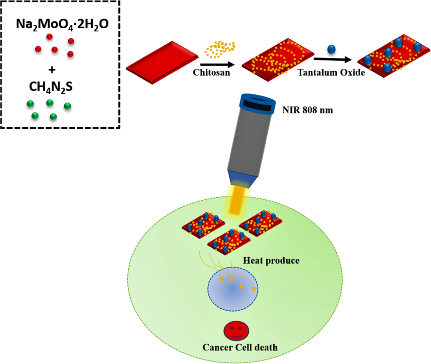
Schematic representation reveals the fabrication route of TaO2-CS-MoS2 nanosheet and their application in cancer PTT.
The inclusive shape and layer structural information of the typical MoS2 nanosheet and TaO2-CS-MoS2 were initially determinate by high resolution transmission electron microscopy (HR-TEM) analysis. Based on the profile shown in 1 A, the TEM image revealed monolayer MoS2 nanosheet exhibit a sheet-like structure with hundreds of nanometers in size. As prepared MoS2 nanosheet possessed similar sheet-like morphology with previous reports (Yang et al., 2019; Vattikuti et al., 2015). Afterward, the detailed TEM analysis was carried out to confirms the formation of TaO2-CS-MoS2 (Fig. 1B). The obtained TEM images clearly shows the small mono-crystalline hexagonal and semispherical TaO2 particles with averaged size of 5–10 nm could be found imperfectly onto surface matrix of MoS2 nanosheet, and thick lucid outer layer around the nanosheet confirms CS coating, describe as CS coated TaO2-MoS2 nanosheet. As well as, their HR-TEM cross-sectional images of lattice fringe shown in Fig. 1C. The images reveal that the well stacked with an intrinsic interlayer lattice fringe distance of MoS2 nanosheets, from which we can assess the averaged interlayer lattice fringes ‘d’ spacing value was about 0.65 nm, corresponding to the layer aligned with the (0 0 2) basal plane (Mishra et al., 2017). Apart from the fringe, the interlayer lattice ‘d’ spacing value was estimated to be 0.27 nm, which is representable to the layer aligned with the (1 0 0) lattice plane of MoS2 nanosheet phase.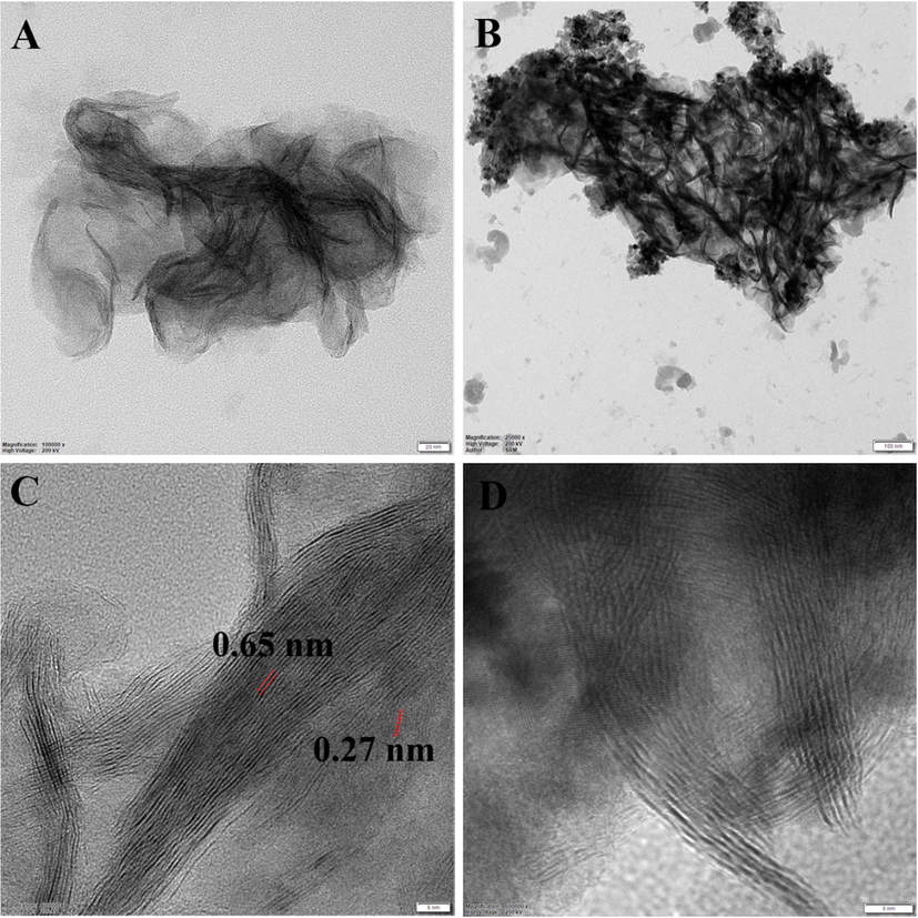
The morphology and structural features of bare MoS2 and TaO2-CS-MoS2 nanosheet were investigated by electron microscopy. (A & C) TEM image of MoS2 and (B &D) TaO2-CS-MoS2 nanosheet.
As shown in Fig. 2, the small spherical-like TaO2 nanoparticles were completely covered the inner sheet-like structure of CS-MoS2 nanosheet.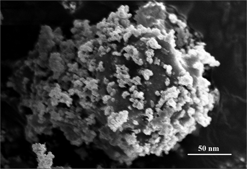
Scanning electron microscopy (SEM) image reveals the morphology of TaO2-CS-MoS2.
In addition, UV-NIR spectrum revealed that the broad absorption bands at 320–280 showing that the prepared composite material has CS and tantalum in each exposed surface of sheet-like MoS2 describes as TaO2-CS-MoS2 nanosheet (Fig. 3A). In Fig. 3b shows the crystal structure of sheet-like MoS2 nanosheet was assessed by the powder X-ray diffraction (XRD) studies that all diffraction peaks can be well indexed and showed at 14°, 32°, and 58° corresponding to the (0 0 2), (1 0 0), and (1 1 0) crystal planes of MoS2 structure, consistent with the corresponding standard card (JCPDS card number 37-1492) (Qiu et al., 2018). In addition, electron microscopic images of Fig. 3C shows the corresponding selected area electron diffraction (SAED) pattern to the different zone axes of crystallographic orientation in each exposed surface of MoS2 and TaO2-CS-MoS2 nanosheet. In Fig. 3b, the presence of three diffraction rings (1 1 0) (1 0 0) and (0 0 2) reflections corresponding to bare MoS2 nanosheet conformed by SAED pattern, it further confirming the nature of these nanosheet (Hu et al., 2018). From the Fig. 3D, the electron diffraction collected from TaO2-CS-MoS2 ensembles indicates a serious of diffraction peaks related to the TaO2 lie in the MoS2 nanosheet plane, the diffraction peaks (white arrows) (1 1 0) (1 0 0) and (0 0 2) reflections corresponding to bare MoS2 nanosheet, and the presence of the geometric diffraction patterns in the SAED confirms thermally induced crystallization confirms that blending of TaO2 and MoS2. It indicates the successfully formation of TaO2-CS-MoS2 nanosheet.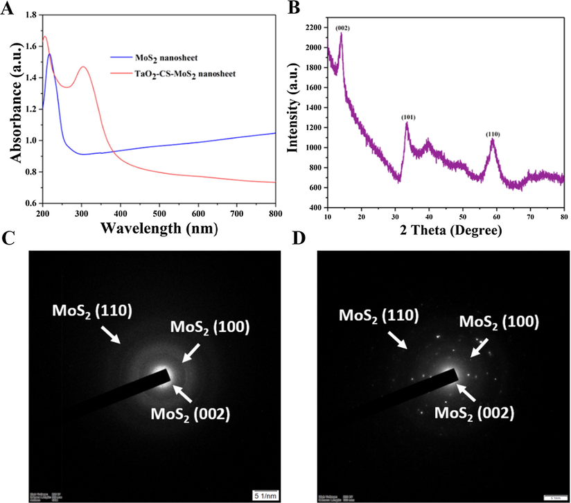
Physiochemical characteristics of MoS2 and TaO2-CS-MoS2 nanosheet. (A) UV-NIR spectrum of MoS2 and TaO2-CS-MoS2 nanosheet (B) XRD pattern of MoS2 nanosheet and SAED pattern of (C) bare MoS2 and (D) TaO2-CS-MoS2 nanosheet.
The absorption spectrum of TaO2-CS-MoS2 shown in Fig. 4A, which showed three absorption bands showing that the prepared composites can strongly absorb the UV light at 280–320 nm. Furthermore, the surface functional groups of bare sheet-like MoS2, and TaO2-CS-MoS2 were evaluated by FTIR spectra (Fig. 4B). Based on the observation and analyses the FTIR spectrum for bare MoS2 nanosheet consisting the broad absorption bands at 3416 cm−1 formed by the stretching vibration of hydroxyls in the MoS2 nanosheet. The absorption band between 1100 cm−1 and 1650 cm−1 is ascribed to the stretching vibrations of the hydroxyl group and Mo-O vibrations, a peak at 900 cm−1 represents to the S-S bond. As shown in FTIR spectra of TaO2 decorated MoS2 nanosheet, the TaO2-CS-MoS2 consisting the major peaks were occurred at 2855, 1100 and 900 cm−1 corresponding to MoS2 and the peaks at 3423, 2356, 539 cm−1, respectively confirmed the presence of TaO2. The broad absorption bands at 3423 cm−1 formed by the stretching vibration of hydroxyls in the TaO2 and CS, the absorption bands at 1634 cm−1 and 1571.05 cm−1 are attributed to the presence of the C⚌O stretching of the amide I band, and bending vibrations of the N—H (N-acetylated residues, amide II band), respectively. It conforms the successfully conjugation of TaO2 on to the CS-MoS2 nanosheet to formulate TaO2-CS-MoS2 nanosheet.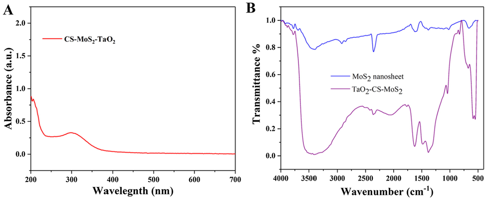
(A) UV–Vis spectrum of TaO2-CS-MoS2 nanosheet and (B) FTIR spectra of MoS2 and TaO2-CS-MoS2 nanosheet.
MTT assay was performed with human normal cell lines such as HBL-100 to investigate the cyto/biocompatibility of formulated nanosheets. Previous reports demonstrated that the formulated MoS2 nanomaterials have a significant cytotoxicity for both normal and cancer cell lines. Therefore, MTT assay of TaO2-CS-MoS2 nanosheets was performed by varying the nanosheets concentrations ranging from 6 to 100 μg/mL for 24 h incubation at 37 °C, as shown in Fig. 5. Even after 24 h of exposure to the highest concentration of nanosheets (100 μg/mL), the viability of the cell population is more than 89.1%, indicating little change in the cell population in the MoS2 and TaO2-CS-MoS2 nanosheet treating HBL-100 cells. Notably, the increase of TaO2-CS-MoS2 concentration enhances the viability of cells indicating that biocompatibility nature (Gao et al., 2016).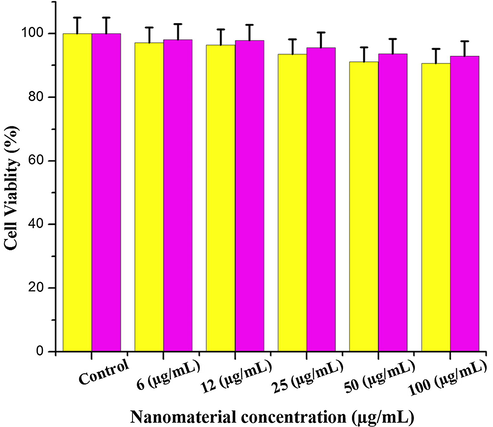
Evaluation of cell viability by MTT assay. The HBL-100 normal breast cells were treated with different nano-formulations at various concentration (6–100 µg/mL) for 24 h at 37 °C.
The photothermal performance of bare MoS2 and TaO2-CS-MoS2 nanosheet (at concentration of 50 and100 μg/mL) in 0.4 mL water at irradiating the suspension with laser light of 808 nm at a power density of 0.5 W/cm2 for 5 min was performed. The temperature was monitored as a function of time, Fig. 6A. Afterwards, the temperature of suspension containing nanosheet modified with TaO2 increased from room temperature of 26 to 42.7 and 47.6 °C, at concentration of 50 and 100 μg/mL of TaO2-CS-MoS2, respectively. The rate of heat generation was higher in the initial period of 2–3 min. It is worth noting that nanosheets prepared with TaO2 improve the heat generation and rise the temperature as about 47.5 °C at 100 μg/mL concentration of TaO2-CS-MoS2 concentration. Similar experiments with bare MoS2 nanosheets and pure milli-Q water showed a temperature increase of only 38.5 °C and 27.8 °C from RT, indicating that TaO2 decoration significantly improves PCE of MoS2 nanosheets at lower power density (0.5 W/cm2). It indicated that TaO2 decoration happened to be on the surface of sheet petals, which in turn, influences the photon absorption ability of MoS2 nanosheets. Heating and cooling cycles of 100 μg/mL of TaO2-CS-MoS2 were monitored continuously for 5 cycles to investigate their potential (photothermal stability) for cancer PTT (Fig. 6B and C). There was no change in heat generation ability of TaO2 decorated nanosheets. Notably, the PCE of nanosheet was maintained at 47.6% at 100 μg/mL of TaO2-CS-MoS2. Such materials have great potential as photothermal agents in cancer PTT as it can easily induce thermal damage to the targeted tissues by increasing the local temperature to >42 °C (hyperthermia) (Chen et al., 2014a,b).
In vitro photothermal-property characterization of TaO2-CS-MoS2 nanosheet. (A) The photothermal-heating curves and (B and C) repeated heating–cooling profiles of different concentrations of TaO2-CS-MoS2 nanosheet in an aqueous solution after 808 nm laser irradiation at 0.5 W/cm−2 for five laser on/off cycles.
To get more detailed information about PTT induced cytotoxicity for MCF-7 cells, MTT assay and apoptosis staining techniques were performed TaO2-CS-MoS2 under laser irradiation at 808 nm for 5 min. Red fluorescent intensity was highly occurred in the cells were treated with TaO2-CS-MoS2 nanosheet, which indicates the treatment effectiveness of TaO2 decorated materials (Fig. 7A). The DAPI staining proved the fragmentation of nuclear segments after the irradiation with TaO2-CS-MoS2 nanosheet (Fig. 7B). Notably, the cell viability was deduced for TaO2-CS-MoS2 nanosheet at 100 μg/mL concentration, when compared to control cells (cells irradiated with laser light), as shown in Fig. 7C. Thus, TaO2-CS-MoS2 nanosheets shown the excellent cytotoxicity against cancer cells under laser light irradiation. It is worth noting that direct irradiation of the MCF-7 cells without nanosheet as a control does not affect the cell viability. It indicated that TaO2 decoration influence the photon absorption ability of MoS2 nanosheet (Chen et al., 2014a,b).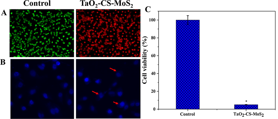
(A) Fluorescent microscopic images of MCF-7 cells staining with acridine orange (green) and propidium iodide (red) after irradiation with 808 nm laser light for 5 min and (B) DAPI (blue) staining and (C) the cell viability was assessed by MTT assay.
ROS generation was studied in the MCF-7 cells treating with bare MoS2 and TaO2-CS-MoS2 nanosheets at a higher concentration (100 μg/mL) for 24 h. After treated, the cells were stained with dichloro-dihydro-fluorescein diacetate (DCFH-DA) to examine ROS generation. (Fig. 8A–C). On the contrary, the fluorescence intensity improved as a function TaO2 concentration in MCF-7 cells indicating the rise of ROS production. ROS intensity was analyzed by fluorescence plate reader (Fig. 8D) in both normal and breast cancer cells after the treating with bare MoS2 and TaO2-CS-MoS2 nanosheets. Notably, the higher fluorescence intensity was observed for MCF-7 cells treated with TaO2-CS-MoS2 nanosheets when compared to bare MoS2 nanosheets. The increased levels of ROS mediated cancer cell death were clearly pointed out in TaO2-CS-MoS2 nanosheet treated MCF-7 breast cancer cells. It proves TaO2 decoration on to MoS2 nanosheet plays a key role in activating intrinsic apoptotic signaling pathways in MCF-7 cancer cells influences ROS generation during the PTT therapy (Murugan et al., 2016; Thapa et al., 2018; Gao et al., 2019).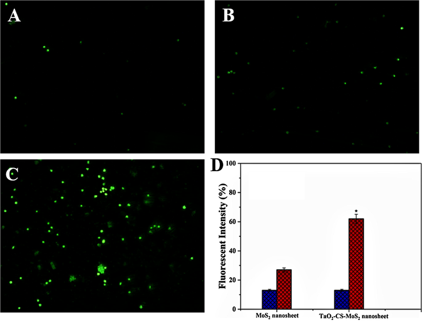
ROS production in DCFH-DA stained MCF-7 cells incubated with MoS2 and TaO2-CS-MoS2 nanosheets with or without laser treatment at 808 nm for 5 min (10× magnification).
4 Conclusion
In summary, we demonstrated the effects of ultra-small sized TaO2 decoration onto the surface of MoS2 nanosheet, in which, the increased concentration of TaO2-CS-MoS2 nanosheet increase the cell viability nature of MoS2 nanosheet in normal HBL-100 breast cells. It indicates TaO2 decoration on to the surface of MoS2 significantly improve the biocompatibility, photo conversion effects, photostability and remarkable photothermal therapeutic effects then bare MoS2 nanosheets. This work opens a new avenue to tune the physio-chemical properties of 2D nanomaterials by metal decoration.
Declaration of Competing Interest
The authors declared that there is no conflict of interest.
References
- Gold nanorods: their potential for photothermal therapeutics and drug delivery, tempered by the complexity of their biological interactions. Adv. Drug Deliv. Rev.. 2012;64(2):190-199.
- [Google Scholar]
- Near-infrared dye bound albumin with separated imaging and therapy wavelength channels for imaging-guided photothermal therapy. Biomaterials. 2014;35(28):8206-8214.
- [Google Scholar]
- Facile and green reduction of covalently PEGylated nanographene oxide via a ‘water-only’route for high-efficiency photothermal therapy. Nanoscale Res. Lett.. 2014;9(1):86.
- [Google Scholar]
- Two-dimensional non-carbonaceous materials-enabled efficient photothermal cancer therapy. Nano Today. 2016;11(3):292-308.
- [Google Scholar]
- Functional nanomaterials for phototherapies of cancer. Chem. Rev.. 2014;114(21):10869-10939.
- [Google Scholar]
- FeSe2-decorated Bi2Se3 nanosheets fabricated via cation exchange for chelator-free 64Cu-labeling and multimodal image-guided photothermal-radiation therapy. Adv. Funct. Mater.. 2016;26(13):2185-2197.
- [Google Scholar]
- Chitosan-based multifunctional nanomedicines and theranostics for targeted therapy of cancer. Med. Res. Rev.. 2018;38(6):2110-2136.
- [Google Scholar]
- Tantalum oxide nanoparticles for the imaging of articular cartilage using X-ray computed tomography: visualization of ex vivo/in vivo murine tibia and ex vivo human index finger cartilage. Angew. Chem.. 2014;53:8406-8410.
- [Google Scholar]
- Titania-coated 2D gold nanoplates as nanoagents for synergistic photothermal/sonodynamic therapy in the second near-infrared window. Nanoscale. 2019;11(5):2374-2384.
- [Google Scholar]
- Hybrid graphene/Au activatable theranostic agent for multimodalities imaging guided enhanced photothermal therapy. Biomaterials. 2016;79:36-45.
- [Google Scholar]
- NIR-responsive carbon dots for efficient photothermal cancer therapy at low power densities. Carbon. 2018;134:153-162.
- [Google Scholar]
- Recent advances in nanomaterials for enhanced photothermal therapy of tumors. Nanoscale. 2018;10(48):22657-22672.
- [Google Scholar]
- Encapsulating tantalum oxide into polypyrrole nanoparticles for X-ray CT/photoacoustic bimodal imaging-guided photothermal ablation of cancer. Biomaterials. 2014;35(22):5795-5804.
- [Google Scholar]
- A tantalum oxide-based core/shell nanoparticle for triple-modality image-guided chemo-thermal synergetic therapy of esophageal carcinoma. Cancer Lett.. 2017;397:61-71.
- [Google Scholar]
- Inorganic nanomaterials for chemo/photothermal therapy: a promising horizon on effective cancer treatment. Biophys. Rev. 2019:1-18.
- [Google Scholar]
- Ultrathin nanosheet assembled Sn0.91Co0.19S2 nanocages with exposed (100) facets for high-performance lithium-ion batteries. Small. 2018;14(5):1702184.
- [Google Scholar]
- Molybdenum disulfide/graphene oxide nanocomposites show favorable lung targeting and enhanced drug loading/tumor-killing efficacy with improved biocompatibility. NPG Asia Mater.. 2018;10(1):e458.
- [Google Scholar]
- Homogenous dispersion of MoS2 nanosheets in polyindole matrix at air-water interface assisted by Langmuir technique. Langmuir. 2017;33(47):13572-13580.
- [Google Scholar]
- Combinatorial nanocarrier based drug delivery approach for amalgamation of anti-tumor agents in breast cancer cells: An improved nanomedicine strategy. Sci. Rep.. 2016;6:34053.
- [Google Scholar]
- Cancer therapeutic Proficiency of dual-targeted mesoporous silica nanocomposite endorses combination drug delivery. ACS Omega. 2017;2(11):7959-7975.
- [Google Scholar]
- Nanoceria decorated flower-like molybdenum sulphide nanoflakes: an efficient nanozyme to tumour selective ROS generation and photo thermal therapy. Chem. Commun.. 2019;55:8017-8020.
- [Google Scholar]
- Two-dimensional cancer theranostic nanomaterials: synthesis, surface functionalization and applications in photothermal therapy. J. Control. Release. 2019;299:1-20.
- [Google Scholar]
- Chemo-photothermal therapy combination elicits anti-tumor immunity against advanced metastatic cancer. Nature Commun.. 2018;9(1):1074.
- [Google Scholar]
- Graphene as cancer theranostic tool: progress and future challenges. Theranostics. 2015;5(7):710.
- [Google Scholar]
- Novel concept of the smart NIR-light–controlled drug release of black phosphorus nanostructure for cancer therapy. Proc. Natl. Acad. Sci.. 2018;115(3):501-506.
- [Google Scholar]
- Peptide-conjugated nano-drug delivery system to improve synergistic molecular chemotherapy for colon carcinoma. ChemistrySelect. 2017;2(27):8524-8534.
- [Google Scholar]
- Nanocomposite materials based on chitosan and molybdenum disulfide. J. Mater. Sci.. 2012;47(15):5861-5866.
- [Google Scholar]
- Biodegradable black phosphorus-based nanospheres for in vivo photothermal cancer therapy. Nat. Commun.. 2016;7:12967.
- [Google Scholar]
- Facile synthesis of ultrathin nickel–cobalt phosphate 2D nanosheets with enhanced electrocatalytic activity for glucose oxidation. ACS Appl. Mater. Interfaces. 2018;10(3):2360-2367.
- [Google Scholar]
- Palladium nanoparticle-decorated 2-D graphene oxide for effective photodynamic and photothermal therapy of prostate solid tumors. Colloids Surf., B. 2018;169:429-437.
- [Google Scholar]
- Synthesis and structural characterization of MoS2 nanospheres and nanosheets using solvothermal method. J. Mater. Sci.. 2015;50(14):5024-5038.
- [Google Scholar]
- Multifunctional two-dimensional nanocomposites for photothermal-based combined cancer therapy. Nanoscale 2019
- [Google Scholar]
- Ultrathin two-dimensional cobalt–organic framework nanosheets for high-performance electrocatalytic oxygen evolution. J. Mater. Chem. A. 2018;6(44):22070-22076.
- [Google Scholar]
- MoS2 nanosheet/MoS2 flake homostructures for efficient electrocatalytic hydrogen evolution. Mater. Res. Express. 2019;6(8):085005.
- [Google Scholar]
- The synthesis of two-dimensional MoS 2 nanosheets with enhanced tribological properties as oil additives. RSC Adv.. 2018;8(17):9564-9573.
- [Google Scholar]
- Recent advances in functional polymer decorated two-dimensional transition-metal dichalcogenides nanomaterials for chemo-photothermal therapy. Chem. – Eur. J.. 2017;24(17):4215-4227.
- [Google Scholar]
- Controllable one-step growth of bilayer MoS2–WS2/WS2 heterostructures by chemical vapor deposition. Nanotechnology. 2018;29(45):455707.
- [Google Scholar]


