

Translate this page into:
Copper oxide nanoparticles from Rabdosia rubescens attenuates the complete Freund’s adjuvant (CFA) induced rheumatoid arthritis in rats via suppressing the inflammatory proteins COX-2/PGE2
⁎Corresponding author. bailb2005@sina.com (Longbin Bai)
-
Received: ,
Accepted: ,
This article was originally published by Elsevier and was migrated to Scientific Scholar after the change of Publisher.
Peer review under responsibility of King Saud University.
Abstract
Rheumatoid arthritis is a common auto-immune disease that affects nearly 1% of the total population worldwide and primarily occurs in the age group of 30–50. The copper oxide nanoparticles (CuONPs) were gained extensive research interests due to their immense biological benefits. This investigation was aimed to synthesize the CuONPs from R. rubescens leaves and examining its anti-arthritic potential against CFA-stimulated arthritis in rats. The formulated CuONPs from R. rubescens was characterized by UV–vis spectroscopy, TEM, XRD, and FT-IR. Arthritis was induced to animals by injecting 0.1 ml of CFA via an intra-planter route. Bodyweight and arthritis score was measured and tabulated and inflammatory markers i.e. IL-10, IL-6, TNF-α, IL-1β, Cox-2 and PGE2 in the serum was examined via ELISA kits. The status of TBARS, antioxidant enzymes, and liver marker enzymes were examined via standard procedures. The hind limb histology was examined microscopically by H&E staining. The UV–vis, XRD, and TEM studies were proved the existence of metallic RR-CuONPs. The RR-CuONPs treated arthritic animals displayed the enhanced bodyweight, reduced paw volume, and arthritic score. RR-CuONPs treatment also improved the antioxidant enzymes and diminished the pro-inflammatory markers. Histopathological investigation proved that the RR-CuONPs were suppressed the bone joints by preventing the inflammatory cell penetration and bone damage. The findings of this investigation were evidenced by the anti-inflammatory and anti-arthritic potentials of fabricated RR-CuONPs against CFA-stimulated arthritis in rats.
Keywords
Arthritis
Copper oxide nanoparticles
R. rubescens
Inflammation
Hind paw
Antioxidant

1 Introduction
The rheumatoid arthritis (RA) is a major class of autoimmune disease that affects more than millions of peoples worldwide and primarily occurs in the age group 30–50. RA is distinguished by the inflammation in joints, synovial breakdown and demolition of articular cartilage, and restricted motor activities which results in irreversible disabilities (Grygielska et al., 2018). The abundant immune regulated pathological anomalies were marked in the RA progression. Among these processes, the pannus formation, synovial hyperplasia, swelling at the joints, synovitis, and joint inflexibility was considered as a distinct sign of RA. The synoviocytes are the prime occupant cells of invasive pannus, which dynamically participate in the inflammatory mechanisms of RA (Okada et al., 2019; Guo et al., 2018).
Inflammation mainly occurs due to the triggering of neutrophils and macrophages, which results in the generation of inflammatory modulators like IL-1 and TNF-α (Dai et al., 2018). RA is an inflammatory rheumatic ailment, which results in the synovial inflammation and obliteration of joint bones. The preceding findings were highlighted that the occurrence of RA is about 0.5–1% worldwide (Pan et al., 2017). In the inflammatory mechanisms, stimulation of inflammatory cells was enhanced via the accretion of pro-inflammatory modulators that occurs in the synovial membrane that is accountable for the increased bone damage (Burmester et al., 2014; Furst and Emery, 2014). The reactive oxygen species (ROS), prostaglandin-E2 (PGE2) and inflammatory mediators like IL-6, IL-1β, and TNF-α are regarded as taking a crucial functions during the pathological progress of RA via severing the cellular pro-inflammatory mechanisms like over accretion of inflammatory modulators, cartilage and bone attrition, leukocyte release, triggering of synovial fibroblast to deliver the pro-inflammatory mediators and deteriorating enzymes. The anti-inflammatory mediators like IL-10 and IL-4 operating the antagonistic functions to decelerate the inflammatory reactions in RA via lessen the accretion of pro-inflammatory mediators like IL-6, IL-1β, and TNF-α. The unevenness between the pro and anti-inflammatory cytokines results in the instigation of auto-immune responses and chronic inflammation as in the RA (El-Gaphar et al., 2018).
It was stated that the complete Freund’s adjuvant (CFA) triggered RA in animal models were displaying the common outcomes with the human RA. The FCA stimulated arthritis exhibits sudden local inflammatory responses (Bihani et al., 2014). Presently, there are few medications were existed in the market to treat the RA like non-steroidal and steroidal drugs, immunosuppressive drugs, and disease-modifying anti-rheumatic drugs (DMARDs). The majority of these treatment approaches for RA may lessen the pain, and reduce the swelling of joints (McInnes and Schett, 2017). Though, a bit high dosage of these drugs often reported with the adverse outcomes to the kidney, stomach and other organs. Hence, the need for alternative herbal-based agents with more efficiency has emerged (Burmester and Pope, 2017; Patel et al., 2019). The curiosity in the metallic nanoparticle research was hurriedly emerged in this decade due to its unique features. The nanoparticles were gained much attention due to their immense applications in numerous fields particularly in medicines (Khashan et al., 2016). The metal oxide nanoparticles were recognized as a disinfecting agent in medical areas, antimicrobials, and cosmetic fillers. The metallic NPs have excellent semiconducting features that increasing their utilization in the microelectronic medical devices (Katwal et al., 2015).
The size of CuONPs determines the effectiveness of these particles and the morphological and structural aspects of the CuONPs may be enhanced via different approaches (Ruiz et al., 2015). To gain the preferred size and morphology of NPs, different fabrication approaches were engaged like sol-gel, electrochemical, sonochemical; chemical precipitation and plant extract mediated formulation (Safa et al., 2016; Kayani et al., 2015). Though in chemical synthesis, the byproducts generated during the bio-reduction of NPs may distress the formation of CuONPs and it was highly toxic to the environment. To exclude the hazards of the chemicals and its byproducts, the eco-friendly approaches were received the greater attention to formulating the metallic NPs. The plant extracts were worked as a reducing, capping and stabilizing agent during the formulation of metallic and metal oxide NPs. The NPs formulation by the green route approach is regarded as an easier, cheap and eco-friendly approach (Sharma et al., 2015).
Rabdosia rubescens is an extensively consumed herbal plant in the conventional Chinese medical system to heal numerous ailments like stomach ache, pharyngitis, and throat infection (Zhao et al., 2013). Preceding findings were highlighted that the plant R. rubescens has anti-inflammatory, antioxidant and antitumor actions (Xu et al., 2009; Bai et al., 2010; Schwarz et al., 2003). The R. rubescens has displayed immense pharmacological actions. The gold nanoparticles formulated form the leaves of R. rubescens were exhibited anticancer activity against human lung cancer (Zhang et al., 2019). The leaf extract of R. rubescens was displayed the excellent anticancer activity against the laryngeal and breast cancer (Kang et al., 2015; Li et al., 2013). The R. rubescens also displayed the anti-stroke activity and antithrombotic activity (Miao et al., 2017; Wang et al., 2015). The current investigation was intended to formulate the copper oxide nanoparticles from the aqueous extract of R. rubescens leaves and inspect its curative potential against CFA-stimulated arthritis in rats.
2 Materials and methods
2.1 Chemicals
Complete Freund’s adjuvant, copper oxide, and other chemicals and reagents were acquired from Sigma Aldrich, St. Louis, Missouri, USA. All the ELISA assay kits used in this work were bought in MyBioSource, San Diego, California, USA. Entire other chemicals and reagents were utilized in this investigation are of diagnostic grade attained from Himedia, Pennsylvania, USA.
2.2 Rabdosia rebescens collection and extract preparation
The fresh and matured leaves of Rabdosia rebescens were collected and dehydrated in a shady place. Then the sample was finely powdered and then utilized for the extraction. The 10 g of leaf powder was suspended in 100 ml of distilled water and heat macerated at 75 °C for 30 min. After that, the suspension was sifted with the help of the Whatman filter paper. Finally, the resulted plant extract was utilized for the formulation of RR-CuONPs.
2.3 Synthesis of copper oxide nanoparticles (RR-CuONPs) from R. rebescens
For synthesizing the RR-CuONPs, the 5 ml of aqueous extract of R.rebescens leaves were added to the 50 ml of copper sulfate (5 mM) solution. Then this reaction solution was placed on the magnetic stirrer at 60 °C for 1 h. The changing of the reaction mixture color to green was denoting the bio-reduction of CuO. Then the suspension was centrifuged at 6000 rpm for 5 min and the pellet was washed thrice with the distilled water. Finally, the gained pellet was dried in an oven at 85 °C for 24 h and then subjected to various characterization techniques.
2.4 Characterization of synthesized RR-CuONPs from R. rebescens
The bio-reduction of CuONPs in the reaction medium was primarily confirmed by the UV–vis spectroscopic technique. The UV–vis absorption range was recorded at the scanning range from 200 to 600 nm. The X-ray diffraction (XRD) pattern of formulated RR-CuONPs was examined via the powder diffractometer with Kα radiation (λ = 1.54059 nm) in the 2θ range from 20° to 80°. The average size of the formulated RR-CuONPs was studied through the transmission electron microscopy (TEM, FEI-Tecnai G220), operating at 200 kV, with a resolution point of 2.04 nm. The functional groups linked with the aqueous extract of R. rebescens leaf and the surface of formulated RR-CuONPs was inspected through the Fourier transform infrared study. Briefly, the fabricated RR-CuONPs were centrifuged at 12,000 rpm for 6mins and the residue was suspended in 10 ml of distilled water and the same was examined by the FT-IR.
2.5 Experimental animals
The male Wistar albino rats weighing about 210–230 g were acquired from the Institutional animal house and caged in polypropylene confines beneath the laboratory situations with 26 ± 1 °C temperature, 65% of air moisture, 12 h of light/dark sequence, and fed with rat pellet food and water ad libitum. All the animals were acclimatized to the lab situations for 7 days before the beginning of experiments. In this investigation, whole animal experiments were done by strictly applying the guidelines mentioned by the International animal ethics committee.
2.6 Experimental design
All rats were arbitrarily alienated into four groups with six animals in every. The group-I rats served as control. Group-II rats are disease control and injected with the 0.1 ml of CFA via the intra-planter route to stimulate the RA. Group-III animals were supplemented with the 10 mg/kg of formulated RR-CuONPs and group-IV rats were treated with the standard drug Diclofenac sodium (5 mg/kg). On the 1st day, all experimental animals except control were injected with the 0.1 ml of CFA via the intra-planter route. Later than the completion of the experimental period (28 days), all animals were anesthetized and killed via cervical displacement. Then the blood and tissue samples were gathered and subjected to the further biochemical and Histopathological investigations.
3 Measurement of arthritis
3.1 Examination of bodyweight and arthritis score
The bodyweight of all investigational animals wasmeasured with the sensitive weighing balance on every 5th day and alterations in the bodyweight were often compared to the initial bodyweight and calculated accordingly. The paw swelling of experimental animals was examined with the aid of the plethysmometer from day 1 to 28th day on the normal interval period (Kim et al., 2016). The arthritis score was investigated via the procedure mentioned by Zhang et al. (2009) (Syed Zameer Ahmed et al., 2019), with few alterations, where the paw wasinspected for the swelling, erythema, etc. by the 20-point scale with maximum arthritis score per animal was fixed at 20.
3.2 Immune organ index
Later than the animals sacrificed, the immune organs i.e. thymus and spleen were expunged suddenly and processed with PBS. Then the weight of the immune organs was weighed and then organ indexes were determined by applying the following formula; organ weight/bodyweight × 103.
3.3 Biochemical assays
The serum was prepared from the blood samples gathered from investigational animals and subjected to the biochemical inspections. The statuses of hematological parameters like red blood cells (RBCs), white blood cells (WBCs), and hemoglobin (Hb) were examined with the automated Hematological analyzer (XT-1800i-Sysmex, Japan) (Shabbir et al., 2016). The level of lipid peroxidation in the RA stimulated animals wasinvestigated via spectrophotometrically by measuring the thiobarbituric acid reactive substances (TBARS) by the procedure described by Niehius and Samuelson, (Niehius and Samuelson, 1968) (Niehius and Samuelson, 1968). The level of superoxide dismutase (SOD) was examined via the procedure of Kakkar et al. (1984). The catalase (CAT) enzyme was studied by thetechnique of Sinha (1972). The glutathione (GSH) status was inspected through the Ellman (1959) procedure.
3.4 Measurement of liver function marker enzymes
The status of glutamic oxaloacetic transaminase (SGOT), glutamic pyruvic transaminase (SGPT), and the alkaline phosphatase (ALP) were examined in the serum of investigational animals. The level of SGOT and SGPT was investigated via the technique defined by Reitman and Frankel (1957). The ALP status was examined through the procedure of Kind and King (1954).
3.5 Measurement of inflammatory markers level
The blood samples were gathered and centrifuged at 6000 rpm for 15 min. Then the resulting serum utilized for the examination of inflammatory mediators status. The levels of IL-10, IL-6, and TNF-α, IL-1β, Cox-2 and PGE2 in the serum of investigational animals were inspected with the aid of commercial ELISA test kits by the guidelines suggested by the manufacturer (MyBioSource, San Diego, California, USA).
3.6 Lymphocyte proliferation index
After the scarification of test animals, the spleen was excised suddenly beneath the sterile situations. Every spleen was pieced and squashed in a 5 ml of RPMI medium with 0.5% of FBS. The squashed spleen was filtered and the suspension was centrifuged at 2000 rpm for 10 min. Then the 5 ml of ACK buffer (Ammonium chloride (8.29 g), Potassium bicarbonate (1 g), and EDTA (32.2 g)) was mixed to eliminate the RBCs. Then the spleen cells were loaded at the 1 × 10⁶cells/ml and maintained for 72 h in a CO2 (5%) chamber. After that, the 25 µl of MTT solution was mixed to every well subsequently 100 μl of DMSO. Finally, the strength of the developed color was examined at 495 nm and the proliferation index was calculated.
3.7 Histopathological analysis of hind limb
After the animal sacrifices, the joints of hind paws were excised for the Histopathological investigations. The gathered tissues were processed with 10% formalin and then decalcified by using the 10% of formic acid solution. Later than the decalcification, the joint tissues were entrenched in the paraffin and sliced with the 5 µm thickness. Then the sliced tissue portions were stained with the hematoxylin and eosin. Finally, the histological alterations of hind limbs of investigational animals were examined beneath the light microscope.
3.8 Statistical analysis
The statistical examination was done in SPSS (version-17) statistical tool. Data are illustrated as mean ± SD. The ANOVA subsequently DMRT investigation was employed to examine the differentiation among the test groups. Data are regarded as statistically significant if p < 0.05.
4 Results
4.1 Characterization of synthesized RR-CuONPs from R. rubescens
4.1.1 UV–visible spectroscopic study of RR-CuONPs
The generation and constancy of the formulated RR-CuONPs were inspected via the UV–vis spectroscopic study. The changing of the color of the reaction solution to green color was denoting the formulation of RR-CuONPs that owing to the surface plasmon excitation. The maximum absorption of the formulated RR-CuONPs was noted at the 356 nm, which proved the formation of RR-CuONPs in the reaction solution (Fig. 1).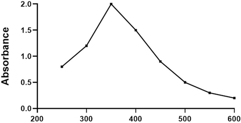
UV–vis spectroscopic analysis of synthesized RR-CuONPs. The highest absorption peak was noted at 356 nm that evidenced the formation of RR-CuONPs in the reaction solution.
4.1.2 TEM and XRD analysis of RR-CuONPs
The mean size and disparity of the formulated RR-CuONPs were examined via the TEM analysis and the microscopic photographs were depicted in Fig. 2. The microscopic images of fabricated RR-CuONPs have displayed the distinct dark spots with defined spherical shape that confirmed the existence of RR-CuONPs in the sample. The RR-CuONPs was possessed the size ranges from 30 to 90 nm (Fig. 2). The XRD examination was revealed the three precise peaks at 40.79, 60.01, and 70.81 with the angle values of 111, 200, and 220 respectively, which evidencing the occurrence of metallic copper oxide nanoparticles with crystalline nature. The maximum peak noted in the study was proved the purest RR-CuONPs (Fig. 2).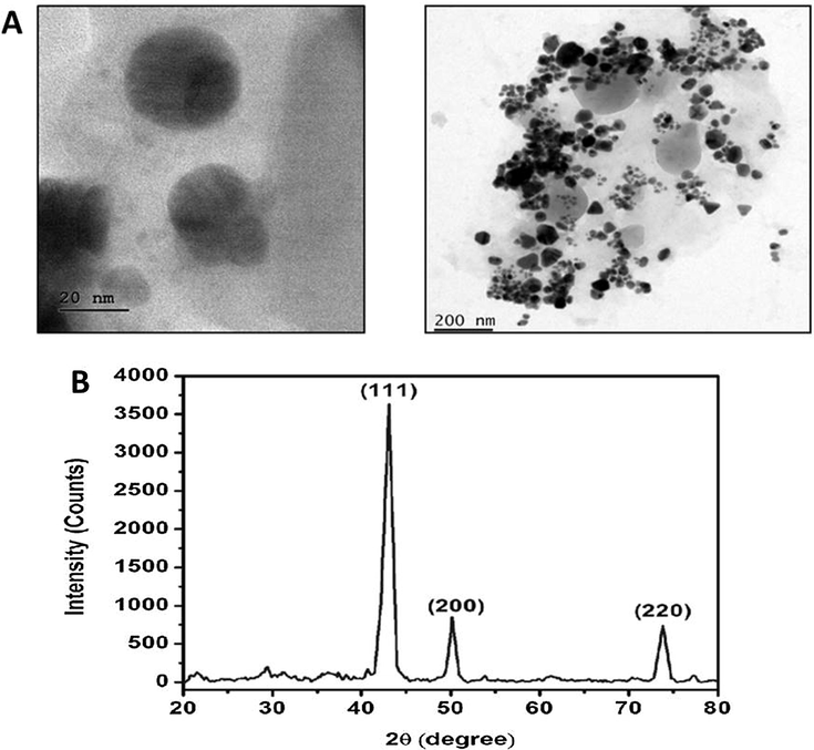
Transmission electron microscopic (TEM) and X-ray diffraction (XRD) analysis of synthesized RR-CuONPs. TEM images revealed that the formulated RR-CuONPs were possessed the mean size ranging from 30 to 90 nm with spherical shape. XRD investigation confirmed the occurrence of metallic copper oxide nanoparticles with crystalline nature.
4.1.3 FTIR spectroscopic study of RR-CuONPs
The frequencies of functional groups that specifically occur on the aqueous extract of R. rubescens leaves were analyzed through the FT-IR spectrophotometry. Fig. 3(A) revealed the FT-IR spectrum of R. rubescens leaf extract possessed the various absorption peaks. The peak at 3268.91 cm−1 is reveals the O—H (hydroxyl) stretching in the leaf extract. The 2920.75 and 2853.24 cm−1 peak indicates the presence of Cu—H stretching. The 1708.03 cm−1, 1621.17, 1406.17, and 1245.12 cm−1 peaks were reveals the C—H and C—O bonds. The 758.00 and 705.15 cm−1 was revealing the existence of Cu—O bonds (Fig. 3A).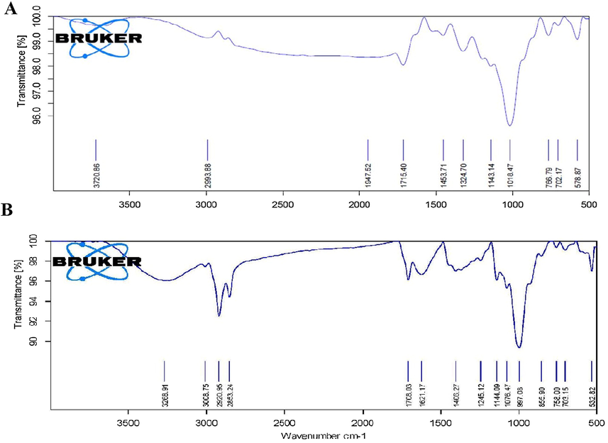
FTIR spectroscopic analysis of aqueous extract of R. rebescens leaves and synthesized RR-CuONPs. FT-IR spectral analysis showed the existence of different active biomolecules like alkynes, alkenes, aromatic rings, phenolic OH that bound on the leaf extract of R. rebescens (A) and the surface of synthesized RR-CuONPs (B).
The active biomolecules that bound on the surface of the fabricated RR-CuONPs were examined via the FTIR study. As mentioned in Fig. 3(B), the interaction of RR-CuONPs with numerous active biomolecules was displayed by different intensive peaks at 3720.86, 2993.88, 1947.52, 1715.40, 1453.71, 1324.70, 1143.14, 1018.47, 766.79, 702.17, and 578.87. The peak noted at the 578.87 was owing to the vibration of Cu—O bonding that occurs in the RR-CuONPs. The peaks at the 3720.86, 2993.88, 1947.52, 1715.40, 1453.71, and 1324.70 were indicating the existence of OH, C—H stretching, C—C triple bonding of alkynes, C⚌C stretch of alkenes, C⚌O, O⚌C stretching of aromatic rings, phenolic OH, and C—C stretching of numerous bio-molecules (Fig. 3B).
4.2 Effect of synthesized RR-CuONPs on hematological parameters of arthritic rats
The blood components i.e. RBCsand Hb were drastically diminished and the status of WBCs augmented in the CFA-triggered arthritic rats while comparing it to the control. Interestingly, the supplementation of fabricated RR-CuONPs (10 mg/kg) to the arthritic rats displayed the appreciable escalation in the status of RBCs, Hb and diminished the WBCs status (Table 1). The standard drug Diclofenac treatment also reversed the CFA-triggered hematological alterations in the arthritic rats, which is comparable to the activity of formulated RR-CuONPs. Values are depicted as mean ± SD of six rats in every test group. Note: ‘#’ denotes the p < 0.05 when compared to the control and ‘*’ denotes the p < 0.05 when compared to the arthritic group.
Groups
RBC(×106/µl)
WBC(×103/µl)
Hb(g/dl)
Group I
7.20 ± 0.28
12.38 ± 1.11
12.32 ± 1.10
Group II
3.11 ± 0.28#
19.70 ± 1.77#
4.99 ± 0.45#
Group III
5.74 ± 0.52*
14.96 ± 1.35*
9.46 ± 0.85*
Group IV
6.83 ± 0.61*
13.40 ± 1.21*
11.86 ± 1.07*
4.3 Effect of RR-CuONPs on the bodyweight and immune organ index in arthritic rats
As depicted in Fig. 4, the bodyweight of the arthritic animals notably decreased and the immune organ weights i.e. thymus and spleen was severely augmented than the control. Conversely, the 10 mg/kg of fabricated RR-CuONPs supplemented arthritic animals displayed the marked enhancement in the bodyweight and appreciably diminished the immune organ indexes while compare to arthritic animals. The RR-CuONPs and standard drug Diclofenac treatment to the arthritic animalsexhibited a similar kind of outcomes, which is comparable to the control animals.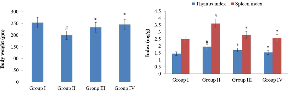
Effect of synthesized RR-CuONPs on the bodyweight and immune organ index in arthritic rats. The bodyweight was severely decreased and the immune organ index was enhanced in the CFA-challenged arthritic rats. Fig. 4 showed that the synthesized RR-CuONPs treated arthritic rats exhibited anappreciable escalation in the bodyweight and diminished immune organ index. Values are depicted as mean ± SD of triplicate values (n = 6). Note: ‘#’ denotes the p < 0.05 when compared to the control and ‘*’ denotes the p < 0.05 when compared to the arthritic group.
4.4 Effect of RR-CuONPs on paw volume and arthritis index score in arthritic rats
The paw volume was severely enlarged and arthritis score also drastically augmented in the CFA-stimulated arthritic rats than the control. The paw volume and arthritis score in the CFA-incited rats exhibited a constant increase, which is noted in 5, 10, 15, 20, and 25th days of investigation. Remarkably, the formulated RR-CuONPs (10 mg/kg) supplementation to the arthritic rats displayed the noticeable diminution in both paw volume and arthritis index scores (Fig. 5). The Diclofenac treatment also suppressed the paw volume and arthritis score in the CFA-challenged arthritic rats. The outcomes of RR-CuONPs and Diclofenac supplemented arthritic animals were similar to each other.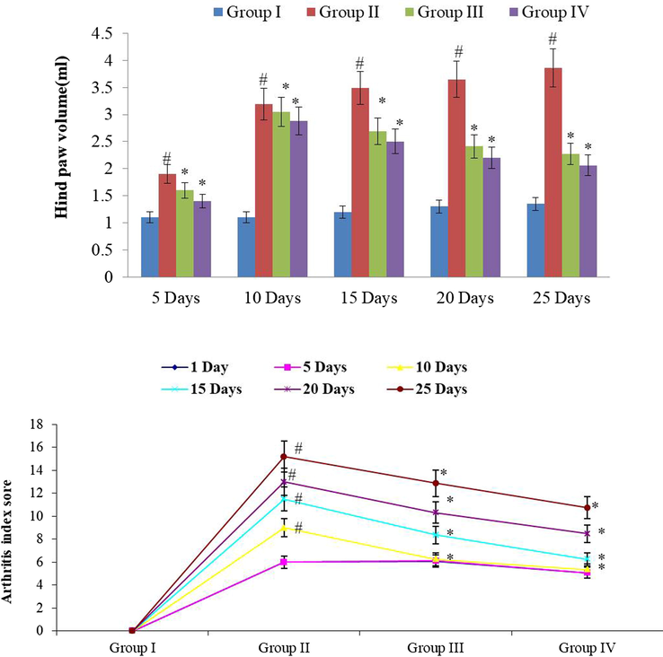
Effect of synthesized RR-CuONPs on paw volume and arthritis index score in arthritic rats. The CFA induced elevation in paw volume and arthritis index score was appreciably inhibited by the synthesized RR-CuONPs. The treatment with RR-CuONPs displayed the marked diminution in both paw volume and arthritis index score. Values are depicted as mean ± SD of triplicate values (n = 6). Note: ‘#’ denotes the p < 0.05 when compared to the control and ‘*’ denotes the p < 0.05 when compared to the arthritic group.
4.5 Effect of RR-CuONPs on liver function marker enzymes in arthritic rats
The CFA-stimulated arthritic rats displayed the severely augmented status of liver function marker enzymes i.e. glutamicoxalo acetic transaminase (SGOT), glutamic pyruvic transaminase (SGPT) and alkaline phosphatase (ALP) while comparing it to control rats. A marked diminution in the status of liver marker enzymes i.e. SGOT, SGPT, and ALP was noted in the serum of RR-CuONPs (10 mg/kg) supplemented arthritic rats (Fig. 6). This outcome was evidenced that the RR-CuONPs were suppressed the liver marker enzymes in the CFA-triggered arthritic rats. The Diclofenac treated arthritic animals also exhibited a similar kind of outcome that is comparable to the RR-CuONPs treatment and control rats.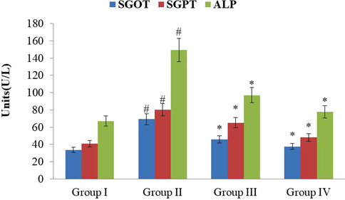
Effect of synthesized RR-CuONPs on liver function marker enzymes in arthritic rats. As mentioned in Fig. 6, the CFA-stimulated arthritic rats showedthe increased levels of liver function marker enzymes like SGOT, SGPT, and ALP than the control rats. But the RR-CuONPs supplemented arthritic animals demonstrated the appreciable diminution in the levels of SGOT, SGPT, and ALP. Values are depicted as mean ± SD of triplicate values (n = 6). Note: ‘#’ denotes the p < 0.05 when compared to the control and ‘*’ denotes the p < 0.05 when compared to the arthritic group.
4.6 Effect of RR-CuONPs on lymphocyte proliferation index in the arthritic rats
Fig. 7 revealed that the spleen cell proliferation index was drastically augmented in the CFA-triggered arthritic rats while comparing it to control. Interestingly, the supplementation of 10 mg/kg of fabricated RR-CuONPs to arthritis stimulated rats were displayed the noticeable diminution in the proliferation index of spleen cells. The Diclofenac treated arthritic rats also suppressed the spleen cell proliferation. The control, RR-CuONPs, and Diclofenac treated arthritic rats were displayed a similar kind of outcomes.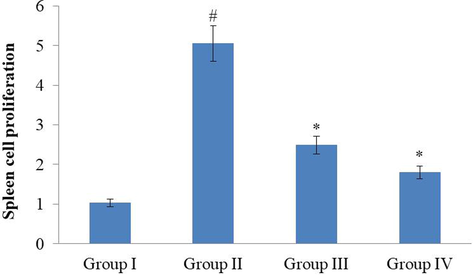
Effect of synthesized RR-CuONPs on lymphocyte proliferation index in the arthritic rats. Thespleen cell proliferation index was increased in the CFA-challenged arthritic rats than the control. On the other hand, the formulated RR-CuONPs treatment to the arthritic animals showed anappreciable reduction in the spleen cellproliferation index. Values are depicted as mean ± SD of triplicate values (n = 6). Note: ‘#’ denotes the p < 0.05 when compared to the control and ‘*’ denotes the p < 0.05 when compared to the arthritic group.
4.7 Effect of RR-CuONPs on inflammatory markers level in the arthritic rats
CFA-stimulated arthritis in rats resulted in the severe escalation of pro-inflammatory modulators like IL-6, IL-1β, TNF-α, Cox-2, PGE-2 and diminished the anti-inflammatory marker i.e. IL-10 in arthritic rats. Interestingly, the formulated RR-CuONPs supplementation (10 mg/kg) to the arthritic rats were displayed the marked reduction in the status of pro-inflammatory markers like TNF-α, IL-6, IL-1β, PGE-2, Cox-2 and also enhanced the anti-inflammatory mediator IL-10 in the arthritic rats (Fig. 8). Diclofenac treatment also diminished the pro-inflammatory modulator status in the serum of arthritic rats. A similar kind of outcome was noted between the RR-CuONPs and Diclofenac treated arthritic rats.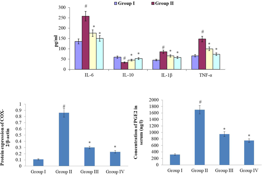
Effect of synthesized RR-CuONPs on inflammatory markers level in the arthritic rats. Fig. 8 showed the CFA challenged arthritis in rats showed the drastic enhancement in the levels of pro-inflammatory modulators like IL-6, IL-1β, TNF-α, Cox-2, PGE-2 and diminished the anti-inflammatory marker i.e. IL-10. The RR-CuONPs treated arthritic animals exhibited anoticeable reduction in the levels of the pro-inflammatory marker. Values are depicted as mean ± SD of triplicate values (n = 6). Note: ‘#’ denotes the p < 0.05 when compared to the control and ‘*’ denotes the p < 0.05 when compared to the arthritic group.
4.8 Effect of RR-CuONPs on antioxidant enzymes status in the arthritic rats
The antioxidant enzymes i.e. SOD, CAT, and GSH were severely diminished and the lipid peroxidation status was augmented in the serum of CFA-stimulated arthritic rats. As mentioned in Fig. 9, the CAT, SOD, and GSH were markedly escalated and the TBARS appreciably diminished in the serum of formulated RR-CuONPs (10 mg/kg) supplemented arthritic animals. The Diclofenac treated arthritic animals also enhanced the antioxidant enzymes. The outcomes of RR-CuONPs and Diclofenac supplemented arthritic animals were similar to each other.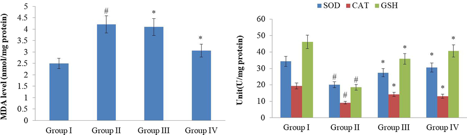
Effect of synthesized RR-CuONPs on antioxidant enzymes status in the arthritic rats. The lipid peroxidation level was increased and the antioxidant enzymes like SOD, CAT, and GSH were drastically reduced in the serum of CFA-induced arthritic rats. Interestingly, the RR-CuONPs treatment to the arthritic animals showed a noticeable improvement in the levels of the antioxidant enzymes. Values are depicted as mean ± SD of triplicate values (n = 6). Note: ‘#’ denotes the p < 0.05 when compared to the control and ‘*’ denotes the p < 0.05 when compared to the arthritic group.
4.9 Effect of RR-CuONPs on the histological analysis of hind limb of arthritic animals
The histological examination of hind limbs of CFA-triggered arthritic rats were displayed the distinct signs of synovial hyperplasia, penetration of inflammatory cells, pannus development and the obliterations of joints and cartilage, which is quite contrasted to the control. Conversely, the supplementation of formulated RR-CuONPs to the arthritic animals were displayed the astounding protection to the cartilages, synovial and bone joints. The RR-CuONPs supplemented arthritic animals exhibited suppressed inflammatory cell penetration, hyperplasia, along with very slight tissue damage while comparing it to arthritic animals (Fig. 10). Diclofenac treatment also provided noticeable protection to the joint bones and cartilages. A similar kind of outcome was noticed between the RR-CuONPs and Diclofenac treated arthritic rats.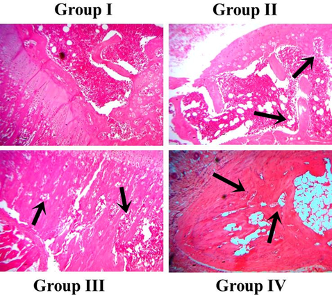
Effect of synthesized RR-CuONPs on histological analysis of hind limb of arthritic animals. The control animals showed normal cellular arrangements and tissue architecture. Conversely, the distinct signs of synovial hyperplasia, inflammatory cell infiltration, pannus formation, and joints and cartilage damages were noted in the CFA-challenged arthritic animals. The treatment with the RR-CuONPs showed an amazing protective activity and it prevented the cartilages, synovial and bone joint damages.
5 Discussion
RA is the foremost type of auto-immune disease and it affects nearly 1% of the total population worldwide (Shabbir et al., 2016). RA often distinguished by the swelling, pain, irregularity and stiffness of joints, and serious functional complications. The copper oxide (CuO) has gained broad attention among the researchers due totheir immense biological benefits. The numerous pharmacological actions of CuO led their wide utilization in a biomedical area (Ahamed et al., 2014; Nations et al., 2015). The findings were evidenced that the CuONPs synthesized from the R. rubescens leaves (RR-CuONPs) were displayed the potential anti-arthritic actions against the CFA-provoked RA in rats.
The numerous preceding findings were evidenced that the CFA-stimulated RA in animal replica was imitating the immunological and pathological processes of human RA along with reduced bodyweight due to severe inflammation and disease harshness (Kong et al., 2013). The retrieval of bodyweight is a general incident that occurs subsequently to the supplementation with the herbals and/or its derivative compounds. In our investigation, the constant fall down in the bodyweight was noted in CFA-triggered arthritic animals than the control. Conversely, the supplementation of 10 mg/kg of formulated RR-CuONPs was appreciably retrieved the bodyweight of arthritic animals. The CFA-challenge to the investigational animals is predicted that to activate a severe inflammation, infiltration of synovial layer, swelling, pain, and deformity of the joints. These outcomes often reported with the human RA with paw edema, joint pain, reduced body weight, and cartilage destruction (Zheng et al., 2014). The intra-planter injection of CFA in the hind paw of investigational animals displays the swelling, pain, edema of joints that denotes the occurrence of harsh inflammation. The inflammatory mechanisms instigating as primary lesions in 4–6 days of CFA challenge, subsequently secondary wounds in later than the 12th day of CFA injection. The joint swelling is the prime signs that display the RA progression in the animals. The enhanced paw edema was often reported due to the rise in the synovial fluids, vascular permeation, and cellular penetration in the inflammatory regions (Alamgeer et al., 2017). In this study, we found that the occurrence of inflammation was continued to 28 days that owing to the persisted edema and cellular invasions. Interestingly, the formulated RR-CuONPs (10 mg/kg) supplemented investigational animals displayed the stunning diminution in the paw volume while comparing it to the arthritic animals. Similarly, the RR-CuONPs treatment also appreciably diminished the arthritic scores in the FCA-challenged animals and guarded the morphological alterations of RA via preventing the inflammatory mechanisms and inhibiting the progression of secondary lesions that owing to the anti-inflammatory and anti-arthritic benefits of fabricated RR-CuONPs.
The pro-inflammatory cytokines and cells from the synovium, lymphocytes, and monocytes are believed to be an imperative modulator of the instigation and prolongation of RA. It was regarded that the IL-6, TNF-α, and IL-1 are the vital pro-inflammatory arbitrators and take a critical function in the pathological progress of RA (Cheng et al., 2015). In this investigation, we noted the drastically escalated statuses of pro-inflammatory modulators in the FCA-triggered arthritic animals. The augmented status of pro-inflammatory mediators has denoting their influences in the pathological mechanisms of arthritis progression (Kim and Moudgil, 2017). Further, the deformation of bone and cartilage, severe inflammatory conditions and oxidative injuries were regarded to exaggerate the ailing severity that ensuing in the stimulation of numerous pro-inflammatory modulators like IL-6, TNF-α, Cox-2, IL-6 and iNOS in both joint tissues as well as serum. The escalated status of IL-1β and TNF-α is participating in the stimulation of synovial fibroblasts, chondrocytes, and osteoclasts to accrete the tissue destructing MMPs. The enhanced generation of pro-inflammatory modulators that further triggers the oxidative stress marker expressions, subsequently enhancing the inflammation and oxidative injuries (Xie et al., 2015; Chimenti et al., 2015).
The excess accretion of TNF-α was further augmenting the expression of IL-6, IL-1β, PGE-2, and intensifies the synovial hyperplasia, accumulate the destructing enzymes, activates stromelysin, collagenase, and osteoclast deformities, which assist to the arthritic corrosions (Obiri et al., 2014). The IL-1β enhances the stimulation osteoclast and accretion of degrading enzymes, subsequently bone damages. Similarly, the IL-6 provokes the bone resorption, immunological mechanisms, and accretion of auto-antibodies. The PGE-2 was accreted via the metabolic process of arachidonic acid through the Cox-2 cascade and it was regarded as a foremost modulator of rheumatoid synovium. The enhanced PGE-2 status was linked with the pain, redness, and swelling of joints, vasodilation, cartilage corrosions, and fluid extravasation (Zhao et al., 2016). In that way, the therapeutic agents, which intends to the anti-arthritic benefits must potentially hamper the expressions of these pro-inflammatory modulators to counteract the arthritic severity. The findings of this investigation were evidenced that the RR-CuONPs supplemented arthritic animals were displayed the marked diminution in the status of anti-inflammatory modulators, which is drastically enhanced by the CFA-challenge.
The CFA-incited arthritic models were also employed to examine the biochemical and hematological parameters. The CFA-challenge to the investigational animals often exhibits the marked diminution in the RBCs and Hb statuses, which denote the anemic condition, which aroused from the reduced erythropoietin, destructions of erythrocytes, declined bone marrow functions, and reduced plasma iron content. These incidents are often lead by inflammatory cytokines like IL-1. Besides, the escalation in the WBCs in the CFA-challenged arthritic animals are denoting the activation of immune systems against the invading pathogens that liberating the TNF-α, and IL-1 (Kim et al., 2016). In general, the hematological alterations in the pathological progress of RA are a marked reduction in the RBCs and Hb along with the drastic escalation of the WBCs that denote the severity of RA (Liu and Wang, 2014). In this exploration, we also noted the drastic diminution in the Hb and RBCs status on CFA-triggered arthritic animals. We found that the supplementation of 10 mg/kg of formulated the RR-CuONPs to the CFA-incited arthritic animals revealed the appreciable escalation in the Hb and RBCs along with diminution in the WBCs status.
The enhanced status of lipid peroxidation, ROS, Cox-2, and PGE-2 are the prime mediators to instigate the oxidative injury to the cells. The cellular GSH cycle was regarded as a first-line barrier to oxidative stress. The escalated ROS, Cox-2, and lipid peroxidation may cause the inequity of enzymatic and non-enzymatic antioxidant mechanisms via trouncing the antioxidant protection systems. This will turn into endangering the intracellular anti-oxidant enzymes normally SOD, which detoxify the superoxides and peroxides, and CAT that detoxifies the hydrogen peroxides (Comar et al., 2013; Mo et al., 2013). Many preceding findings were highlighted that the excess accretion of reactive substances and free radicals are influenced in numerous pathological processes of RA. The augmented ROS and free radicals during the RA progression result in the reduced statuses of antioxidant enzymes i.e. SOD, GSH, and SOD that further leads to oxidative stress and cell necrosis (Shi et al., 2015). The findings of this investigation were proved that the fabricated RR-CuONPs (10 mg/kg) supplemented arthritic animals were displayed an appreciable escalation in the status of antioxidant enzymes i.e. CAT, SOD, and GSH and declined the TBARS status, which proved the antioxidant potential of formulated RR-CuONPs (Fig. 9).
The noticeable escalation of the SGOT, SGPT, and ALP statuses in the serum was often displayed in the RA stimulated investigational animal models. The severe augmentation in the serum biochemical markers during the progression of RA in investigational animals was linked with severe bone injuries (Xu et al., 2017). Interestingly, the marked diminution in the SGOT, SGPT, and ALP was noted in the serum of fabricated RR-CuONPs supplemented arthritic rats. On the whole, the findings of this exploration were proved the anti-inflammatory and anti-arthritic potentials of formulated RR-CuONPs against the RA.
6 Conclusion
The synthesized CuONPs from the R. rubescens has displayed an appreciable anti-inflammatory and anti-arthritic activity against the CFA-stimulated RA in rats. The supplementation of RR-CuONPs to the arthritic animals were exhibited the marked protection against the CFA-challenge through its enhanced anti-inflammatory and antioxidant actions. The RR-CuONPs treatment escalated the antioxidant enzymes status, diminished the pro-inflammatory modulators level, and prevented the histological alterations and bone damages in the arthritic rats. Thereby, it was concluded that the RR-CuONPs can be utilized as a promising therapeutic agent to treat the RA. Though, additional investigations were needed in the future to recognize the precise action of RR-CuONPs against the CFA-provoked RA in investigational animals.
Acknowledgments
This project was supported by Researchers Supporting Project number (RSP-2019/5) King Saud University, Riyadh, Saudi Arabia.
Declaration of Competing Interest
The authors declare that they have no known competing financial interests or personal relationships that could have appeared to influence the work reported in this paper.
References
- Synthesis, characterization, and antimicrobial activity of copper oxide nanoparticles. J. Nanomater.. 2014;5:519-524.
- [Google Scholar]
- Anti-arthritic activity of aqueous-methanolic extract and various fractions of BerberisorthobotrysBien ex Aitch. BMC Complement. Altern. Med.. 2017;17:371.
- [Google Scholar]
- Flavonoids from Rabdosia rubescens exert anti-inflammatory and growth inhibitory effect against human leukemia HL-60 cells. Food Chem.. 2010;122:831-835.
- [Google Scholar]
- Anti-arthritic activity of methanol extract of Cyathoclinepurpurea (whole plant) in Freund’s complete adjuvant-induced arthritis in rats. Biomed. Aging Pathol.. 2014;4:197-206.
- [Google Scholar]
- Emerging cell and cytokine targets in rheumatoid arthritis. Nat. Rev. Rheumatol.. 2014;10:77-88.
- [Google Scholar]
- Novel treatment strategies in rheumatoid arthritis. The Lancet. 2017;389:2338-2348.
- [Google Scholar]
- Anti-inflammatory and anti-arthritic effects of GugeFengtong Formula: in vitro and in vivo studies. Chin J. Nat. Med.. 2015;13:842-853.
- [Google Scholar]
- The interplay between inflammation and metabolism in rheumatoid arthritis. Cell Death Dis.. 2015;6:e1887
- [Google Scholar]
- Oxidative state of the liver of rats with adjuvant-induced arthritis. Free Radic. Biol. Med.. 2013;58:144-153.
- [Google Scholar]
- Synergistic effect of glucosamine and vitamin E against experimental rheumatoid arthritis in neonatal rats. Biomed. Pharmacother.. 2018;105:835-840.
- [Google Scholar]
- Effect of losartan in complete freund’s adjuvant –induced arthritis in rats. Iranian J. Pharmaceut. Res.. 2018;17:1420-1430.
- [Google Scholar]
- Rheumatoid arthritis pathophysiology: update on emerging cytokine and cytokine-associated cell targets. Rheumatol.. 2014;53:1560-1569.
- [Google Scholar]
- The impact of nutrition and generally available products such as nicotine and alcohol on rheumatoid arthritis – Review of the literature. Reumatolog.. 2018;56:121-127.
- [Google Scholar]
- Rheumatoid arthritis: pathological mechanisms and modern pharmacologic therapies. Bone Res.. 2018;6:15.
- [Google Scholar]
- A modified spectroscopic assay of superoxide dismustase. Indian J. Biochem. Biophys.. 1984;21:130-132.
- [Google Scholar]
- Inhibition of caspase-9 by oridonin, a diterpenoid isolated from Rabdosia rubescens, augments apoptosis in human laryngeal cancer cells. Int. J. Oncol.. 2015;47(6):2045-2056.
- [Google Scholar]
- Electrochemical synthesized copper oxide nanoparticles for enhanced photocatalytic and antimicrobial activity. J. Ind. Eng. Chem.. 2015;31:173-184.
- [Google Scholar]
- Characterization of copper oxide nanoparticles fabricated by the sol-gel method. J. Electron. Mater.. 2015;44:3704-3709.
- [Google Scholar]
- Synthesis and antibacterial activity of CuO nanoparticles suspension induced by laser ablation in liquid. Arab. J. Sci. Eng.. 2016;41:301-310.
- [Google Scholar]
- Immunomodulation of autoimmune arthritis by pro-inflammatory cytokines. Cytokine. 2017;98:87-96.
- [Google Scholar]
- Evaluation of anti-inflammatory potential of the new Ganghwaljetongyeum on adjuvant-induced inflammatory arthritis in rats. Evid. Based Complement Alternat. Med.. 2016;1230294
- [Google Scholar]
- Evaluation of anti-inflammatory potential of the new Ganghwaljetongyeum on adjuvant-induced inflammatory arthritis in rats. Evid. Based Complement Alternat. Med.. 2016;1230294
- [Google Scholar]
- Estimation of plasma phosphatase by determination of hydrolysed phenol with aminoantipyrine. J. Clin. Path.. 1954;7:322-326.
- [Google Scholar]
- The suppressive effects of Saposhnikoviadivaricata(Fangfeng) chromone extract on rheumatoid arthritis via inhibition of nuclear factor-κB and mitogen activated proteinkinases activation on collagen-induced arthritis model. J. Ethnopharmacol.. 2013;148(3):842-850.
- [Google Scholar]
- Reversal effects of Rabdosia rubescens extract on multidrug resistance of MCF-7/Adr cells in vitro. Pharm. Biol.. 2013;51(9):1196-1203.
- [Google Scholar]
- Therapeutic effect of Captopril on rheumatoid arthritis in rats. Asian Pac. J. Trop. Med.. 2014;7(12):996-999.
- [Google Scholar]
- Pathogenetic insights from the treatment of rheumatoid arthritis. The Lancet. 2017;389:2328-2337.
- [Google Scholar]
- Effects of the Rabdosia rubescens total flavonoids on focal cerebral ischemia reperfusion model in rats. Saudi Pharmaceut. J.. 2017;25(4):607-614.
- [Google Scholar]
- Topical antiinflammatory and analgesic activities of standardized pomegranate rind extract in comparison with its marker compound ellagic acid in vivo. J. Ethnopharmacol.. 2013;148:901-908.
- [Google Scholar]
- Subchronic and chronic developmental effects of copper oxide (CuO) nanoparticles on Xenopuslaevis. Chemosphere. 2015;2015(135):166-174.
- [Google Scholar]
- Formation of malondialdehyde from phospholipid arachidonate during microsomal lipid per-oxidation. Eur. J. Biochem.. 1968;6:126-130.
- [Google Scholar]
- Xylopiaaethiopica(Annonaceae) fruit extract suppresses Freund's adjuvant-induced arthritis in Sprague-Dawley rats. J. Ethnopharmacol.. 2014;152:522-531.
- [Google Scholar]
- Genetics of rheumatoid arthritis: 2018 status. Ann Rheumatic Dis.. 2019;78:446-453.
- [Google Scholar]
- Anti-rheumatoid arthritis effects of traditional Chinese herb couple in adjuvant-induced arthritis in rats. J. Ethnopharmacol.. 2017;205:1-7.
- [Google Scholar]
- Cost-effectiveness of early treatment with originator biologics or their biosimilars after Methotrexate failure in patients with established rheumatoid arthritis. Adv. Ther.. 2019;36:2086-2095.
- [Google Scholar]
- A colorimetric method for the determination of serum glutamic oxaloacetic and glutamic pyruvic transaminase. Am. J. Clin. Pathol.. 1957;28(1):56-63.
- [Google Scholar]
- Short-term effects on antioxidant enzymes and long-term genotoxic and carcinogenic potential of CuO nanoparticles compared to bulk CuO and ionic copper in mussels Mytilusgalloprovincialis. Mar. Environ. Res.. 2015;111:107-120.
- [Google Scholar]
- Investigating on photocatalytic performance of CuO micro and nanostructures prepared by different precursors. Desalin. Water Treatment.. 2016;57:6723-6731.
- [Google Scholar]
- Pancreatic cancer in vitro toxicity mediated by Chinese herbs SPES and PCSPES: implications for monotherapy and combination treatment. Cancer Lett.. 2003;189:59-68.
- [Google Scholar]
- Discovery of new benzothiazine derivative as modulator of pro- and anti-inflammatory cytokines in rheumatoid arthritis. Inflammation. 2016;39(6):1918-1929.
- [Google Scholar]
- Green synthesis of CuO nanoparticles with leaf extract of Calotropisgigantea and its dyesensitized solar cells applications. J. Alloys Compds.. 2015;632:321-325.
- [Google Scholar]
- The protective effects of chronic intermittent hypobaric hypoxia pretreatment against collagen-induced arthritis in rats. J. Inflamm.. 2015;12:23.
- [Google Scholar]
- Therapeutic effect of Parmotrema tinctorum against complete Freund’s adjuvant-induced arthritis in rats and identification of novel Isophthalic ester derivative. Biomed. Pharmacother.. 2019;112:108646
- [Google Scholar]
- Aqueous extract of Rabdosia rubescens leaves: forming nanoparticles, targeting P-selectin, and inhibiting thrombosis. Int. J. Nanomed.. 2015;10:6905-6918.
- [Google Scholar]
- The effects of interleukin-1beta in modulating osteoclast-conditioned medium's influence on gelatinases in chondrocytes through mitogen-activated protein kinases. Int. J. Oral Sci.. 2015;7:220-231.
- [Google Scholar]
- Multiple-modulation effects of oridonin on the production of proinflammatory cytokines and neurotrophic factors in LPS-activated microglia. IntImmunopharmacol.. 2009;9:360-365.
- [Google Scholar]
- Antiarthritic activity of qi-wu rheumatism granule (a Chinese herbal compound) on complete freund’s adjuvant-induced arthritis in rats. Evid. Based Complement. Altern. Med. 2017:1-13.
- [Google Scholar]
- Rabdosia rubescens Linn: green synthesis of gold nanoparticles and their anticancer effects against human lung cancer cells A549. Artificial Cell Nanomed. Biotech.. 2019;47(1):2171-2178.
- [Google Scholar]
- Advanced phytochemical analysis of herbal tea in China. J. Chromatogr. A. 2013;1313:2.
- [Google Scholar]
- Anti-inflammatory effects of pcoumaric acid in LPS-stimulated RAW264.7 cells: involvement of NF-κB and MAPKs Pathways. Med. Chem.. 2016;6:327-330.
- [Google Scholar]
- Therapeutic effects of standardized Vitex negundo seeds extract on complete Freund's adjuvant induced arthritis in rats. Phytomed.. 2014;21:838-846.
- [Google Scholar]


