

Translate this page into:
Green decorated of gold nanoparticles over kaolin mediated by plant extract and investigation of its anti-bile duct cancer properties in the in vitro condition
⁎Corresponding authors. wbhuang348912@126.com (Wenbin Huang)
-
Received: ,
Accepted: ,
This article was originally published by Elsevier and was migrated to Scientific Scholar after the change of Publisher.
Peer review under responsibility of King Saud University.
Abstract
In the current research, kaolin-decorated Au nanoparticles were produced by a green medication method by using Strawberry root’s extract as a natural coating and reducing component for capping and stabilizing Au NPs over the kaolin. The Au NPs/kaolin material was thoroughly characterized using XRD, FE-SEM, EDX-elemental mapping, TEM, and ICP. Characterization data confirmed the well-supporting of Au NPs over the kaolin. TEM images displayed the spherical shaped gold nanoparticles averaging 20–40 nm in size over the kaolin surface. The cytotoxicity and anti-bile duct cancer properties of Au NPs/kaolin were investigated through the utilization of MTT assay on three bile duct cancer cell lines, namely OCUCHLM1, EGI1, and GBCSD. Au nanoparticles (Au NPs) combined with kaolin exhibited dose-dependent anti-bile duct cancer properties and significantly reduced cell viability in OCUCHLM1, EGI1, and GBCSD cell lines. Importantly, these effects were observed without causing any cytotoxicity in the normal cell line (HUVECs). The OCUCHLM1 cell line exhibited the most significant anti-bile duct cancer effects when treated with Au NPs/kaolin, surpassing the results observed in other cell lines. Upon the successful validation of the aforementioned findings in the clinical trial investigations, Au NPs/kaolin could potentially serve as an effective chemotherapeutic agent in combating bile duct cancer.
Keywords
Gold nanoparticles
Kaolin
Strawberry
Bile duct cancer
MTT assay

1 Introduction
In recent years, precision-engineered nanomaterials have emerged as a prominent player in the field of treatment of several cancers, offering immense potential for effective anticancer treatments. Enhancing the distribution of medications to precise locations within the body at specific intervals and optimal levels is achievable (Shait Mohammed et al., 2021; Mitchell et al., 2021). Various reports have indicated diverse therapeutic advantages of NP-based therapy, which can function as antiproliferative agents, anti-permeability agents, antitumor agents, antiangiogenic agents, and molecular probes (Tabrez et al., 2022; Saeed et al., 2019; Gerosa et al., 2020).
Gold nanoparticles (AuNPs) have garnered significant scientific interest in recent years due to their ease of synthesis, making them a prominent nanotherapeutic option. AuNPs are being utilized more and more in the field of medicine due to their remarkable biocompatibility, which stems from their chemical and physical stability as well as their ease of functionalization with biologically active molecules (Elahi et al., 2018). AuNPs can directly bind and engage with a diverse array of molecules present on their surface, such as fluorescent dyes, nucleic acids (RNA or DNA), enzymes, antibodies, medications, and proteins (Hu et al., 2020). AuNPs are recognized for their effective use in nanomedicine, displaying strong anti-cancer properties attributed to their distinct physicochemical and bio-reactive characteristics (Albarwary et al., 2021). The remarkable clinical applications of AuNPs are attributed to their unique characteristics, including radioactivity, localized surface plasmon resonance, and high X-ray absorption coefficient (Bai et al., 2020). AuNPs can function as delivery systems, therapeutics, and molecular sensors for medications and imaging agents (Siddique and Chow, 2020). Functionalized AuNPs have shown considerable progress due to their ability to be effectively taken up by cells. Nevertheless, factors like grafting density, molecular weight, and the type of ligand can also impact cellular uptake (Lu et al., 2020). AuNPs-induced cytotoxicity is affected by various factors such as penetrating ability, tissue absorption, tissue distribution, cell type, and size, as well as other influences (Zhang et al., 2011). As a result of the influence of surface energy transfer, AuNPs possess strong quenching abilities toward fluorescence donors, rendering them an excellent foundation for fluorescent nanoprobes (Siddique and Chow, 2020). Despite the lack of pharmaceutical approval for AuNPs, numerous studies have indicated their potential use in cancer treatment and targeting tumors (Priya and Iyer, 2020; Ali et al., 2017; Adnan et al., 2022).
In recent years, clay minerals as natural supports have garnered significant attention in research due to their low cost, abundance, high chemical and thermal stability, their environmental and economic advantages in comparison with other carbonaceous support materials (Fatimah et al., 2022; Zou et al., 2022). Kaolin is one of the most common clay composite, has been investigated and because of the mentioned properties and presents a nanolayered structure, interesting in being used as efficient support (Wang et al., 2011; Kanakaraju et al., 2022).
Modification of kaolinite can be easily happen due to the layered structure to produce novel nanocomposites (Dou et al., 2021; Unuabonah et al., 2017; Yiga et al., 2022; David et al., 2020). So, in this regard, we modified kaolin with Strawberry root’s extract to make a natural capping and reducing support for immobilizing gold nanoparticles (Au NPs/kaolin). Also, the anti-bile duct cancer efficacies of Au NPs/kaolin nanocomposite were determined in the in vitro condition.
2 Experimental
2.1 Preparation of Strawberry root extract
The Strawberry root extract was obtained by heating 1.0 g of the powdered roots into 20 mL of pure water for 0.5 h at 80 °C. The plant suspension was then filtered over Whatman-1 paper, the pale brown extract was used for the next run.
2.2 Bio-fabrication of Au NPs/kaolin
0.5 g raw kaolin clay powder is dispersed in 200 mL deionized water and stirred for about 25 min. Next, 10 mL of the freshly prepared Strawberry root extract was put into the mixture and stirred for about 60 min with a magnetic stirrer. Afterward, 10 mL of HAuCl4 (0.05 M) solution was added into the suspension and stirred for 2 h at 100 °C. Finally, the solid product (Au NPs/Kaolin) was isolated by filtration and washed with H2O and dried in oven. Amount of Au was 0.074 mmol/g in the produced composite that determined by ICP-OES.
2.3 Anticancer efficacies of Au NPs/kaolin
The OCUCHLM1, EGI1, and GBCSD bile duct cancer cells were cultured and sustained in a 75 cm2 flask by DMEM medium. The cultures were placed at 37 °C, containing 5 % CO2, with high humidity. Culture medium was added every other day until an appropriate confluency of approximately 90 %. Gibco Company was the source of all cell culture reagents and medium. The cell studies were conducted repeatedly on multiple occasions. The effect of Au NPs/Kaolin nanocomposite on OCUCHLM1, EGI1, and GBCSD bile duct cancer cells viability was measured by MTT assay. The OCUCHLM1, EGI1, and GBCSD bile duct cancer cells were promptly placed into a 96-well plate with a volume of 1 × 10⁴ cells/well. The cells were let to adhere to the wells bottom by incubating them for 24 h. Following this, varying doses of Au NPs/Kaolin nanocomposite were introduced to the cells and incubated for another 24 h. Subsequently, the culture medium within the wells was expelled, and the wells underwent three rounds of washing with PBS. After adding a new medium to all wells, 001 mL of MTT dye (0.5 mg/mL) was introduced into each well. The cells were subsequently placed in an incubator at 37 °C for 4 h. Following the incubation, 0.1 ml of DMSO was introduced into each well to facilitate the formazan dye dissolution produced post-removal of the medium. The well absorbance was determined using a microplate reader at 570 nm (Alafaleq et al., 2022). The viable cells percentage was determined by utilizing the formula provided in the equation.
2.4 Statistical analysis
The experiment was conducted multiple times, with a minimum of three repetitions for each substance, dilution, and adjacent time. Each repetition was performed at least in triplicate. For the data analysis, one-way ANOVA was employed with a post-test using Dennett’s method. A P≤0.01 significance level was deemed notable in this research.
3 Results and discussion
The Au NPs/Kaolin was synthesized by a green and eco-friendly way by using Strawberry root extract as an efficient coating and reducing agent. The morphology and microstructure of the produced sample were studied by using TEM and FE-SEM images. TEM images provide evidence of the fluffy morphology of the desired material (Fig. 1 and Fig. 2). The efficient loading of Au NPs on the surface of modified kaolin with good dispersion is evident from the TEM images. From the TEM images, the Au NPs were spherical with particle sizes ranging from 20 to 40 nm.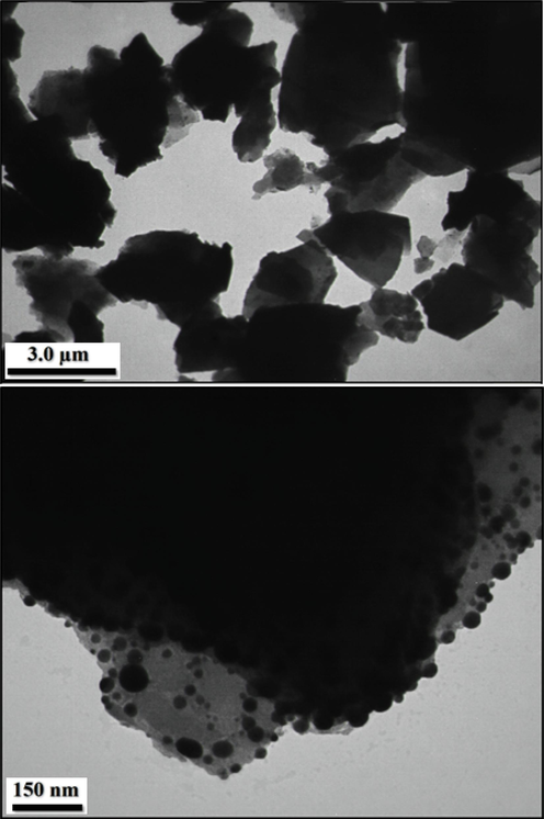
TEM images of Au NPs/Kaolin.
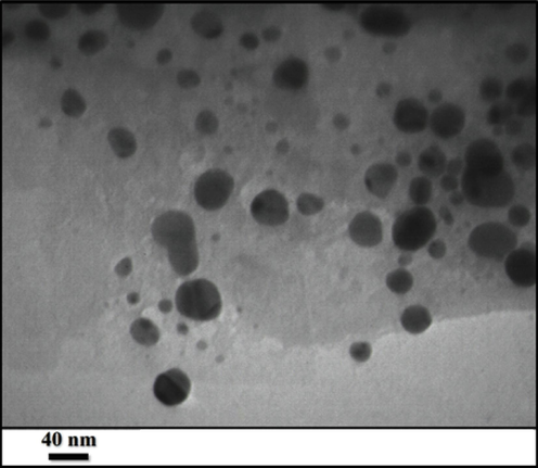
TEM image of Au NPs/Kaolin.
The FE-SEM images of Au NPs/Kaolin showed that the prepared composite has layered structures with rough surface which well confirmed the successful functionalization (Fig. 3).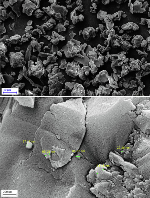
FE-SEM images of Au NPs-Kaolin.
EDX pattern (Fig. 4) shows that the produced Au NPs-Kaolin nanocomposite are mainly composed of Al, Si, O and Au, with the presence of carbon peak perhaps due to the extract component that coated on the surface. Elemental mapping of Au NPs-Kaolin nanocomposite showed well distributed Si, Al, O, C and Au across the surface (Fig. 5).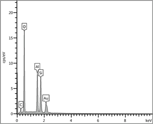
EDX profile of the Au NPs/Kaolin.
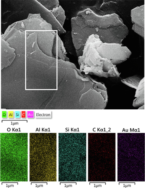
EDS-elemental mapping of Au NPs/Kaolin.
The crystal structure of Au NPs/Kaolin was investigated by XRD and the results presented in Fig. 6. First, in the XRD pattern of the prepared sample, we observed a major characteristic peak at 26.7°, which corresponds to the quartz structure of Kaolin. Also, another major diffraction peak appeared at 2θ = 21.08° is also assigned to the kaolin structure (Ma et al., 2007). The peak intensities of gold nanoparticles are low, because of the lower concentration of gold (Kurtan et al., 2016). Next, the diffraction peaks at 38.8°, 42.3°, 64.8°and 77.6° can be attributed to the characteristic peaks of (1 1 1), (2 0 0), (2 2 0) and (3 1 1), respectively, which indicates pure Au NPs of the fcc (Ai et al., 2011).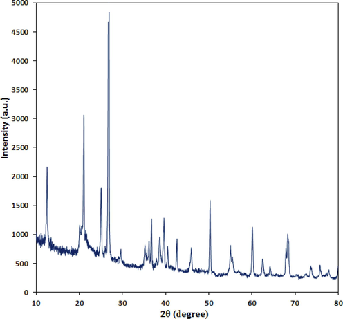
XRD pattern of Au NPs/Kaolin.
Numerous research studies have demonstrated that the bioactive components found in plant extracts exhibit cytotoxic effects on both normal and cancerous cells (Kettmann et al., 2004; Senff-Ribeiro et al., 2004; Zangeneh et al., 2019; Zangeneh et al., 2019). The bioactive elements can migrate to the nanoparticles surface in the green synthesis course. As a result, metal nanoparticles are commonly employed in research related to anticancer properties. The growth of OCUCHLM1, EGI1, and GBCSD bile duct cancer cells was suppressed by Au NPs/Kaolin nanocomposite synthesized using Strawberry root’s aqueous extract when treated with 1–1000 µg/mL. Upon examination of the morphological images of the cancer cells, it is evident that the cells exposed to concentrations 62–1000 µg/mL of Au NPs/Kaolin nanocomposite exhibit characteristics of apoptosis, including plasma membrane blebbing and cell shrinkage. Furthermore, within the concentration range of 100–1000 µg/mL, the cells tend to disperse and suspend in the medium, causing them to separate and rise above the surface of the well (Kettmann et al., 2004; Senff-Ribeiro et al., 2004). The concentration-dependent cytotoxicity of OCUCHLM1, EGI1, and GBCSD bile duct cancer cells exposed to Au NPs/Kaolin nanocomposite for 24 h was confirmed by the results obtained from the MTT assay. As the concentration of Au NPs/Kaolin nanocomposite increased, the viability (%) gradually reduced from 100 % to around 0 % (Table 1; Figs. 7-10). The findings were corroborated by research indicating that NPs produced using Allium saracilum exhibited cytotoxic effects on MCF-7 and HeLa cells at 0.5 mg/mL and 0.25 mg/mL, respectively (Zangeneh et al., 2019). Zangeneh MM et al. (2019) found in a separate investigation that the concentration of 592 µg/mL of NPs produced using Falcaria vulgaris extract decreased the HUVEC cells cell viability to 50 % (Zangeneh et al., 2019). The Au NPs/Kaolin nanocomposite produced using Strawberry root’s extract exhibit higher cytotoxicity in contrast to the findings of the previous research. The enhanced cytotoxic impact observed in our investigation is likely attributed to bioactive substances derived from the Strawberry root’s extract on the Au NPs/Kaolin nanocomposite. The nanocomposite exhibited a decrease in cell viability and anti-human laryngeal carcinoma potentials when tested on bile duct cancer (OCUCHLM1, EGI1, and GBCSD) cell lines, while showing no cytotoxic effects on normal cell lines (Figs. 5-9). The OCUCHLM1, EGI1, and GBCSD bile duct cancer cells exhibited IC50 values of 42, 54, and 63 µg/ml, respectively, when exposed to the iron nanoparticles. The most effective outcome in combating laryngeal cancer in humans was observed with Au NPs/Kaolin nanocomposite when tested against the OCUCHLM1 cell line.
Au NPs/kaolin nanocomposite (µg/mL)
IC50 against HUVEC
−
IC50 against OCUCHLM1
42 ± 0b
IC50 against EGI1
54 ± 0a
IC50 against GBCSD
63 ± 0a
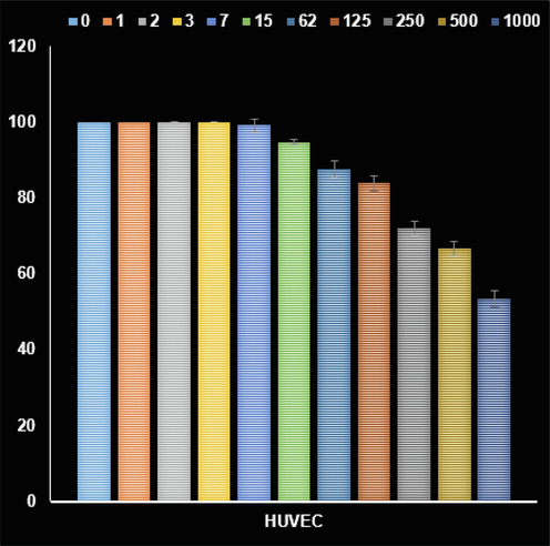
The activities of Au NPs/kaolin nanocomposite on the normal cell viability (%).
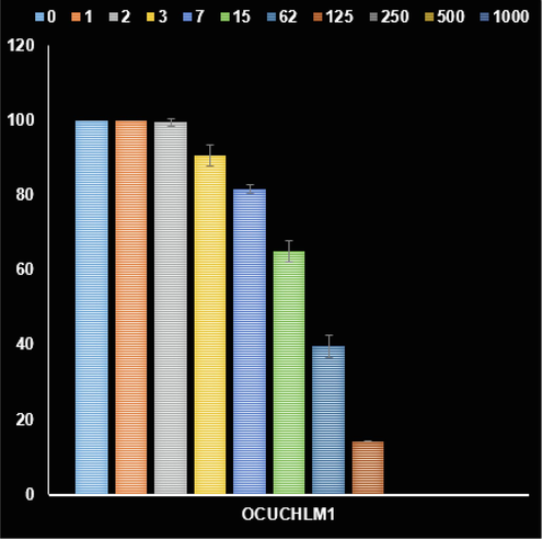
The activities of Au NPs/kaolin nanocomposite on the OCUCHLM1 cancer cell viability (%).
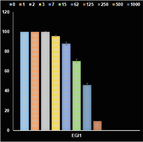
The activities of Au NPs/kaolin nanocomposite on the EGI1 cancer cell viability (%).
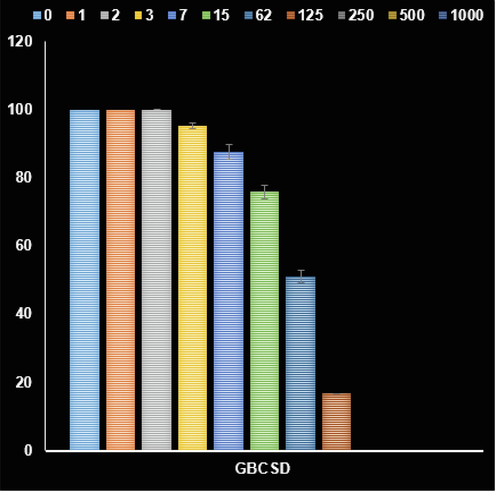
The activities of Au NPs/kaolin nanocomposite on the GBCSD cancer cell viability (%).
The toxic effects of AuNPs have been documented in numerous cancer cell lines, including MCF-7, Caco-2, MDA-MB-231, and Hep2 cancer cells (Adnan et al., 2022; Majoumouo et al., 2020; Jeyarani et al., 2020). The toxicity of gold nanoparticles is linked to their functional groups, surface charge, and size (Kus-Liśkiewicz et al., 2021). AuNPs, due to their smaller size, exhibit heightened toxic effects, enhanced cellular uptake, profound infiltration into specific tissues, and widespread tissue distribution (Peng and Liang, 2019). Based on the aforementioned information, we noted a notable cytotoxic effect of the produced Au NPs/kaolin nanocomposite at extremely low levels in three cell types. The reduced size and diverse surface properties of Au NPs/kaolin nanocomposite may account for the observed significant cytotoxicity.
4 Conclusions
In summary, we reported the synthesis, characterization and anti-oral cancer activity of Au NPs-decorated over Strawberry root extract-modified kaolin by a simple and green method. The structure, morphology and physicochemical properties of the synthesized Au NPs/Kaolin were characterized by different analytical techniques. The FE-SEM images of Au NPs/Kaolin revealed that the composite has layered structures with a rough surface, providing strong evidence of successful functionalization. The research examined the potential anti-bile duct cancer properties of Au NPs/kaolin nanocomposite produced using an aqueous extract from Strawberry root, marking the first investigation of its kind. This method allows us to avoid expensive physical methods requiring costly equipment, as well as chemical methods using harmful substances to the environment and living organisms. It was additionally confirmed that the nanoparticles displayed a suppressive influence on the proliferation of OCUCHLM1, EGI1, and GBCSD bile duct cancer cells, with the degree of effect differing based on the dosage given. Additionally, due to the cytotoxic effects of Au NPs/kaolin nanocomposite derived from Strawberry root on OCUCHLM1, EGI1, and GBCSD bile duct cancer cells, there is potential for these cells to be utilized as an effective chemotherapeutic treatment for laryngeal cancer.
Funding
To explore the mechanism of metastasis in different sites of pancreatic cancer based on multi-omics studies, NO. LHGJ20230445.
Declaration of competing interest
The authors declare that they have no known competing financial interests or personal relationships that could have appeared to influence the work reported in this paper.
References
- Microwave-assisted synchronous nanogold synthesis reinforced by kenaf seed and decoding their biocompatibility and anticancer activity. Pharmaceuticals. 2022;15(2):111.
- [Google Scholar]
- Catal. Commun.. 2011;14:68e73.
- Alafaleq, Nouf Omar, Alomari, Alya, Khan, Mohd Shahnawaz, Shaik, Gouse M., Hussain, Afzal, Ahmed, Faheem, Hassan, Iftekhar, M. Alhazza, Ibrahim, Alokail, Majed S., Alenad, Amal Majed H., Jabir, Nasimudeen R. and Tabrez, Shams. “Anticancer potential of gold nanoparticles (AuNPs) using a battery of in vitro tests” Nanotechnology Reviews, vol. 11, no. 1, 2022, pp. 3292-3304.
- The efficiency of AuNPs in cancer cell targeting compared to other nanomedicine technologies using fuzzy PROMETHEE. J. Healthc. Eng.. 2021;2021:e1566834
- [Google Scholar]
- Nuclear membrane-targeted gold nanoparticles inhibit cancer cell migration and invasion. ACS Nano. 2017;11(4):3716-3726.
- [Google Scholar]
- The basic properties of gold nanoparticles and their applications in tumor diagnosis and treatment. Int. J. Mol. Sci.. 2020;21(7):E2480.
- [Google Scholar]
- Thermal and hydrothermal alkaline modification of kaolin for the adsorptive removal of lead (II) ions from aqueous solution. SN Appl. Sci.. 2020;2:1134.
- [Google Scholar]
- Dou, Y.; Tu, F.; Wu, Y.; Wang, X.; Lu, G.; Zhao, L. Facile preparation of Kaolin supported silver nanoparticles mediated by Thymbra spicata extract and investigation of the anti-human lung cancer properties. J. Saudi Chem. Soc. 2021, 25, No. 101303.
- Recent biomedical applications of gold nanoparticles: A review. Talanta. 2018;184:537-556.
- [Google Scholar]
- Clay-supported metal oxide nanoparticles in catalytic advanced oxidation processes: A review. Nanomaterials. 2022;12:825.
- [Google Scholar]
- Multifunctional gold nanoparticles: A novel nanomaterial for various medical applications and biological activities. Front. Bioeng. Biotechnol.. 2020;8:990.
- [Google Scholar]
- Biomimetic gold nanoparticles for its cytotoxicity and biocompatibility evidenced by fluorescence-based assays in cancer (MDA-MB-231) and non-cancerous (HEK-293) cells. J. Photochem. Photobiol. B: Biol.. 2020;202:111715
- [Google Scholar]
- Kanakaraju, D.; Anak Kutiang, F. D.; Lim, Y. C.; Goh, P. S. Recent progress of Ag/TiO2 photocatalyst for wastewater treatment: Doping, co-doping, and green materials functionalization. Appl. Mater. Today 2022, 27, No. 101500.
- In vitro cytotoxicity of berberine against HeLa and L1210 cancer cell lines. Pharm. Int. J. Pharma. Sci.. 2004;59:548-551.
- [Google Scholar]
- Appl. Surf. Sci.. 2016;376:16-25.
- Biocompatibility and cytotoxicity of gold nanoparticles: Recent advances in methodologies and regulations. Int. J. Mol. Sci.. 2021;22(20):10952.
- [Google Scholar]
- Cellular uptake of gold nanoparticles and their movement in 3D multicellular tumor spheroids: Effect of molecular weight and grafting density of poly(2-hydroxyl ethyl acrylate) Macromol. Biosci.. 2020;20(1):1900221
- [Google Scholar]
- J. Hazard Mater.. 2007;145:417e423.
- Synthesis of biogenic gold nanoparticles from Terminalia mantaly extracts and the evaluation of their in vitro cytotoxic effects in cancer cells. Molecules. 2020;25(19):4469.
- [Google Scholar]
- Engineering precision nanoparticles for drug delivery. Nat. Rev. Drug Discov.. 2021;20(2):101-124.
- [Google Scholar]
- Progress in research on gold nanoparticles in cancer management. Med. (Baltim.). 2019;98(18):e15311
- [Google Scholar]
- Antiproliferative effects on tumor cells of the synthesized gold nanoparticles against Hep2 liver cancer cell line. Egypt Liver J.. 2020;10(1):15.
- [Google Scholar]
- Antiangiogenic properties of nanoparticles: a systematic review. Int. J. Nanomed.. 2019;14:5135-5146.
- [Google Scholar]
- Senff-Ribeiro A Echevarria A Silva E Franco C Veiga S -thiadiazolium mesoionic compound (MI-D) on cell lines of human melanoma. Cytotoxic effect of a new 1 . 2004;91:297–304.
- Prospective of nanoscale metal organic frameworks [NMOFs] for cancer therapy. Semin. Cancer Biol.. 2021;69:129-139.
- [Google Scholar]
- Gold nanoparticles for drug delivery and cancer therapy. Appl. Sci.. 2020;10(11):3824.
- [Google Scholar]
- Biosynthesis of ZnO N.P.s from pumpkin seeds’ extract and elucidation of its anticancer potential against breast cancer. Nanotechnol. Rev.. 2022;11(1):2714-2725.
- [Google Scholar]
- Unuabonah, E. I.; Adewuyi, A.; Kolawole, M. O.; Omorogie, M. O.; Olatunde, O. C.; Fayemi, S. O.; Günter, C.; Okoli, C. P.; Agunbiade, F. O.; Taubert, A. Disinfection of water with new chitosan-modified hybrid clay composite adsorbent. Heliyon 2017, 3, No. e00379.
- Characterization and thermal behavior of kaolin. J. Therm. Anal. Calorim.. 2011;105:157-160.
- [Google Scholar]
- Thermal and alkali modification of kaolin for potential utilization as filler material in fiber-reinforced polylactic acid composites. J. Therm. Anal. Calorim.. 2022;147:11077-11091.
- [Google Scholar]
- Ethnomedicinal plant-extract-assisted green synthesis of iron nanoparticles using Allium saralicum extract, and their antioxidant, cytotoxicity, antibacterial, antifungal and cutaneous wound-healing activities. Appl. Organomet. Chem.. 2019;34:e5254.
- [Google Scholar]
- Falcaria vulgaris leaf aqueous extract mediated synthesis of iron nanoparticles and their therapeutic potentials under in vitro and in vivo condition. Appl. Organomet. Chem.. 2019;33:e5246.
- [Google Scholar]
- Size-dependent in vivo toxicity of PEG-coated gold nanoparticles. Int. J. Nanomed.. 2011;6:2071-2081.
- [Google Scholar]
- Application of aluminosilicate clay mineral-based composites in photocatalysis. J. Environ. Sci.. 2022;115:190-214.
- [Google Scholar]


