

Translate this page into:
In vitro cytotoxic potential of cow dung and expired tomato sauces-derived carbon nanodots against A-375 human melanoma cell line
⁎Corresponding author. ansahu.phe@iitbhu.ac.in (Alakh N Sahu)
-
Received: ,
Accepted: ,
This article was originally published by Elsevier and was migrated to Scientific Scholar after the change of Publisher.
Peer review under responsibility of King Saud University.
Abstract
Abstract
Synthesis of carbon nanodots (CNDs) derived from biomass such as cow dung and expired tomato sauces. Fabricated without additional passivating agents. Exhibited good physicochemical and optical properties. Significantly inhibited the proliferation of A-375 cells in a dose-dependent manner. Revealed free radicals scavenging potential.
Abstract
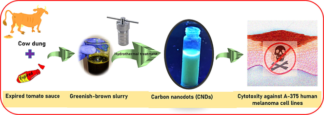
Converting biowaste into a functional product is put to the test by the growing amount of biowaste in the world and the environmental problems it causes. In this research study, we synthesized, characterized, and evaluated bluish-green luminescent carbon nanodots (CNDs) from cow dung and expired tomato sauces via a hydrothermal synthesis method at 160 °C for 8 h. The carbon nanodots were fabricated without additional passivating agents and exhibited good physicochemical and optical properties. The intrinsic properties of carbon nanodots were characterized using various spectral techniques. First, we evaluated the cytotoxic potential of carbon nanodots against A-375 human melanoma cell lines. This study revealed that carbon nanodots exhibited potent cytotoxicity and significantly inhibited the proliferation of A-375 cells in a dose-dependent manner. Next, we demonstrated these carbon nanodot's free radical scavenging potential by employing 2,2-diphenyl-1-picrylhydrazyl (DPPH) assay. The bluish-green fluorescent carbon nanodots fabricated using a green synthesis approach have broad potential for biological applications.
Keywords
Green chemistry
Biomass
Carbon nanodots
Nanoparticles
Melanoma
Fluorescence

1 Introduction
The need for research in the areas of solid waste management is crucial for the implementation of low-cost methods for the exploitation of solid waste (Khan et al., 2022). Synthesis of waste-derived Carbon-based nanomaterials such as carbon nanodots (CNDs) could be beneficial in the field of green chemistry applications (Chen et al., 2022; Murugesan et al., 2023; Tewari et al., 2023). The startling increase of bio-waste on the earth leads to perilous environmental problems. One feasible solution could be to transform biowaste into a functional product.
CNDs were discovered in 2004 while purifying single-walled carbon nanotubes (SWCNTs) using preparative electrophoresis (Pramanik et al., 2018). CNDs are a new type of fluorescent ultra small-carbon nanomaterial with particles smaller than 10 nm (Mintz et al., 2021). They have numerous applications in bioimaging, drug delivery, biosensing, disease diagnosis, synthetic chemistry, and materials science (Naik et al., 2021, 2024). These nanomaterials are water soluble, have low toxicities, and are inexpensive to produce. They also have tunable fluorescence emission and excitation, are photochemically and physicochemically stable, and have high biocompatibility. CNDs can also be employed for the delivery of natural products in order to overcome various limitations (Naik et al., 2023a, 2023b). As a result, the production, properties, and applications of CNDs have received a great deal of attention. Future research will focus on developing various morphologies, sizes, and target-specific carbon nanodots (Lou et al., 2023; Raval et al., 2023; Z. Song et al., 2023b; Zhang et al., 2023).
Hydrothermal treatment is a promising technique for converting biomass into novel carbon materials for various potential applications (Naik et al., 2021, 2023a). This process is a carbon nanomaterial synthesis technology that involves the thermochemical degradation of biomass in the presence of water (water/biomass ratios can range from 5:1 to 75:1) at elevated temperature and pressure and has found widespread application in various fields. It has been used to synthesize new carbon-based materials from biomass carbon precursors such as sweet pepper, garlic, nut husks, papaya juice, rice husks, and other biomass materials (Chen et al., 2023; Mohapatra et al., 2023).
CNDs are fabricated by modifying the surfaces of carbon nanoparticles with organic and polymeric molecules (surface passivation). CNDs preparation methods include laser ablation, combustion/thermal microwave-assisted heating, electrochemical oxidation, and supported synthesis; however, some of these techniques necessitate complex equipment and treatment processes. Because of their low cost and simple operational steps, hydrothermal, solvothermal, and microwave-assisted synthesis methods are highly valued. Natural sources for CNDs synthesis are advantageous because they are convenient, cost-effective, simple, and readily available in nature (Chen et al., 2023; Raval et al., 2023).
Green nanotechnology provides tools for converting biological systems to environmentally friendly approaches to nanomaterial synthesis while avoiding any associated toxicity. Green chemistry methods use biological sources instead of a large number of toxic chemicals and extreme environment for the production of these nanoparticles.
The core concept of this study is as follows: best out of waste. Through this work, we intend to develop CNDs from biomass such as cow dung and expired tomato sauce. As such, the expired tomato sauce has no value in our daily life, but by converting them into potential CNDs, we intend to augment its usage and sustainable value. Furthermore, based on the rich content of minerals and carbon, we hypothesized that cow dung and expired sauces could serve as excellent green and natural precursors for synthesizing CNDs without using any additional passivating agent. The current hydrothermal method is cost-effective, easy to use, and less cumbersome than other methods.
2 Materials and methods
DPPH (1,1-diphenyl-2-picrylhydrazyl), MTT (3-(4,5-dimethylthiazol-2-yl)-2,5-diphenyltetrazolium bromide), DAPI (4′,6-diamidino-2-phenylindole), methanol, dichloromethane, formic acid, and quinine sulfate were purchased from Sigma-Aldrich Co. LLC (St. Louis, MO, US). All chemicals were of analytical grade and hence used directly without further purification. Deionized water was obtained from a Millipore water purification system. Cow dung was collected from Gaushala of Banaras Hindu University, Varanasi, Uttar Pradesh, and expired tomato sauce (Kissan, India) was collected from Home. Fetal bovine serum (FBS) was procured from Sigma, trypsin-EDTA (trypsin-ethylenediamine tetraacetic acid) and Pen Strep (Penicillin-Streptomycin) were procured from Gibco, Dulbecco's modified Eagle’s medium (DMEM) was purchased from Himedia, MT, and dimethylsulfoxide (DMSO) and ethanol were purchased from SRL (Mumbai, India). Plastic wares such as T-25 flasks and 96-well plates were provided by Genetix Biotech Pvt. Ltd (Mumbai, India).
2.1 Synthesis of carbon nanodots
The hydrothermal method, a well-established technique for preparing CNDs, is favored by many material chemists due to its simplicity, affordability, non-toxicity, and involvement of green chemistry principles (Anastas and Warner, 2005). Hydrothermal treatment was used to synthesize CNDs from expired tomato sauce and cow dung. CNDs were made by boiling 10.0 g each of tomato sauce and cow dung mixture (ratio 1:1) in 150 mL of water until the volume was reduced to 100 mL. The prepared mixture was then filtered, placed in a Teflon-lined hydrothermal autoclave, and heated at 160 °C for 8 h. After cooling to room temperature, CNDs were centrifuged at 11000 rpm for 15 min before being purified through a 0.22 µm membrane syringe filter. They were kept at 4 °C for further characterization and biological testing (Naik et al., 2020a).
2.2 Characterization and stability studies of CNDs
The physicochemical and optical properties of the fabricated CNDs were deduced by using HR-TEM (Tecnai G2 20 TWIN, FEI, USA), XPS (K-Alpha, Thermo Fisher Scientific, USA), FT-IR (Nicolet iS5, Thermo Fisher Scientific, USA), fluorescence spectrophotometry (HORIBA, France), and UV–visible spectrophotometry. The thermal stability, photostability, and colloidal dispersion stability of the CNDs were tested by employing a Shimadzu (Asia Pacific) TGA-50 thermogravimetric analyzer, UV–vis spectrophotometer (Cary 60 UV–vis, Agilent, USA), and Malvern Zetasizer Pro, respectively. An average of three measurements (each with 15 runs) was considered for statistical analysis.
2.3 Cell lines and cell culture
The A-375 cell line was obtained from the cell repository at the National Centre for Cell Science (NCCS), Pune, India. The cells were grown in 25 cm2 flasks with DMEM supplemented with 10 % FBS, penicillin (100 units/mL, and streptomycin (100 μg/mL) and incubated at 37 °C in a humidified 5 % CO2 chamber (TermoHepa class 100).
2.4 Cell viability assay
A cell viability assay was performed to determine the cytotoxic activity of the test compound (fabricated CNDs) against melanoma cells as per the method published elsewhere (Naik et al., 2022). First, A-375 cells were seeded at a density of 1x10⁴ cells/well in a 96-well plate overnight and incubated with various concentrations (10–1000 µg/mL) of the test compound for 24 h. After treatment, the cells were incubated with 10 μL of MTT solution (5 mg/mL) for 2 h, followed by the addition of 100 μL of DMSO to read the absorbance at 570 nm using a microplate reader.
2.5 Cell morphology assay
Cell morphology assay was performed to examine the antiproliferative ability of CNDs (Naik et al., 2022). First, A-375 cells were seeded at a density of 1x10⁴ cells/well in a 96-well plate overnight and incubated with various concentrations (10–1000 µg/mL) of the test compound for 24 h. After treatment, the cells were washed with PBS and photographed under a phase contrast microscope.
2.6 Free radical scavenging potential
The radical scavenging potential of the CNDs was assessed using the DPPH assay as per the previously reported method with slight modifications (Naik et al., 2022). The sample (CNDs) and antioxidant standard concentrations (Vitamin C) that induced 50 % inhibition of the DPPH radicals (IC50) were computed.
3 Results and discussion
3.1 Formation of CNDs
Researchers frequently overlook the significance of carbon sources in waste biomass in favor of simply focusing on carbon nanodots derived from plants and animals. When making CNDs, biomass is an excellent source of carbon. For the synthesis of CNDs for various biomedical and environmental applications, diverse biomass, including banana peel (Sul et al., 2023), chicken feathers (Imran Din et al., 2023), and rice husk (N. Song et al., 2023a), etc, has been employed as a carbon source in previous reports.
Cow dung, also known as cow pats, cow pies, or cow manure, is the feces of domestic cattle that is the undigested plant matter after passing through the animal's digestive system. Cow dung is abundant in a variety of bacteria, minerals, and other byproducts. It has a pH range of roughly 7.1–8.0 and is primarily made up of water (80 %), undigested remnants (14.4 %), and bacteria (5.6 %). The resulting fecal matter is mineral-rich. The color ranges from green to blackish, and it darkens quickly when exposed to air. It is commonly used as manure and is usually a dark brown color (agricultural fertilizer). Cow dung can dry out and remain on the pasture if it is not recycled into the soil by species such as earthworms and dung beetles, resulting in an unpalatable grazing area for livestock (Gupta et al., 2016). A one-step hydrothermal treatment was used to successfully prepare such CNDs, which resulted in a yellow or light brown color. The hydrothermal method, which utilizes organic compounds as carbon sources and water as the solvent, is a trendy, environmentally acceptable, and economical way to synthesize CNDs (Suresh et al., 2023; Varatharajan et al., 2023).
A variety of applications, including diagnosis, catalysis, drug delivery, bioimaging, chemical sensing, environmental monitoring, and solar cells, can benefit from the excellent photostability, high fluorescence quantum yield, excellent biocompatibility, low cytotoxicity, and high photocatalytic activity of CNDs synthesized from waste biomass (Mansi et al., 2023).
3.2 Characterization
As shown in Fig. 1 (a & b), uniformly dispersed carbon nanodots with near-spherical morphology were visible in HR-TEM photomicrographs. Fig. 1a inset shows size distribution of CNDs. The size distribution of nanodots revealed the particles ranging from 1.03 to 1.99 nm, with an average particle size of the CNDs was 1.50 nm. Such a very low size distribution of CNDs was reported previously by our research group (Mohapatra et al., 2023; Naik et al., 2023a, 2022, 2020a, 2020b).![Morphology of carbon nanodots (CNDs) at different magnifications. [a] High-resolution transmission electron microscopy (HR-TEM) photomicrographs of CNDs at 200 nm scale. Inset depicts size distribution of CNDs [b] HR-TEM photomicrograph of CNDs at 20 nm scale.](/content/184/2024/17/2/img/10.1016_j.arabjc.2023.105576-fig2.png)
Morphology of carbon nanodots (CNDs) at different magnifications. [a] High-resolution transmission electron microscopy (HR-TEM) photomicrographs of CNDs at 200 nm scale. Inset depicts size distribution of CNDs [b] HR-TEM photomicrograph of CNDs at 20 nm scale.
CNDs exhibit good optical adsorption in the UV zone in the range of 260 to 320 nm with the spectrum extending to the visible region. An absorption signal for the π-π* transition of aromatic sp2 domains and a peak for the n-π* transition of surface functional moieties, such as carboxyl, hydroxyl, carbonyl, and ester groups, are usually present in pristine CNDs. The formation of CNDs was implied by the bluish-green luminescence under UV light. As depicted in Fig. 2a, the aqueous solution of CNDs exhibited two absorption peaks at 275 nm and 320 nm, which were attributed to the π–π* transition of C = C bonds and the n–π* transition of C = O bonds, respectively (Mohapatra et al., 2023b). Doping heteroatoms into a carbon nanomaterials can significantly alter their absorption wavelength due to the shift in the π-π* energy level. The wide absorption spectra of CNDs are owed by surface defects (Runprapan et al., 2023).![Physicochemical characterizations of carbon nanodots (CNDs). [a] UV–visible absorption spectrum of CNDs, [b] Excitation and Photoluminescent spectra of the CNDs, [c] Fourier Transformed Infrared (FT-IR) spectrum of CNDs, and [d] Selected area electron diffraction pattern (SAED) of CNDs.](/content/184/2024/17/2/img/10.1016_j.arabjc.2023.105576-fig3.png)
Physicochemical characterizations of carbon nanodots (CNDs). [a] UV–visible absorption spectrum of CNDs, [b] Excitation and Photoluminescent spectra of the CNDs, [c] Fourier Transformed Infrared (FT-IR) spectrum of CNDs, and [d] Selected area electron diffraction pattern (SAED) of CNDs.
The PL characteristics of CNDs are significantly influenced by their composition, particle size, shape, and internal structure. The major phenomena responsible for PL emission of CNDs are molecular and molecule-like states, edge and surface defects along with surface defect states (Gude et al., 2016). As shown in the fluorescence spectra in Fig. 2b, when excited at 357 nm, CNDs exhibited significant fluorescence at 400–530 nm and a maximum peak at approximately 432 nm. CNDs possess wide fluorescence bandwidth compared to quantum dots (QDs), which is due to the presence of random chemical structures and diverse PL centers (Mohapatra et al., 2023b).
FT-IR spectroscopy is one of the valuable techniques for identifying surface functional groups. It measures the absorption of electromagnetic radiation with wavelengths within the infrared region. The majority of CND surfaces are rich in carbonyl, carboxylic acid, and hydroxyl groups since the synthesis involves partially oxidation of the carbon precursors (Roy et al., 2022). The FTIR spectrum (Fig. 2c) was recorded to ascertain the chemical composition of the CNDs. Peaks at 3429 cm−1 and 2947 cm−1 were attributed to O–H/N–H stretching bands and C–H stretching vibrations, respectively. Peaks at 1598 cm−1, 1400 cm−1, and 1109 cm−1 were attributed to the presence of N–H bending of primary amine, aromatic C–C stretching, and C-O stretching, respectively. These results indicated that CNDs were passivated with amine, hydroxyl, and carbonyl groups. Cow dung consists of various ammonium compounds along with bacterial byproducts, whereas tomato sauce contains steroidal glycoalkaloids (tomatine), lycopene, α- carotene, β-carotene, and amino acids (Patel et al., 2023), which becomes a rich source of nitrogen in the synthesis of CNDs.
In the fields of nanostructure chemistry, biology, along with solid state physics, HR-TEM is one of the most highly informative research techniques. It is feasible to gather crucial information about the symmetry of the crystal lattice and structural flaws of the material under study by concurrently observing the electron diffraction pattern and the images of the sample's nanostructure (Bonnamy and Oberlin, 2016). As shown in Fig. 2d, the SAED pattern of CNDs consists of halo concentric rings, demonstrating its amorphous character.
The elemental composition of the CNDs was investigated using photoelectron X-ray spectroscopy (XPS). As shown in Fig. 3a, the XPS spectra of the CNDs revealed three peaks related to carbon, nitrogen, and oxygen with binding energies of 285 eV, 399 eV, and 531 eV, respectively. The high-resolution C 1 s spectrum of the CNDs (Fig. 3b) depicted major peaks at 284.5 eV and 287.6 eV, attributed to C–C bonds and C-N bonds, respectively.![X-ray Photoelectron Spectroscopy (XPS) results of carbon nanodots (CNDs) [a] XPS survey spectra of the CNDs, XPS high-resolution survey scan of [b] C1 s [c], N1 s, and [d] and O1 s region. (B.E.- Binding energy).](/content/184/2024/17/2/img/10.1016_j.arabjc.2023.105576-fig4.png)
X-ray Photoelectron Spectroscopy (XPS) results of carbon nanodots (CNDs) [a] XPS survey spectra of the CNDs, XPS high-resolution survey scan of [b] C1 s [c], N1 s, and [d] and O1 s region. (B.E.- Binding energy).
The high-resolution XPS spectrum of N 1 s for the CNDs (Fig. 3c) revealed a major peak at 400.1 eV attributed to C–N bonds. As shown in Fig. 3d, the O 1 s spectrum depicted one prominent peak at 531.2, corresponding to C = O bonds. The XPS survey spectrum of CNDs depicts the effect of surface functionalization. These self-passivated biomass-derived CNDs contain characteristic C, N, and O peaks around 285, 399, and 531 eV, which came from the cow dung and tomato sauce. Similar results have been reported in other biomass-derived CNDs studies (Pricilla et al., 2023; Roy et al., 2022; Sharma et al., 2020). Utilizing cow dung and tomato sauce can result in the integration of N into the carbon nanodots as pyridinic N and graphitic N, which can support the electron system (Kumara et al., 2023; Onfray and Thiam, 2023; Stepacheva et al., 2023). Thus, for the purpose of characterizing and analyzing the surface chemistry of CNDs, XPS is one of the valuable analytical technique. The elemental content, oxidation states of the elements, and electrical structure of CND can all be better understood utilising such photoelectric effect-based technique (Mokhena et al., 2020).
3.3 Stability studies of CNDs
The photostability of the CNDs was investigated by exposing them to continuous exposure to UV light at 254 nm for 120 min. As shown in Fig. 4a, no photobleaching was observed after 120 min of continuous exposure. When viewed with the naked eye, the fluorescence behavior was found to be unaffected. This illustrates the good photostability of these CNDs. Zeta potential (ZP) is helpful in predicting the stability of nanomaterials. ZP measurement aids in surface charge estimation, which in turn aids in comprehending CNDs' physical stability . As shown in Fig. 4b, the ZP of CNDs was found to be −4.05 mV. ZP of colloidal dispersion varies from − 30 to + 30 mV. A very low ZP of CNDs will offer low repulsive force and stability.![Stability studies of carbon nanodots (CNDs). [a] Photostability studies, [b] Zeta potential, and [c] Thermogravimetric analysis (TGA) thermogram.](/content/184/2024/17/2/img/10.1016_j.arabjc.2023.105576-fig5.png)
Stability studies of carbon nanodots (CNDs). [a] Photostability studies, [b] Zeta potential, and [c] Thermogravimetric analysis (TGA) thermogram.
Thermogravimetric analysis (TGA) is used for investigating the thermal stability of CNDs. By tracking the weight change that takes place during the sample's continuous heating as a function of time, this technique is incredibly effective in examining their thermal stability, quantity of volatile compounds, and purity (Zhang and Wu, 2023). The thermal stability of the CNDs was determined by weight loss with respect to temperature in a controlled environment. The thermal degradation is demonstrated by the TGA curve (Fig. 4c). The thermogram exhibited a three-step degradation pattern, with an initial weight loss at 100 °C due to the elimination of water molecules and weakly associated functional groups with CNDs. A substantial gradual weight loss (93 %) in the range of 200–845 °C might be due to the degradation of the tightly bound surface functional groups of CNDs. Beyond 845 °C, the curve leveled off.
3.4 Biological applications of CNDs
3.4.1 Effect of CNDs on melanoma cell proliferation
Melanoma is one of the most aggressive and deadly forms of skin cancer, with a high mortality rate worldwide (Long et al., 2023). Various drug candidates have been well explored for its treatment, but the increasing mortality rate is still a major health issue. The development of acquired resistance and systemic toxicity frequently limit the long-term efficacy of therapy. Furthermore, to overcome the side effects and toxicity of conventional treatment regimens, carbon nanomaterials-based nanomedicine and targeted therapy are considered to be potential for the treatment of cancer nowadays (Brindhadevi et al., 2023; Parihar et al., 2023; Runprapan et al., 2023). Recently, carbon nanodots came into the limelight as promising anti-cancer agents for biomedical applications (Naik et al., 2022, 2020a). In the present study, we evaluated the cytotoxic potential of CNDs against human melanoma cell line. The colorimetric MTT assay was employed to assess the cytotoxicity of CNDs. In this assay, metabolically active cells cleave MTT to produce a purple formazan product. The amount of formazan produced is a measure of % of viable cells (Jubeen et al., 2022). The outcomes of MTT assay revealed the strong anti-cancer potential of CNDs against A-375 cells and significantly (p < 0.05) inhibited the proliferation of A-375 cells in a dose-dependent manner. In addition, the decreased number of viable cells and increased number of apoptotic cells confirm the anti-proliferative attribute. The overall findings of the current study revealed that CNDs could be used as therapeutic molecules against melanoma. The proliferative ability of A-375 cells was significantly reduced with increasing concentrations (10–1000 µg/mL) of CNDs. (Fig. 5a). The obtained half-maximum inhibitory concentrations (IC50) value was found to be 84.32 μg/mL. The results revealed the cytotoxic potential of CNDs to A-375 melanoma cells.![Cytotoxicity study of carbon nanodots (CNDs) against A375 melanoma cells. [a] Dose-dependent effect of CNDs on the proliferation of melanoma cell lines, [b] Phase contrast microscopic images of A-375 cells treated with 50 and 200 μg/mL CNDs at 10X magnification.](/content/184/2024/17/2/img/10.1016_j.arabjc.2023.105576-fig6.png)
Cytotoxicity study of carbon nanodots (CNDs) against A375 melanoma cells. [a] Dose-dependent effect of CNDs on the proliferation of melanoma cell lines, [b] Phase contrast microscopic images of A-375 cells treated with 50 and 200 μg/mL CNDs at 10X magnification.
The chemotherapeutic effects of various biomass-derived CNDs have been reported by numerous researchers to date. Using the Sulforhodamine B (SRB) assay, the carbogenic nanodots (CgNDs) and graphitic nanodots (GNDs) were evaluated against the human non-small cell lung cancer cell line (NSCLC, A549). The reported IC50 for the CgNDs and GNDs, were 356.5 and 220.3 µg/mL, respectively (Emam et al., 2023). In another study, Cashew nut skin waste-derived CNDs were evaluated in vitro for bioimaging purposes against HCT-116 cells due to their negligible cytotoxic potential (Kishore et al., 2023).
3.4.2 Effect of CNDs on cell morphology
The morphology analysis (Fig. 5b) showed morphological changes, including shrinkage, irregular shape, and detachment from the microplate well surface, compared to the control. In addition, as we increased the concentration of CNDs, an increase in the number of apoptotic cells and a decrease in the number of viable cells were also noted. This effect may be induced by interactions between CNDs and proteins and other essential cell structures like the mitochondria, or nucleus, membrane, which alter both the effective surface charge of proteins and the cell apoptosis rate (Chen and Tseng, 2017; Zhang et al., 2015). The CNDs may be used as adjuvant therapy for melanoma. However, thorough in vivo studies employing animal cancer models may be utilized to reveal the detailed anti-cancer mechanism of CNDs against melanoma.
3.4.3 Antioxidant activity
An important risk factor in the development of many chronic diseases is oxidative stress. Free radicals and other reactive oxygen species are known to play a role in the pathogenesis of diseases such as atherosclerosis, Parkinson's disease, Alzheimer's disease, diabetes, and asthma. Reactive oxygen species are also responsible for human aging (Liu et al., 2023). Electron transfer to CNDs cores and the hydrogen donor behavior of surface functional groups can contribute to antioxidant activity, which is a remarkable attribute of CNDs (Kumar et al., 2020; Li et al., 2023).
One of the most widely used techniques to evaluate free radical scavenging potential is the DPPH-based assay. The free radical DPPH, which contains nitrogen, is deep purple in color and turns yellow when it comes into contact with an antioxidant. After incubation of CNDs with DPPH solution, a decrease in absorbance at 517 nm was noticed. The dose-dependent scavenging activity was observed for Vitamin C (ascorbic acid) (Fig. 6a) with an IC50 value of 6.821 μg/mL. As shown in Fig. 6b, the free radical scavenging potential of CNDs increased from 28.86 % to 93.41 % as the concentration of CNDs increased from 10 to 100 µg/mL with IC50 value of 29.324 μg/mL. CNDs from various natural precursors have shown their free radical scavenging potential (Mohapatra et al., 2023b; Naik et al., 2023a).![1,1-diphenyl-2-picrylhydrazyl (DPPH) free radical scavenging activity. [a] Scavenging activity of Vitamin C and [b] Scavenging activity of carbon nanodots (CNDs).](/content/184/2024/17/2/img/10.1016_j.arabjc.2023.105576-fig7.png)
1,1-diphenyl-2-picrylhydrazyl (DPPH) free radical scavenging activity. [a] Scavenging activity of Vitamin C and [b] Scavenging activity of carbon nanodots (CNDs).
4 Conclusion
In this study, we synthesized CNDs from cow dung and expired tomato sauce via the hydrothermal method. The characterization of CNDs was performed by using HR-TEM, XPS, FTIR, absorption spectroscopy, and fluorescence spectroscopy. CNDs exhibited encouraging cytotoxic potential against A-375 melanoma cells. The CNDs also showed free radical scavenging potential against DPPH. However, as part of the future scope of this study, detailed in vivo studies are required to reveal the mechanistic aspects of the anti-cancer and antioxidant activities of these CNDs. Such approaches may pave the realistic and affordable way to reduce down the bio-waste.
Funding
Not applicable.
Ethical approval
Not applicable.
Consent to participate
Not applicable.
Consent to publish
Not applicable.
Author contributions
GGN contributed to the study conception and design. Material preparation, data collection, and analysis were performed by GGN, RM, TM, DM, RP, SS, PKP, AP, NSJ, SS, MK and MDH. All the authors have read and approved the final manuscript. SKY, ASP, and ANS contributed to conceptualization, supervision, investigation, project administration, reviewing, and editing functions.
Acknowledgments
The authors would like to thank the Ministry of Education, New Delhi and the Central Instrumentation Facility at the Indian Institute of Technology (BHU) for their support. ANS would also like to thank the Uttar Pradesh Council of Science and Technology for providing funding (CST/D-1163) for developing CNDs for oral cancer. ANS is thankful to the Department of Biotechnology (DBT), Ministry of Science & Technology, Government of India, New Delhi, India, for providing funding for exploring phytochemical and pharmacological evaluations of bioactivity-guided fractions of medicinal plants (Sanction order No. BT/PR25498/NER/95/1223/2017). The authors would like to extend their sincere appreciation to the Researchers Supporting Project Number (RSP2023R301), King Saud University, Riyadh, Saudi Arabia.
Declaration of competing interest
The authors declare that they have no known competing financial interests or personal relationships that could have appeared to influence the work reported in this paper.
References
- The incorporation of hazard reduction as a chemical design criterion in green chemistry. Chem. Health Saf.. 2005;12:9-13.
- [CrossRef] [Google Scholar]
- Chapter 4 – transmission electron microscopy. In: Inagaki M., Kang F., eds. Materials Science and Engineering of Carbon. Butterworth-Heinemann; 2016. p. :45-70.
- [Google Scholar]
- Carbon nanomaterials: types, synthesis strategies and their application as drug delivery system for cancer therapy. Biochem. Eng. J.. 2023;192:108828
- [CrossRef] [Google Scholar]
- Design and applications of carbon dots-based ratiometric fluorescent probes: a review. Nano Res.. 2023;16:1064-1083.
- [CrossRef] [Google Scholar]
- Self-assembly of monodisperse carbon dots into high-brightness nanoaggregates for cellular uptake imaging and iron(III) sensing. Anal. Chem.. 2017;89:11348-11356.
- [CrossRef] [Google Scholar]
- Waste-derived catalysts for water electrolysis: circular economy-driven sustainable green hydrogen energy. Nano-Micro Lett.. 2022;15:4.
- [CrossRef] [Google Scholar]
- Functionalized starch for formulation of graphitic carbon nanodots as viricidal/anticancer laborers. Biocatal. Agric. Biotechnol.. 2023;47:102577
- [CrossRef] [Google Scholar]
- Molecular origin of photoluminescence of carbon dots: aggregation-induced orange-red emission. Phys. Chem. Chem. Phys.. 2016;18:28274-28280.
- [CrossRef] [Google Scholar]
- Current status of cow dung as a bioresource for sustainable development. Bioresour. Bioprocess.. 2016;3:28.
- [CrossRef] [Google Scholar]
- Novel and facile synthesis of carbon quantum dots from chicken feathers and their application as a photocatalyst to degrade methylene blue dye. J. Chem.. 2023;2023:e9956427.
- [Google Scholar]
- Anticancer potential of novel 5-Fluorouracil co-crystals against MCF7 breast and SW480 colon cancer cell lines along with docking studies. Arab. J. Chem.. 2022;15:104299
- [CrossRef] [Google Scholar]
- Current solid waste management strategies and energy recovery in developing countries – state of art review. Chemosphere. 2022;291:133088
- [CrossRef] [Google Scholar]
- Eco-friendly synthesis of functionalized carbon nanodots from cashew nut skin waste for bioimaging. Catalysts. 2023;13:547.
- [CrossRef] [Google Scholar]
- Carbon quantum dots and reduced graphene oxide modified self-assembled S@C3N4/B@C3N4 metal-free nano-photocatalyst for high performance degradation of chloramphenicol. J. Mol. Liq.. 2020;300:112356
- [CrossRef] [Google Scholar]
- Synthesis, properties and potential applications of photoluminescent carbon nanoparticles: a review. Anal. Chim. Acta. 2023;1268:341430
- [CrossRef] [Google Scholar]
- Carbon quantum dots as ROS-generator and -scavenger: a comprehensive review. Dyes Pigm.. 2023;208:110784
- [CrossRef] [Google Scholar]
- Reactive oxygen species-responsive polymer drug delivery systems. Front. Bioeng. Biotechnol.. 2023;11
- [Google Scholar]
- Thermally enhanced and long lifetime red TADF carbon dots via multi-confinement and phosphorescence assisted energy transfer. Adv. Mater.. 2023;35:2211858.
- [CrossRef] [Google Scholar]
- Chapter 24 – Synthesis and applications of carbon dots from waste biomass. In: Kailasa S.K., Hussain C.M., eds. Carbon Dots in Analytical Chemistry. Elsevier; 2023. p. :319-328.
- [Google Scholar]
- A deep investigation into the structure of carbon dots. Carbon. 2021;173:433-447.
- [CrossRef] [Google Scholar]
- Bioengineered dual fluorescent carbon nano dots from Indian long pepper leaves for multifaceted environmental and health utilities. Environ Sci Pollut Res. 2023;30:52182-52208.
- [CrossRef] [Google Scholar]
- T.C. Mokhena M.J. John M.A. Sibeko V.C. Agbakoba M.J. Mochane A. Mtibe T.H. Mokhothu T.S. Motsoeneng M.M. Phiri M.J. Phiri P.S. Hlangothi T.G. Mofokeng 2020. Nanomaterials: types, synthesis and characterization, M. Srivastava N. Srivastava P.K. Mishra V.K. Gupta Nanomaterials in Biofuels Research, Clean Energy Production Technologies. Springer, Singapore, 115–141. https://doi.org/10.1007/978-981-13-9333-4_5.
- Fluorescence resonance energy transfer-based sensor with silver-conjugated orange peel waste-derived carbon dots for melamine detection. Carbon Lett 2023
- [CrossRef] [Google Scholar]
- Pink fluorescent carbon dots derived from the phytomedicine for breast cancer cell imaging. ChemistrySelect. 2020;5:6954-6960.
- [CrossRef] [Google Scholar]
- Multi-functional carbon dots from an ayurvedic medicinal plant for cancer cell bioimaging applications. J. Fluoresc.. 2020;30:407-418.
- [CrossRef] [Google Scholar]
- Applications of natural product-derived carbon dots in cancer biology. Nanomedicine (lond). 2021;16:587-608.
- [CrossRef] [Google Scholar]
- Asparagus racemosus root-derived carbon nanodots as a nano-probe for biomedical applications. J Mater Sci. 2022;57:20380-20401.
- [CrossRef] [Google Scholar]
- From phytomedicine to photomedicine: quercetin-derived carbon nanodots—synthesis, characterization and healthcare applications. J. Mater. Sci.. 2023;58:13744-13761.
- [CrossRef] [Google Scholar]
- Chapter 9 - Phytopharmaceuticals and herbal drugs: prospects and safety issues in the delivery of natural products. In: Singh M.R., Singh D., eds. Phytopharmaceuticals and Herbal Drugs. Academic Press; 2023. p. :215-248.
- [CrossRef] [Google Scholar]
- Nip in the bud: can carbon/quantum dots be a prospective nano-theranostics against COVID-19? Bull. Mater. Sci. 2024
- [CrossRef] [Google Scholar]
- Biomass-derived carbon-based electrodes for electrochemical sensing: a review. Micromachines. 2023;14:1688.
- [CrossRef] [Google Scholar]
- Carbon nanomaterials-based electrochemical aptasensor for point-of-care diagnostics of cancer biomarkers. Mater. Today Chem.. 2023;30:101499
- [CrossRef] [Google Scholar]
- Physiological functions, pharmacological aspects and nutritional importance of green tomato- a future food. Crit. Rev. Food Sci. Nutr.. 2023;1–29
- [CrossRef] [Google Scholar]
- Biomass-derived Carbon dots and their coated surface as a potential antimicrobial agent. Biomass Conv. Bioref. 2023
- [CrossRef] [Google Scholar]
- Chapter 3 – An overview of optical, physical, biological, and catalytic properties of carbon dots. In: Kailasa S.K., Hussain C.M., eds. Carbon Dots in Analytical Chemistry. Elsevier; 2023. p. :31-41.
- [Google Scholar]
- Preparation of turmeric-derived sulfur-functionalized carbon dots: antibacterial and antioxidant activity. J. Mater. Sci.. 2022;57:2941-2952.
- [CrossRef] [Google Scholar]
- Role of defects of carbon nanomaterials in the detection of ovarian cancer cells in label-free electrochemical immunosensors. Sensors. 2023;23:1131.
- [CrossRef] [Google Scholar]
- Green synthesis of multipurpose carbon quantum dots from red cabbage and estimation of their antioxidant potential and bio-labeling activity. Appl. Microbiol. Biotechnol.. 2020;104:7187-7200.
- [CrossRef] [Google Scholar]
- Preparation of biomass carbon dots for foam stabilizer of foamed concrete. Constr. Build. Mater.. 2023;364:129853
- [CrossRef] [Google Scholar]
- A molecular engineering strategy for achieving blue phosphorescent carbon dots with outstanding efficiency above 50%. Adv. Mater.. 2023;35:2207970.
- [CrossRef] [Google Scholar]
- Plant-biomass-derived carbon materials as catalyst support. A Brief Review. Catalysts. 2023;13:655.
- [CrossRef] [Google Scholar]
- Preparation of chitosan/gelatin-based functional films integrated with carbon dots from banana peel for active packaging application. Int. J. Biol. Macromol.. 2023;246:125600
- [CrossRef] [Google Scholar]
- Green hydrothermal synthesis of carbon dot-silver nanocomposite from Chondrococcus hornemanni (marine algae): an application of mosquitocidal, anti-bacterial, and anti-cancer (MDA-MB-231 cells) Biomass Conv. Bioref. 2023
- [CrossRef] [Google Scholar]
- Development of fluorescent epoxy composite with carbon-based nanomaterial additives derived from agricultural waste. J. Vinyl Addit. Technol. 2023
- [CrossRef] [Google Scholar]
- Hydrothermal synthesis of orange fluorescent carbon dots and their application in fabrication of warm WLEDs and fluorescent ink. Phys. B Condens. Matter. 2023;654:414703
- [CrossRef] [Google Scholar]
- Mitochondria-targeting nanoplatform with fluorescent carbon dots for long time imaging and magnetic field-enhanced cellular uptake. ACS Appl. Mater. Interfaces. 2015;7:10201-10212.
- [CrossRef] [Google Scholar]
- High-speed electro-optic modulation in topological interface states of a one-dimensional lattic. Light: Sci. Appl. 2023
- [CrossRef] [Google Scholar]
- Creation and stabilization of carbon dots in silica-confined compartments with high thermal stability. Chem. Commun.. 2023;59:1665-1668.
- [CrossRef] [Google Scholar]


