

Translate this page into:
Magnetic properties, anticancer and antibacterial effectiveness of sonochemically produced Ce3+/Dy3+ co-activated Mn-Zn nanospinel ferrites
⁎Corresponding authors. malmessiere@iau.edu.sa (M.A. Almessiere), yaslimani@iau.edu.sa (Y. Slimani) slimaniyassine18@gmail.com (Y. Slimani)
-
Received: ,
Accepted: ,
This article was originally published by Elsevier and was migrated to Scientific Scholar after the change of Publisher.
Peer review under responsibility of King Saud University.
Abstract
Some new types of Ce3+ and Dy3+ co-doped manganese-zinc nanospinel ferrites (CDMZNSFs) of the form (Mn0.5Zn0.5)[Fe2-2xCexDyx]O4 (with 0.0 ≤ x ≤ 0.1) were sonochemically produced and characterized. The structure, morphology, optical and magnetic properties of these NSFs were determined as a function of co-dopant (Ce3+ and Dy3+) contents. The direct optical band gap energies of the studied NSFs were ranged from 1.54 to 1.85 eV. The measurements of magnetization versus magnetic field of the prepared NSFs disclosed a superparamagnetic (SPM) behavior at room temperature (RT). The measurements of temperature-dependent magnetizations revealed a transition from superparamagnetic (SPM) state above blocking temperature TB to a ferromagnetic (FM) state below TB. The saturation magnetization and TB decreased with the increase in co-dopant contents. In addition, the bactericidal (on the gram-positive and gram-negative bacterial strains) and anti-cancerous effectiveness of these NSFs were assessed. The cancer cells' growth inhibitory action of these NSFs was tested against both normal (HEK-293) and cancerous (HCT-116) human cells. After 48 h of treatment of the cancerous cells with the NSFs, their population was significantly dropped as shown by the MTT assay, indicating the selective inhibition of the cancer cells growth by the proposed NSFs. Conversely, the non-cancerous cells (HEK-293) population remained unaffected. The IC50 values of the NSFs-treated cancerous cells (HCT-116) were in the range of 0.74–2.35 µg/mL. The results of the MIC and MBC assays revealed the reasonable antibacterial efficacy (growth inhibitory activity) of these NSFs when tested against the E. coli and S. aureus bacterial strains. It is established that the proposed Ce3+/Dy3+ co-activated CDMZNSFs may be beneficial for the anti-cancerous and bactericidal applications.
Keywords
Nanospinel ferrites
Sonochemistry
Anti-cancerous application
Bactericidal application
Magnetic properties
Morphology

1 Introduction
The novel spinel ferrites became the most active research areas due to numerous manufacturing such as data storage, radar absorbing, telecommunication, negligible eddy current losses, microwave, permanent magnets, refractory materials, sensors, ferro-fluids, hydrogen production, decomposition of alcohols, oxidative dehydrogenation, electro-catalytic activity and corrosion resist (Joulaei et al., 2019; Kiani et al., 2019; Eskandari et al., 2019; Etminan et al., 2018; Moradi et al., 2018; Naghikhani et al., 2018). Lately, the Mn-Zn spinel ferrites (MZSFs, the soft ferromagnetic ceramic materials) due to their exclusive electrical, magnetic and optical properties have widely been used in the communication, home appliances, computer, cores of inductors, transformers, recording heads, hyperthermia devices, cancer cells treatment, magnetic resonance imaging (MRI) and so forth (Hu et al., 2010; Xing et al., 2012; Ahmed et al., 2007; Parekh et al., 2006). In-depth studies disclosed that all these unusual characteristics of the MZSFs are decided by preferred distributions of the Fe3+ and Fe2+ cations into the tetrahedral and octahedral sub-lattice sites in the crystal structure (Ahmed et al., 2007), where the syntheses techniques of these ferrites play a significant role.
The crystal unit cell of the MZSFs encloses sixty-four tetrahedral lattice positions (A-sites) and thirty-two octahedral lattice positions (B-sites), where the metal cations occupy eight A-lattice sites and sixteen B-lattice sites, leaving seventy-two positions vacant. Consequently, the unoccupied lattice sites in crystalline unit cells of the MZSFs can accommodate various metal ions such as the trivalent lanthanides, causing a modification in their structural, optical, and magnetic characteristics (Modak et al., 2009). The present study is motivated by the possibilities where rare earth ions co-doped new types of low dimensional MZSFs with improved traits can be composed for novel applications.
To fulfill the ever-increasing demand of the high-quality MZSFs, numerous syntheses strategies have been adopted such as the co-precipitation (Mathur et al., 2007), polyol process (Topfer and Angermann, 2011), hydrothermal (Akhtar et al., 2019), solid-state reaction (Kogias and Zaspalis, 2016), sonochemical (Almessiere et al., 2020; Rehman et al., 2020), auto-combustion (Baykal et al., 2015) and so on. Recent studies showed that using the ultrasonic waves at high intensity some good quality MZSFs nanostructures (doped and undoped) with customized properties can be achieved. Amongst all these techniques the sonochemical strategy is advantageous due to its ability to form nanocomposites of uniform sizes rapidly with less energy consumption and fast reaction rates (Asgharzadehahmadi et al., 2016; Pinjari and Pandit, 2011). On top, the sonochemical technique being relatively simple, accurate, and economic, it can produce contaminant-free Mn-Zn nanospinel ferrites (MZNSFs) at a large scale in short duration compared to the conventional approaches. In this view, we used this approach to prepare Ce3+/Dy3+ co-activated MZNSFs (CDMZNSFs).
According to Rezlescu et al. (1998); the trivalent rare earth impurities due to their large ionic radii and exceptional 4f shell electronic configurations can be the excellent substituents in the lattice structures of the soft MZNSFs, thereby improving their structural, optical, magnetic and electrical traits. The rare earth ions substitutions in the MZNSFs leads to the development of a strong exchange coupling between the 4f and 3d electrons, contributing to the significant magneto-crystalline anisotropy in the spinel structures and physical properties improvement (Islam et al., 2013; Song et al., 2010; Almessiere et al., 2019; Ateia et al., 2020). Mohd. Hashim and et al. investigated the Structural, optical, elastic and magnetic features of CoCexDyxFe2-2xO4 (x = 0.0, 0.01, 0.02, 0.03, 0.04, 0.05) were fabricated by sol gel auto combustion technique. They found that the blocking temperatures at x = 0.00, 0.03 and 0.05 higher than room temperature and reductions with growing the Ce and Dy ratio. The higher values of saturation magnetism and Hyperfine field for x = 0.01 composition may be due to the all Dy3+ ions occupy at octahedral site while the Ce3+ ions might be occupied at tetrahedral site. As the doping amount increases, the saturation magnetism decreases noticeably, and this attributable to the replacement of iron by these doping ions (Hashim et al., 2020). While A.A. Kadam and et al. studied the Structural, morphological, electrical and magnetic characteristics of dysprosium doped Ni0.8Co0.2Fe2-xDyxO4 ferrite prepared by simple ceramic process. The samples showed a reduction in dielectric constant, loss tangent and ac conductivity with frequency. Moreover, low coercive field suggested that these ferrites can be used in magnetic shielding devices (Kadam et al., 2013). Le-Zhong Li and et al. explored the structural and electromagnetic propertiesNi0.4Zn0.6 − xCexFe2O4 (0 ≤ x ≤ 0.20) were synthesized via the sol-gel combing with the solid reaction procedure. They observed that the saturation magnetism (Ms) initially increases with increasing amount of Ce substitution due to the increase of density and reduction of porosity, and decreases when Ce substitution excessed 0.15 that contributes to the increase of the net magnetic moment in tetrahedral site and porosity and the presence of CeO2 phase. The initial permeability firstly increases at room temperature with the Ce substitution content increasing and reach the maximum at x = 0.15. However, it sharply drops when the amount of Ce replacement is 0.20 because the porosity increasing and impurity phase appearance (Li et al., 2019). Shirsath et al. (2011) reported Ce substituted NiFe2O4 ferrite prepared by solid-state reaction. Ce4+ increased the pore mobility in NiFe2O4 ferrite by creating excess cation vacancies affecting overall grain growth and bulk density. Moreover, higher magnetocrystalline anisotropy associated with rare earth Ce4+ ions promoted the enhancement in coercivity.
In this perception, we used the sonochemical method to synthesize some new types of MZNSFs with Ce3+ and Dy3+ co-substitutions for the first time. As-prepared CDMZNSFs were characterized using diverse tools to determine the role of varying Ce3+ and Dy3+ contents on their structural, morphological, optical and magnetic features. In addition, the anticancer and anti-bacterial efficacy of the produced CDMZNSFs was tested against the human cells (normal HEK-293 and cancerous HCT-116) and bacterial strains (Escherichia coli and Staphylococcus aureus).
2 Experimental
2.1 Synthesis and characterizations of Ce3+and Dy3+ co-doped MZNSFs
A series of MZNSFs co-activated with Ce3+/Dy3+ impurities were produced using the sonochemical route. First, the high purity chemical reagents (from Merck) of [MnCl2·4H2O] (manganese II chloride), [Zn(NO3)2] (zinc nitrate), [Dy(NO3)3·H2O] (dysprosium III nitrate hydrate), [Fe(NO3)3·9H2O] (Iron (III) nitrate nonahydrate) and [Ce(NO3)3·6H2O] (cerium nitrate hexahydrate) were mixed in deionized water (DIW). The pH of the resultant mixture was adjusted to 11 via the addition of 2 M of NaOH solution before being irradiated ultrasonically for 30 min (UZ SONOPULS Ultrasonic Homogenizer HD 2070 operated at the frequency of 20 kHz and power of 70 W). Next, the solution containing the product was rinsed using DIW followed by the drying at 60 °C for nearly 24 h to achieve the solid samples. Finally, samples were characterized at room temperature using different analytical instruments.
The X-ray diffraction (XRD) analyses of the produced CDMZNSFs were performed (Rigaku Benchtop Miniflex X-ray Diffractometer operated with Cu-Kα radiation) to determine their crystalline structures and phase compositions. The surface morphologies of the prepared NSFs were evaluated using the scanning electron microscopy (SEM, FEI Titan ST). The elemental compositions of the samples were studied using the energy dispersive X-ray (EDX) spectroscopy (attached to SEM). The shape and size distributions of the CDMZNSFs were recorded using a transmission electron microscope (TEM, FEI Morgagni 268). The UV–Visible diffuse reflectance spectra (in the wavelength range of 200–800 nm) of the samples were obtained. The room temperature (RT, 300 K) magnetic measurements of the prepared samples were examined by using Quantum Design Physical Property Measurement System (PPMS DynaCool) coupled with a vibrating sample magnetometer (VSM) head in the applied magnetic field (H) range of ±7 Tesla. In addition, the measurements of magnetization versus temperature under zero-field-cooling (ZFC) and field-cooling (FC) modes were performed in the range of 300–10 K.
2.2 Anticancer activities
2.2.1 In vitro cytotoxicity assay
The colorectal carcinoma cells of human (HCT-116) were used to inspect the cancer cells inhibitory action of the as-prepared CDMZNSFs. Meanwhile, the normal (healthy) embryonic kidney cells of human (HEK-293) were used to assess the cytotoxicity of the CDMZNSFs. The earlier referred (Asgharzadehahmadi et al., 2016) protocols were used for the cells culture. The studied cells were cultivated at 37 °C in a carbon dioxide (CO2) incubator inside the DMEM medium (Dulbecco's Modified Eagle's Medium) composed of the L-glutamine, fetal bovine serum, penicillin (antibiotic), selenium chloride and streptomycin. Upon reaching the confluence of 70–80%, the MTT [3-(4,5-dimethylthiazol-2-yl)-2,5-diphenyl tetrazolium bromide] assay (Molecules, New Zealand) was performed on the cultured cells to determine the anti-cancer viability of the proposed CDMZNSFs-treated at various doses (2.0 to 40 μg//mL) where the control group was devoid of NSFs. After treating the cells culture for a period of 48 h, the culture media was isolated and supplemented with the MTT solution of concentration 10 mg/mL (Sigma-Aldrich, USA) followed by further incubation of the plates enclosing cultures. Again, the cells culture medium was isolated and every well was supplemented with dimethyl sulfoxide (DMSO) in which MTT created Formazan crystallites. Later, the plates enclosing the CDMZNSFs-treated cells culture were analyzed (at wavelength of 570 nm) using the micro-plate readers (Bio-Rad Laboratories, USA). The mean and standard errors of both normal and CDMZNSFs-treated groups were scrutinized using the GraphPad Prism (GraphPad Software).
2.2.2 Nuclear staining by 4′,6-diamidino-2-phenylindole, DAPI) fluorescent dye
The DAPI (4′,6-diamidino-2-phenylindole) fluorescent dye (1.0 μg/mL) was used to stain the CDMZNSFs-treated cancer cells to discern their effects on the cell nuclei. The normal cells (HCT-116) in group-I were treated with CDMZNSFs and group-II was not treated. After 48 h of treatments using the CDMZNSFs, both non-cancerous (HCT-116) and cancerous (HEK-293) human cells were treated with 4% of paraformaldehyde prepared in phosphate buffer saline (PBS). Thereafter, the cells’ membranes were permeabilized using Triton X-100-PBS treatment for 5 min. Finally, the DAPI staining of the cells was conducted in the darkroom. The nuclear morphologies of the cells were examined via a confocal scanning microscope (Zeiss, Germany) (Pinjari and Pandit, 2011).
2.3 Bactericidal activities
2.3.1 Synthesis of inoculum and stock solutions
To evaluate the antibacterial activities of the CDMZNSFs, the NSFs were homogenized using sterile distilled water to get the solution. The NSFs were added to the sterile Luria Bertaini (LB) broth at the concentration range of 16 to 0.5 mg/mL. The inoculum of Escherichia coli (ATCC35218, gram-negative) and Staphylococcus aureus (ATCC29213, gram-positive) bacteria was prepared using the LB broth after the incubation at 35 ± 2 °C for overnight. The cell densities (2.5 × 10⁶ CFUs/mL) of the freshly cultured bacterial strains were adjusted by the addition of sterile Luria Bertaini broth (Rezlescu et al., 1998).
2.3.2 Minimal inhibitory concentration (MIC)
The MIC values of the prepared CDMZNSFs solutions were determined using the broth dilution method after supplementing the freshly adjusted bacterial inoculum (density of 2.5 × 10⁵ CFU/mL) with incubation in air for 24 h at 35 ± 2 °C. The solution without bacterial treatment was considered as the negative control. The MIC record was chosen as the minimal drug content at which no growth was visually seen, indicating the absence of turbidity.
2.3.3 Minimum antibacterial concentration (MBC)
After the MIC of evaluation of the CDMZNSFs solutions, the aliquot of suspension was withdrawn without causing any turbidity followed by the inoculation on freshly made Mueller Hinton Agar (MHA) plate with further incubation for 24 h at 35 ± 2 °C. The MBC of the drug was recorded as the lowermost content without bacterial cells or below 3 CFU on the MHA plate.
2.3.4 Morphology characterization of CDMZNSFs-treated bacteria
The effect of CDMZNSFs on the morphologies of the bacterial cells (gram positive and negative) was examined via SEM imaging. Both bacterial strains (106 CFU/mL) were treated at the concentration obtained as MIC value of the particular NSFs with the overnight incubation at 37 ± 2 °C wherein the bacteria without NSFs treatment were taken as the control. Upon completing the incubation, the bacterial cells were centrifuged and washed repeatedly using the PBS. Thereafter, the bacterial cells were treated for 4 h using the glutaraldehyde (2.5%) followed by the fixing via osmium tetroxide (1%). Then, the resultant samples were rinsed and dried using ethanol at different concentrations. Next, the bacterial cells were put on the Al stubs and dried inside the desiccator followed by the Au coating for the SEM measurements (Ateia et al., 2020).
3 Results and discussion
3.1 Structural analyses of CDMZNSFs
The formation of phases in the CDMZNSFs (0.0 ≤ x ≤ 0.1) was observed via the XRD powder pattern as shown in Fig. 1. The reflection planes for all the samples were found belong to the typical pure single phase of the MZNSFs correspond to h k l planes of (2 2 0), (3 1 1), (2 2 2),(4 0 0), (4 2 2) (5 1 1) and (4 4 0) respectively without any other phase of the metals oxides according to card number [96-230-0619] and [96-101-0131]. The presence of intense diffraction peaks confirmed the presence of high crystallinity. The most intense peaks (3 1 1) of all the samples were broadened, indicating a decrease in the average crystallite size. The Rietveld refinement using (Match3! and fullproof) was used to estimate the lattice constant (a) and average crystallite size as listed in Table 1.The values of lattice constant (a) and unit cell volume (V) were found increase with increasing the co-dopant (Ce3+/Dy3+) concentration which was due to their larger ionic radii Ce3+ (1.01 Å), Dy3+ (0.91 Å) than the Fe3+ ions (0.64 Å), generating a deformation in the spinel ferrite lattice to recompense the developing strain and stress (Parekh et al., 2006). The crystallite size was estimated via Scherrer formula by considering the most intense plane (3 1 1) and it was found in the range from 8.6 to 18.5 nm (Table 1).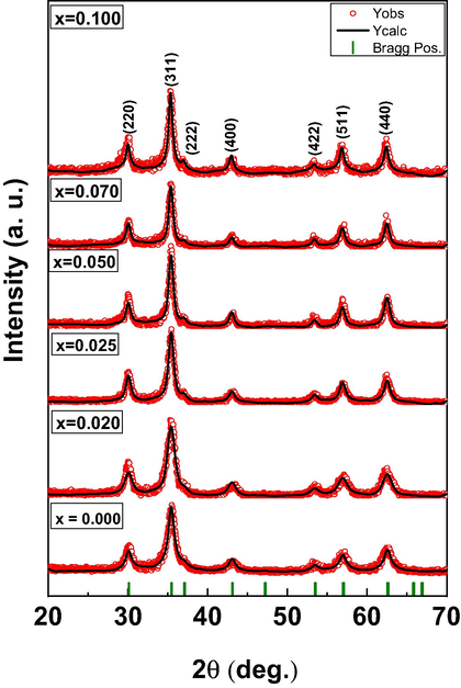
The XRD patterns of the studied CDMZNSFs (0.0 ≤ x ≤ 0.1).
x
A (Å)
V (Å)3
DXRD ± 0.04 (nm)
χ2
RBragg
0.000
8.3977
592.2151
18.5
1.2
13.2
0.020
8.4018
593.0809
11.3
1.1
10.4
0.025
8.4027
593.2673
10.8
1.3
12.7
0.050
8.4053
593.8287
10.4
1.2
15.9
0.070
8.4069
594.1721
9.6
1.0
13.9
0.100
8.4167
596.2524
8.6
1.1
17.8
The cations distribution in the CDMZNSFs crystalline unit cells was determined by analyzing the XRD data via the Bertaut technique as shown in Table 2 (Shirsath et al., 2013, 2014). The majority of the Fe3+ occupied the octahedral sites (B-sublattice position) with few at the tetrahedral sites (A-sublattice position). Approximately 70–84% of the Mn3+ occupied the A-sites and 30–16% was distributed on the B-sites consistent with the preferential occupation at the tetrahedral sites (Shirsath et al., 2018). The Zn2+ showed the strong preferential occupation towards the tetrahedral A-sites as expected. Meanwhile, the Ce3+/Dy3+ substituents preferentially occupied the octahedral B-sites only due to their larger ionic radii compared to the host metallic ions. A close investigation of XRD patterns confirmed the single phase formation of the prepared samples without forming the secondary phases of DyFeO3, CeFeO3, CeO and DyO. Further, lattice constant increased throughout the substitution range of Ce and Dy ions. Thus, it can be concluded that Ce and Dy ions enters into to the crystal lattice of CDMZNSFs and occupied octahedral B-site because of their larger ionic radii.
x
Tetrahedral position (A-sites)
Octahedral position (B-sites)
0.000
Mn0.35Zn0.5Fe0.15
Mn0.15Fe1.85
0.020
Mn0.35Zn0.5Fe0.15
Mn0.15Dy0.02Ce0.02Fe1.81
0.025
Mn0.36Zn0.5Fe0.14
Mn0.14Dy0.025Ce0.025Fe1.8
0.050
Mn0.38Zn0.5Fe0.12
Mn0.12Dy0.05Ce0.05Fe1.78
0.070
Mn0.4Zn0.5Fe0.1
Mn0.1Dy0.07Ce0.07Fe1.75
0.100
Mn0.42Zn0.5Fe0.08
Mn0.08Dy0.1Ce0.1Fe1.72
3.2 Morphologies of CDMZNSFs
Fig. 2 displays the FESEM micrographs of the prepared CDMZNSFs (x = 0.000, 0.020, 0.050 and 0.100), which were comprised of the nonuniformly aggregated spherical nanocrystallites with the average size around 20 nm. Fig. 3 shows the TEM images of the studied CDMZNSFs (x = 0.025 and 0.100) which were contained of strong agglomerated nearly spherical nanocrystallites, supporting the FESEM observations. Moreover, the HRTEM images of CDMZNSFs (x = 0.025 and 0.100) are consisted of well resolved two-dimensional lattice spaces with of 0.25 nm, 0.29 nm and 0.19 nm corresponding to lattice plane (3 1 1), (2 2 0) and (3 3 1), respectively for Mn-Zn spinel ferrites. The SAED pattern displayed the well-separated dotted rings indicating the crystalline nature of the particles. The first six rings of the SAED patterns were labelled as, (2 2 0), (3 1 1), (4 0 0), (4 2 2), (5 1 1) and (4 4 0). The maximum intensity was obtained for (3 1 1) reflection which is consistent with the XRD pattern as shown above by Fig. 1. Fig. 4 illustrates the EDX spectra of the achieved CDMZNSFs, verifying the appropriate elemental compositions (Mn, Zn, Dy, Fe, Ce and O) of the samples. EDS color mapping analysis (Fig. 4) confirmed that the Ce and Dy ions are uniformly distributed without forming a cluster. Thus, this is a further evidence that Ce and Dy ions indeed are in the crystalline grain and not on the grain boundary.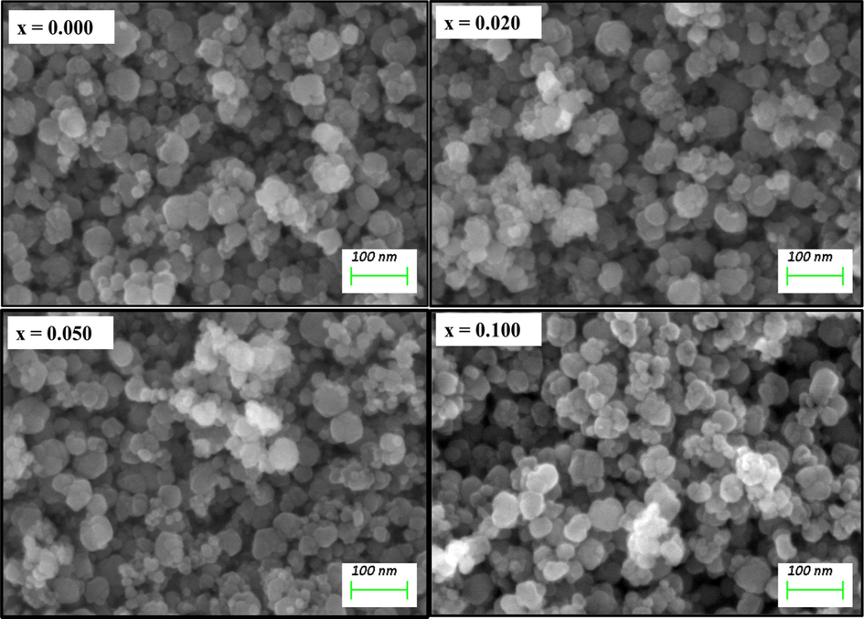
The SEM images of the synthesized CDMZNSFs (x = 0.000, 0.020, 0.050 and 0.100).
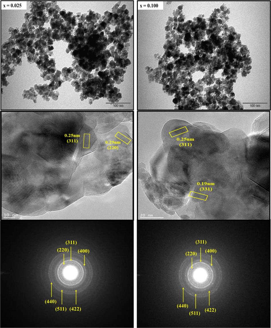
The TEM, HR-TEM and SAED pattern of the synthesized CDMZNSFs (x = 0.025 and 0.100).
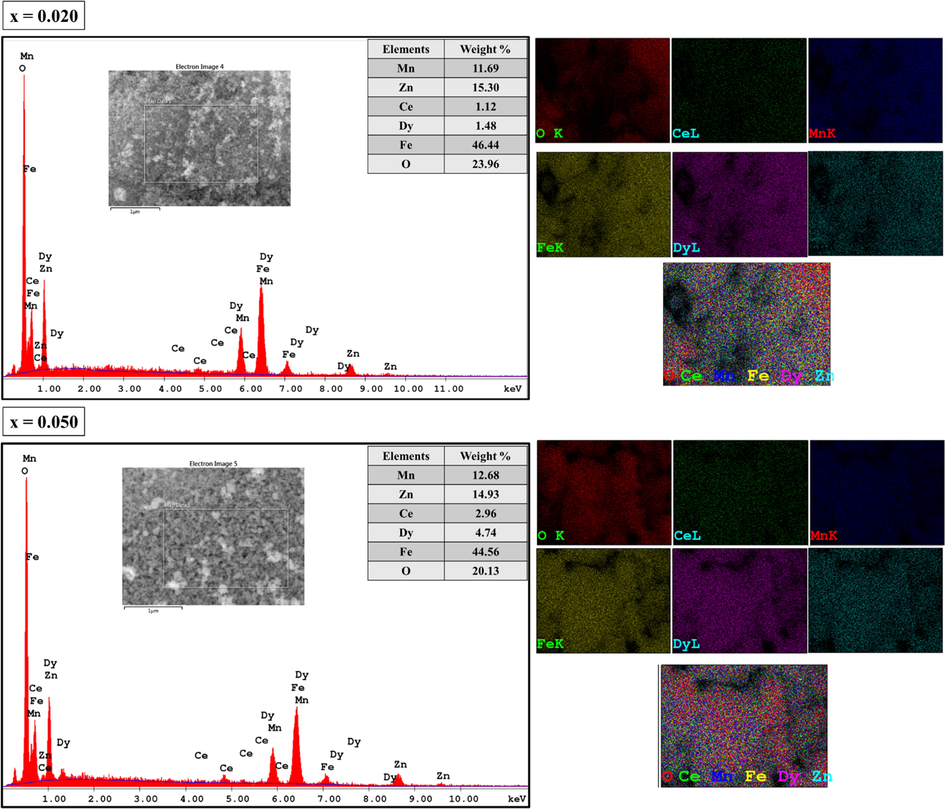
The EDX spectra and corresponding elemental maps of the synthesized CDMZNSFs (Inset: measured weight% of various elements).
3.3 Optical band gap energy
Optical band gap energy (Eg) is a decisive property of semiconductors for optoelectronic applications. UV–vis diffuse reflectance spectroscopy (DRS) is one of the most employed methods for the determination of the optical bandgap of nanoparticle sized semiconducting substances. Fig. 5 shows the percent diffuse reflectance (DR %) spectra of the synthesized CDMZNSFs (Baykal et al., 2015; Güner et al., 2015). Spectra were recorded by a UV–vis spectrophotometer equipped with an integrating sphere between 200 nm and 800 nm of wavelength. The spectral peaks have appeared in the wavelength region between 200 and 500 nm with the intensity values in a narrow range of 13.40–15.70 %. Above 500 nm, we observe a sharp increase at reflectance intensities and among all the NSFs Mn0.5Zn0.5Ce0.10Dy0.10Fe2O4 showed a maximum reflectance intensity of 28.8% corresponding to 800 nm.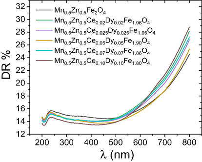
The diffuse reflectance spectra of the prepared CDMZNSFs.
The DR spectral data were analyzed using the radiative transfer model introduced by Kubelka-Munk (K-M). In this model, the function
is defined as (Blitz, 1998):
Fig. 6 shows the Tauc plots of all the prepared CDMZNSFs. The value of Eg for the undoped MZNSFs was found to be 1.65 eV. The estimated values of Eg for the Dy3+/Ce3+ co-doped MZNSFs were in the range of 1.65–1.77 eV, indicating the remarkable sensitiveness of the direct band gap energy of the NSFs on the co-dopant contents. All the prepared samples were semiconducting in nature. Some of the literature reports on the Mn0.5Zn0.5Fe2O4 NSFs prepared using hydrothermal and sonication assisted MW irradiation techniques revealed the corresponding Eg values of 1.98 and 1.99 eV (Nam et al., 2018; Ashok et al., 2019). In addition, other reports on the citric acid assisted sol-gel and thermal decomposition method grown Mn1-xZnxFe2O4 MZNSFs (where x from 0.2 to 0.8) showed the Eg values in between 1.86 and 2.80 eV (Baykal et al., 2015, 2014; Demir et al., 2014). Present work is the first report of the Eg values for the Dy3+/Ce3+ co-substituted MZNSFs.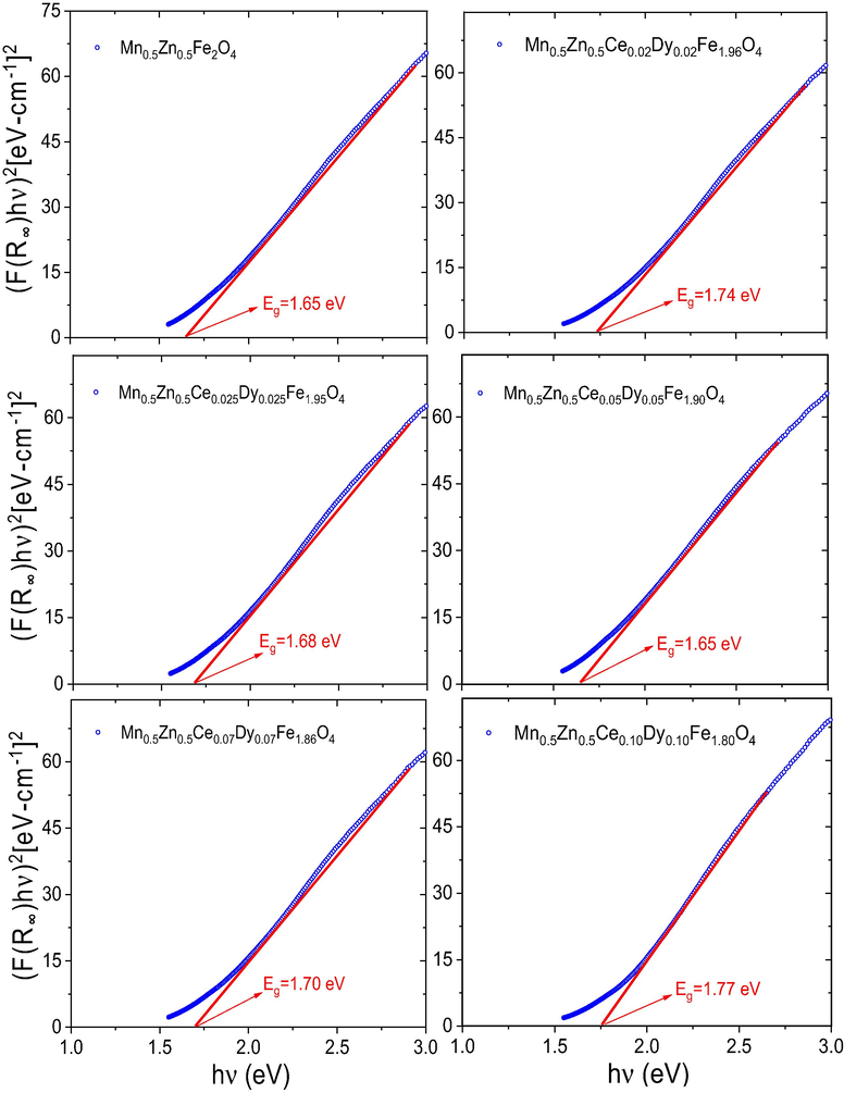
Tauc plots for all the obtained CDMZNSFs showing the estimated value of the optical band gap energies.
3.4 Magnetic properties
3.4.1 Magnetization versus magnetic field (M-H) hysteresis loops
Fig. 7 displays the RT hysteresis loops of all the prepared CDMZNSFs, which revealed superparamagnetic (SPM) behaviors. The measured M-H curves of all the samples revealed S-shape without any coercivity (Hc) and remanence (Mr). This behavior was due to the nucleation of the tiny NSF crystallites that could easily orient via the thermal activation, thereby overcoming the magnetic anisotropy (Chand et al., 2011). Essentially, the mean size of these crystallites was below the critical size (25 nm), thereby disclosing the predominant SPM phase (Lazarević et al., 2013; Almessiere et al., 2019; Zhang et al., 2009). This result was consistent with the XRD analyses that yield DXRD values below 15 nm for various samples, confirming the crystallites sizes less than the critical limit (Table 1).![RT magnetic hysteresis loops (M-H) of all the prepared (Mn0.5Zn0.5)[Fe2-2xCexDyx]O4 NSFs (0.0 ≤ x ≤ 0.1).](/content/184/2020/13/10/img/10.1016_j.arabjc.2020.08.017-fig7.png)
RT magnetic hysteresis loops (M-H) of all the prepared (Mn0.5Zn0.5)[Fe2-2xCexDyx]O4 NSFs (0.0 ≤ x ≤ 0.1).
Since the magnetization of the prepared NSFs did not saturate completely (Fig. 7) despite the high magnetic field, the law of approach to saturation (LAS) at the higher field regions was applied in the M against 1/H2 plot (Fig. 8) for the selected undoped sample that enabled to estimate the values of the saturation magnetization (Ms) (Stoner and Wohlfarth, 1948; Almessiere et al., 2019).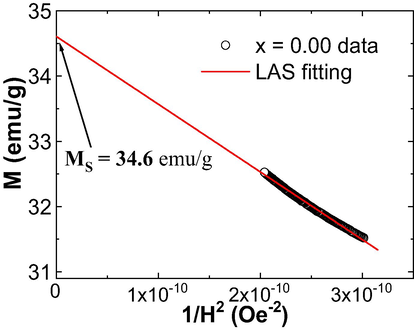
An example of the M against 1/H2 plot of the undoped CDMZNSFs. The solid line shows the LAS fit.
Fig. 9 illustrates the variations in the Ms and magneton numbers (
) as a function of the Ce3+/Dy3+ contents. The
values were calculated via (Almessiere et al., 2019a, 2019b):
![Variations of Ms and n B values, for (Mn0.5Zn0.5)[Fe2-2xCexDyx]O4 NSFs (0.0 ≤ x ≤ 0.1), with respect to the content of Ce-Dy co-substituting elements.](/content/184/2020/13/10/img/10.1016_j.arabjc.2020.08.017-fig9.png)
Variations of Ms and
values, for (Mn0.5Zn0.5)[Fe2-2xCexDyx]O4 NSFs (0.0 ≤ x ≤ 0.1), with respect to the content of Ce-Dy co-substituting elements.
The values of Ms and for the undoped (x = 0.0) NSFs was 34.6 emu/g and 1.46 μB, respectively. The observed value of the Ms is lower than that of its bulk counterpart form (Ms ≈ 140 emu/g) (Isfahani et al., 2009), which is mainly ascribed to the existence of the tinier crystallites that encountered the surface structural disorders, development of the magnetic inactive layers and spins canting (Coey, 1971; Slimani et al., 2019; Almessiere et al., 2019). Nevertheless, the value of Ms obtained here is higher than those recorded in other MZNSFs produced using co-precipitation (Parvatheeswara Rao et al., 2006), low-temperature solid-state reaction (Amiri et al., 2011), hydrothermal precipitation (Rath et al., 1999); evaporation and auto-combustion (Ranjith Kumar et al., 2013) and sol-gel combustion (Azadmanjiri, 2007) methods. In comparison to the pristine sample, the values of Ms of the NSFs are decreased with the rise in Ce3+ and Dy3+ contents.
Generally, the magnetic properties can be affected by various factors including the variations in the crystallites morphology (size and shape), the magnetic moments of the substituent and host ions, the strength of the super-exchange interactions, presence of disorder and inhomogeneities, etc. (Almessiere et al., 2019; Nairan et al., 2016). Several studies acknowledged the proportionality in the evolutions of crystallytes/grains size and Ms value (Almessiere et al., 2019; Nairan et al., 2016). Predominantly, the value of Ms increases with the increase of the crystallites/grains size and vice versa. As found in XRD part, the values of DXRD are decreased with the increase in Ce3+ and Dy3+ contents, which are in line with the variations in Ms values. Furthermore, the dissimilarities in the ionic radii of Ce3+ (1.01 Å), Dy3+ (0.91 Å) and Fe3+ (0.64 Å) have a tendency to induce structural disorder, lattice strains and alteration in the electronic states into the prepared spinel ferrites, which in turn will affect on the magnetic parameters (Slimani et al., 2018). Additionally, the reduction in the magnetic moment ( ) values signifies the weakening in the super-exchange interaction (Gabal et al., 2013; Almessiere et al., 2019). The variations of the as function of Ce3+ and Dy3+ contents showed similar trend as that of Ms, indicating a weakening of the super-exchange interactions with Ce-Dy substitution.
To further elucidate the magnetic properties of the produced spinel ferrites, the Néel model was employed. The net magnetization (M) of the present spinel ferrites in terms of the A and B sublattice magnetizations (
and
) yields:
The magnetic moments of Mn2+, Zn2+, Fe3+, Ce3+ and Dy3+ ions are equal to 5, 0, 5, 2.5 and 10.5 μB, respectively. Firstly, by assuming only the replacement of some of the Fe3+ (5 μB) by the Dy3+ (10.5 μB), an enhancement in the magnetization should be observed, but this is not the case here. In the present study, the value of Ms decreased with the rise in the content of substituting elements. This could be due to the further substitution of some of the Fe3+ ions by the Ce3+ ions that display a low magnetic moment of 2.5 μB. The observed tendency in magnetization can be therefore explained based on the distribution of different cations in the present ferrite crystal structure (i.e. occupation of Mn2+, Zn2+, Fe3+, Ce3+, and Dy3+ ions in A and B sites). The cations distribution in the current study showed that the Mn2+ and Fe3+ ions occupy both A and B-sites, Zn2+ ions occupy only A site, and Ce3+ and Dy3+ ions preferentially to occupied only the B site (Thakur et al., 2020; Mohd Hashim et al., 2018). Consequently, the magnetic moment in the A site is increased with the rise in Dy3+ and Ce3+ contents, thus reducing the net magnetization of the proposed CDMZNSFs.
3.4.2 Magnetization against temperature (M-T)
Fig. 10 shows the measured ZFC and FC curves (M against T variation) of the produced CDMZNSFs. A splitting and irreversibility are observed in ZFC and FC curves, which is a characteristic of spinel ferrites. In comparison to undoped sample, both MZFC and MFC values are significantly decreased with the increase in Dy3+ and Ce3+contents, which is in line with the M-H results. In addition, the MFC values increased with the decrease in the temperature and reached a maximum value at a certain particular temperature before being dropped. Generally, SPM materials disclose a continuous increase in in the MFC values with the increase in temperature (Peddis et al., 2010). Conversely, an observed plateau-like behavior or a decrement in the MFC value at lower temperatures indicates a transformation in magnetic behavior. Several reports suggested that the emergence of the peak at a particular temperature in the MZFC versus T curves might be associated to the called blocking temperature (TB). The blocking temperature (TB) is a temperature at which the magnetic anisotropy barrier is overcome by the thermal activation, which lead to the fluctuations in magnetization (Pawar et al., 2016). With the increase in the temperature below TB, the B-site antiferromagnetic interaction becomes stronger. As a consequence, it resulted in a strong spin frustration and a decrease in magnetization. Above TB, the thermal energy is higher than the magnetic energy barrier, thus the superparamagnetism appears by following the Curie-Weiss law. Accordingly, the magnetic system is undergoing to a transition from the ferromagnetic (FM) state (under TB) to the superparmagnetic (SPM) state (above TB). In the present study, various NSFs disclosed a wide peak at TB in the MZFC against T curves, indicating a SPM to FM state transition below TB. It is clearly noticed that the Ce-Dy substitution with MnZn spinel ferrite significantly influences on the value of TB. Indeed, the value of TB shifted from 79.5 K (for the undoped sample) to the lower temperatures with the increase in the content of co-substituting Ce and Dy elements. The values of TB for the (Mn0.5Zn0.5)[Fe2-2xCexDyx]O4 NSFs with 0.00, 0.02, 0.025, 0.05, 0.07, and 0.10 of Ce3+/Dy3+ are discerned to be 79.5, 76.9, 74.8, 74.1, 57.4 and 45.3 K, respectively. Actually, the theory proposed by Néel describes the dependence of the blocking temperature (TB) on particles size and magneto-crystalline anisotropy constant (K) (Pawar et al., 2016; Humbe et al., 2018). They are related by the following expression:
![ZFC-FC magnetizations of the prepared (Mn0.5Zn0.5)[Fe2-2xCexDyx]O4 NSFs (0.0 ≤ x ≤ 0.1) performed under an applied magnetic field of 100 Oe.](/content/184/2020/13/10/img/10.1016_j.arabjc.2020.08.017-fig10.png)
ZFC-FC magnetizations of the prepared (Mn0.5Zn0.5)[Fe2-2xCexDyx]O4 NSFs (0.0 ≤ x ≤ 0.1) performed under an applied magnetic field of 100 Oe.
3.5 Anticancer effectiveness
The anticancer activities of the produced CDMZNSFs on the human cells were evaluated via the MTT assay where the inhibitory concentration (IC50) was measured. The 48 h post-treated HCT-116 (cancerous) cells by the CDMZNSFs revealed appreciable inhibition activities, confirming their anti-cancerous benefits for the drug formulation. Table 3 shows the values of IC50 obtained by treating the cancel cells by different CDMZNSFs. To verify the cytotoxicity of the proposed CDMZNSFs, they were tested on the healthy human kidney cells (HEK-293). The 48 h post-treated HEK-293 (noncancerous) cells by the CDMZNSFs did not show any cytotoxic effect, indicating the feasibility of using these NSFs for cancer treatments without damaging the healthy cells.
x
IC50 (µg/mL) for HCT-116
IC50 (µg/mL) for HEK-293
0.000
0.74
Zero inhibitory action
0.020
0.85
Zero inhibitory action
0.025
0.84
Zero inhibitory action
0.050
0.60
Zero inhibitory action
0.070
0.45
Zero inhibitory action
0.100
2.35
Zero inhibitory action
3.5.1 Cytotoxicity due to nuclear disintegration
Fig. 11 shows the CDMZNSFs-treated HCT-116 (cancer) cells and untreated cells (control group) morphologies after staining by DAPI for 48 h. The control (Fig. 11 (a)) did not reveal any inhibition activity. However, the CDMZNSFs-treated (Fig. 11 (a) with the dose of 0.74 µg/mL and Fig. 11 (b) with the dose of 2.35 µg/mL) HCT-116 (cancer) cells exhibited stronger inhibition action (indicated by arrow heads at 200× magnified) compared to the control group, confirming the nuclear staining-mediated loss of the cells.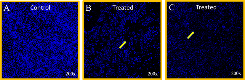
The cell morphologies after DAPI staining (48 h) for the (a) untreated control group, (b) CDMZNSFs-treated (x = 0.00 dose of 0.74 µg/mL) cancer cells and (c) CDMZNSFs-treated (x = 0.10 dose of 2.35 µg/mL) cancer cells. Arrow heads shows the nuclear staining-mediated decay of the cells.
Present findings indicated that the proposed CDMZNSFs were selective in targeting the colon cancer cells without causing any damage to the noncancerous (normal or healthy) cells, suggesting their prospects for cancer therapeutic purposes. It was acknowledged that the magnetic nanoparticles are beneficial for the drugs delivery and other diagnoses (Qiaoqiao et al., 2019). Furthermore, some reports showed the death of cancerous cells with strong nuclear fragmentations and disintegrations when treated with magnetic nanoparticles, indicating their promise for cancer therapy (Javad et al., 2016; Mytych et al., 2015). In short, the proposed CDMZNSFs revealed strong inhibitory actions on the cancerous cells, indicating their potential for cancer cures.
3.6 Bactericidal activity
3.6.1 MIC/MBC evaluation
The bactericidal efficacy of the as-synthesized CDMZNSFs (at various concentrations from 16 to 0.5 mg/mL) against the E. coli and S. aureus bacteria was assessed by the values of MIC/MBC. The values of MIC/MBC for the CDMZNSFs-treated E. coli bacterial strains were discerned to be 8/16 (for x = 0.00), 8/16 (for x = 0.02), 8/16 (for x = 0.025), 8/16 (for x = 0.05), 4/16 (for x = 0.07) and 4/8 mg/mL (for x = 0.1) as shown in Fig. 12(a). The values of MIC/MBC for the CDMZNSFs-treated S. aureus bacterial strains were observed to be 8/16 (for x = 0.00), 8/16 (for x = 0.02), 8/16 (for x = 0.025), 8/16 (for x = 0.05), 8/16 (for x = 0.07) and 4/8 mg/mL (for x = 0.1) as shown in Fig. 12(b). These MIC/MBC indicated the possibility of bactericidal improvement by controlling the Ce3+ and Dy3+ doping contents in the MZNSFs with the lowest one for x = 0.1. The antibacterial activity of the CDMZNSFs-treated gram-negative bacteria was slightly higher than the one obtained for the gram-positive bacterial strains that was ascribed to the compositional dissimilarities in their cells wall structures (Rehman et al., 2019; Fekri et al., 2019). The bactericidal potential of various metal ions doped Zn, Cu, Ni and Mn nanoparticles reported in the literature (Asma M et al., 2019; Elsa et al., 2016) are good agreement with the present findings on CDMZNSFs, indicating their prospects for sundry biomedical applications.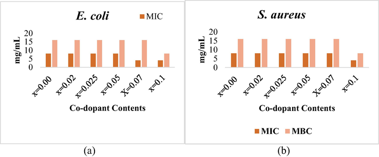
The measured values of MIC/MBC for the CDMZNSFs-treated (a) E.coli, and (b) S. aureus bacteria.
3.6.2 Morphologies of CDMZNSFs-treated bacteria
The structural alteration in the E. coli and S. aureus caused by the CDMZNSFs treatment were examined via SEM imaging. The untreated E. coli and S. aureus bacterial cells were observed to be normal rod and spherical in shapes with uniform cell surface, respectively (Fig. 13 (a). E. coli and S. aureus cells treated with x = 0.00 and x = 0.02 showed a mild damage. However, when treated with the CDMZNSFs of x = 0.025 and x = 0.05, the cells were found to be significantly affected (Fig. 13 b, c, d, e). Again, after treating with the CDMZNSFs of x = 0.07 and x = 0.1, the cells were observed to be significantly damaged with a fewer number of cells visible. This damage of the cells with distortion and deformation of the cell surface clearly indicated the loss of integrity of membrane due to the impact of the CDMZNSFs (Fig. 13 e, f). The cells damage and count of the E. coli bacteria was lower compared to the S. aureus, which are due to their dissimilar structural compositions of the cells wall (Elsa et al., 2016; de Toledo et al., 2018). It is affirmed that the morphogenesis of both gram negative and gram-positive bacteria was appreciably affected by the CDMZNSFs treatment. This observation was attributed to the efficient attachment and penetration of the CDMZNSFs through the cell membrane, thereby destructing the bacterial cells.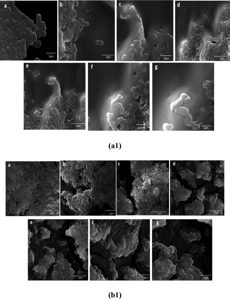
SEM micrographs of the CDMZNSFs-treated bacteria (a1) E. coli (b1) S. aureus. a) untreated cells(control), b) x = 0.00, c) 0.02, d) 0.025, e) 0.05, f) 0.07 and g) 0.01.
4 Conclusion
This paper evaluated the structure, morphology, optical, magnetic, bactericidal, and anticancer traits of the new series of Ce3+/Dy3+ co-activated CDMZNSFs for the first time. These samples were sonochemically prepared and analyzed in-depth. The prepared NSFs manifested a transition from SPM state to FM state below TB. Both Ms and TB values were reduced with the rise in Ce3+/Dy3+ contents. The variations in various magnetic parameters of the CDMZNSFs as ascribed to the variations in crystallites size and shape, the magnetic moments of substituting ions and host ions, and the cations distribution in the crystal lattice. It was shown that by adjusting the Ce3+/Dy3+ co-substituent contents the overall properties of the CDMZNSFs could be tailored. The produced CDMZNSFs revealed appreciable bactericidal (when tested against E. coli and S. aureus bacteria) and anti-cancerous (tested against cancerous HCT-116 human cells) efficacy. The cancer cells growth was significantly inhibited (with IC50 values ranged from 0.74 to 2.35 µg/mL) due to the CDMZNSFs treatment (for 48 h), whereas the normal HEK-293 cells growth was not affected. On top, these CDMZNSFs did not show any cytotoxicity. The MTT assay of the CDMZNSFs-treated cancerous cells disclosed a notable drop in their population, indicating the selective inhibition (DAPI staining for 48 h) of the cancer cells growth by the prepared NSFs. However, the populations of the HEK-293 cells were not affected by the CDMZNSFs treatment. The MIC and MBC displayed realistic bactericidal activity of these NSFs against gram negative and gram-positive bacterial strains. It was established that the proposed Ce3+/Dy3+ co-substituted CDMZNSFs can be beneficial for the anti-cancerous and bactericidal applications. In addition, the proposed CDMZNSFs composition may contribute to the development of biomedical and pharmaceutical applications. It is worth to perform more careful studies to validate these disclosures and claim, which is underway. As a future study, microwave absorption properties of Ce3+/Dy3+ co-substituted CDMZNSFs will be investigated.
Acknowledgment
This study is supported by the Institute for Research and Medical Consultations (Project application No. 2018-IRMC-S-2) and by the Deanship for Scientific Research (Project application No. 2020-164-IRMC) of Imam Abdulrahman Bin Faisal University (IAU – Saudi Arabia).
Declaration of Competing Interest
The authors declare there is no conflict of interest.
References
- Enhancement of the physical properties of rare-earth-substituted Mn–Zn ferrites prepared by flash method. Ceram. Int.. 2007;33:49-58.
- [Google Scholar]
- Synthesis of Mn0.5Zn0.5SmxEuxFe1.8-2xO4 nanoparticles via hydrothermal approach induced anti-cancer and anti-bacterial activities. Nanomaterials. 2019;9(11):1635.
- [Google Scholar]
- Magneto-optical properties of rare earth metals substituted Co-Zn spinel nanoferrites. Ceram. Int.. 2019;45:3449-3458.
- [Google Scholar]
- Sonochemical synthesis of Eu3+ substituted CoFe2O4 nanoparticles and their structural, optical and magnetic properties. Ultrason. Sonochem.. 2019;58:104621
- [Google Scholar]
- Ce–Nd Co-substituted nanospinel cobalt ferrites: An investigation of their structural, magnetic, optical, and apoptotic properties. Ceram. Int.. 2019;45:16147-16156.
- [Google Scholar]
- Structural, optical and magnetic properties of Tm3+ substituted cobalt spinel ferrites synthesized via sonochemical approach. Ultrason. Sonochem.. 2019;54:1-10.
- [Google Scholar]
- Effect of dysprosium substitution on magnetic and structural properties of NiFe2O4 nanoparticles. J. Rare Earths. 2019;37:871-878.
- [Google Scholar]
- Structural, magnetic, optical properties and cation distribution of nanosized Co0.7Zn0.3TmxFe2-xO4 (0.0 ≤ x ≤ 0.04) spinel ferrites synthesized by ultrasonic irradiation. Ultrason. Sonochem.. 2019;58:104638
- [Google Scholar]
- Magneto-optical properties of rare earth metals substituted Co-Zn spinel nanoferrites. Ceram. Int.. 2019;45:3449-3458.
- [Google Scholar]
- Correlation between microstructure parameters and anti-cancer activity of the [Mn0.5Zn0.5](EuxNdxFe2-2x)O4 nanoferrites produced by modified sol-gel and ultrasonic methods. Ceram. Int.. 2020;46:7346-7354.
- [Google Scholar]
- Magnetic properties and microwave absorption in Ni–Zn and Mn–Zn ferrite nanoparticles synthesized by low-temperature solid-state reaction. J. Magn. Magn. Mater.. 2011;323:730-734.
- [Google Scholar]
- Sonochemical reactors: Review on features, advantages and limitations. Renew. Sustain. Energy Rev.. 2016;63(May)
- [Google Scholar]
- Structural, optical and magnetic properties of Zn1-xMnxFe2O4 (0 ≤ x ≤ 0.5) spinel nano particles for transesterification of used cooking oil. J. Alloy. Compd.. 2019;780:816-828.
- [Google Scholar]
- Synthesis of an activatable tetra-substituted nickel phthalocyanines-4 (3H)-quinazolinone conjugate and its antibacterial activity. Adv. Pharmacol. Sci. 2019 Volume 2019 |Article ID 5964687 | 10 pages
- [Google Scholar]
- Comparative study on the physical properties of rare-earth-substituted nano-sized CoFe2O4. Appl. Phys. A. 2020;126(91)
- [Google Scholar]
- Preparation of Mn–Zn ferrite nanoparticles from chemical sol–gel combustion method and the magnetic properties after sintering. J. Non-Cryst. Solids. 2007;353:4170-4173.
- [Google Scholar]
- Effect of Zinc substitution on magneto-optical properties of Mn1-xZnxFe2O4/SiO2 nanocomposites. Ceram. Int.. 2014;40:13401-13408.
- [Google Scholar]
- Magnetic and optical properties of Cu1-xZnxFe2O4 nanoparticles dispersed in a silica matrix by a sol-gel auto-combustion method. Ceram. Int.. 2015;41:231-239.
- [Google Scholar]
- Synthesis and magneto-optical properties of triethylene glycol stabilized Mn1-xZnx Fe2O4 nanoparticles. J. Alloy. Compd.. 2015;619:5-11.
- [Google Scholar]
- Magnetic and optical properties of Cu1-xZnxFe2O4 nanoparticles dispersed in a silica matrix by a sol–gel auto-combustion method. Ceram. Int.. 2015;41:231-239.
- [Google Scholar]
- Md Amir, A Manikandan, Magneto Optical Properties and Hyperfine Interactions of Cr3+ Ion Substituted Copper Ferrite Nanoparticles. J. Inorg. Organo. Polym. Mater.. 2018;28:2533-2544.
- [Google Scholar]
- Blitz, John P., 1998. Diffuse Reflectance Spectroscopy, Modern Techniques in Applied Molecular Spectroscopy, John Wiley & Sons, Inc., 1998. ISBN 0-471-12359-5 © (Chapter 5)
- Investigations on MnxZn1-xFe2O4 (x = 0.1, 0.3 and 0.5) nanoparticles synthesized by sol-gel and co-precipitation methods. Ind. J. Eng. Mater. Sci.. 2011;18:385.
- [Google Scholar]
- Noncollinear spin arrangement in ultrafine ferrimagnetic crystallites. Phys. Rev. Lett.. 1971;27:1140-1142.
- [Google Scholar]
- Iron oxide magnetic nanoparticles as antimicrobials for therapeutics. Pharm. Dev. Technol.. 2018;23:316-323.
- [Google Scholar]
- Magnetic and optical properties of Mn1-xZnxFe2O4 nanoparticles. J. Inorg. Organometall. Polym. Mater.. 2014;24:729-736.
- [Google Scholar]
- Synthesis of manganese dioxide nanoparticles using co-precipitation method and its antimicrobial activity. Int. J. Modern Sci. Technol.. 2016;1:17-22.
- [Google Scholar]
- Preparation of a new magnetic and photo-catalyst CoFe2O4–SrTiO3 perovskite nanocomposite for photo-degradation of toxic dyes under short time visible irradiation. Compos. B Eng.. 2019;176:107343
- [Google Scholar]
- Preparation of tin ferrite–tin oxide by hydrothermal, precipitation and auto-combustion: photo-catalyst and magnetic nanocomposites for degradation of toxic azo-dyes. J. Mater. Sci.: Mater. Electron.. 2018;29:1766-1776.
- [Google Scholar]
- Evaluation of antibacterial and antifouling properties of silver-loaded GO polysulfone nanocomposite membrane against Escherichia coli, Staphylococcus aureus, and BSA protein. React. Funct. Polym.. 2019;140:136-147.
- [Google Scholar]
- Influence of Al-substitution on structural, electrical and magnetic properties of Mn–Zn ferrites nanopowders prepared via the sol–gel auto-combustion method. Polyhedron. 2013;57:105-111.
- [Google Scholar]
- Magneto-optical properties of Mn3+ substituted Fe3O4 nanoparticles. Ceram. Int.. 2015;41:10915-10922.
- [Google Scholar]
- Structural, optical, elastic and magnetic properties of Ce and Dy doped cobalt ferrites. J. Alloy. Compd.. 2020;834:155089
- [Google Scholar]
- Heat treatment effects on microstructure and magnetic properties of Mn-Zn ferrite powders. J. Magn. Magn. Mater.. 2010;322:173-177.
- [Google Scholar]
- Rietveld refinement, morphology and superparamagnetism of nanocrystalline Ni0.70−xCuxZn0.30Fe2O4 spinel ferrite. Ceram. Int.. 2018;44:5466-5472.
- [Google Scholar]
- Magnetic properties of nanostructured MnZn ferrite. J. Magn. Magn. Mater.. 2009;321:152-156.
- [Google Scholar]
- Study of the structural, magnetic and electrical properties of Gd-substituted Mn–Zn mixed ferrites. J. Alloy. Compd.. 2013;559:174-180.
- [Google Scholar]
- Induction of apoptosis by green synthesized gold nanoparticles through activation of caspase-3 and 9 in human cervical cancer cells. Avicenna J. Med. Biotechnol.. 2016;8:75.
- [Google Scholar]
- Investigation of magnetic, mechanical and flame retardant properties of polymeric nanocomposites: Green synthesis of MgFe2O4 by lime and orange extracts. Compos. B Eng.. 2019;176:107345
- [Google Scholar]
- Structural, morphological, electrical and magnetic properties of Dy doped Ni–Co substitutional spinel ferrite. J. Magn. Magn. Mater.. 2013;329:59-64.
- [Google Scholar]
- A novel magnetic MgFe2O4–MgTiO3 perovskite nanocomposite: Rapidphoto-degradation of toxic dyes under visible irradiation. Compos. B Eng.. 2019;175:107080
- [Google Scholar]
- Temperature stable MnZn ferrites for applications in the frequency region of 500 kHz. Ceram. Int.. 2016;42:7639-7646.
- [Google Scholar]
- Nanodimensional spinel NiFe2O4 and ZnFe2O4 ferrites prepared by soft mechanochemical synthesis. J. Appl. Phys.. 2013;113:187221
- [Google Scholar]
- Effects of Ce substitution on the structural and electromagnetic properties of NiZn ferrite. J. Magn. Magn. Mater.. 2019;475:1-4.
- [Google Scholar]
- Study of low-temperature sintered nano-crystalline Mn-Cu-Zn ferrite prepared by co-precipitation method. Modern Phys. Lett. B. 2007;21:1425-1430.
- [Google Scholar]
- XRD, HR-TEM and magnetic properties of mixed spinel nanocrystalline Ni-Zn-Cu-ferrite. J. Alloy. Compd.. 2009;473:15-19.
- [Google Scholar]
- Influence of rare earth ion doping (Ce and Dy) on electrical and magnetic properties of cobalt ferrites. J. Magn. Magn. Mater.. 2018;449:319-327.
- [Google Scholar]
- Rapid photo-degradation of toxic dye pollutants: green synthesis of mono-disperse Fe3O4–CeO2 nanocomposites in the presence of lemon extract. J. Mater. Sci.: Mater. Electron.. 2018;29:11065-11080.
- [Google Scholar]
- Gold nanoparticles promote oxidant-mediated activation of NF-κB and 53BP1 recruitment-based adaptive response in human astrocytes. BioMed Res. Int. 2015
- [Google Scholar]
- Simple and green synthesis of CuFe2O4–CuO nanocomposite using some natural extracts: photo-degradation and magnetic study of nanoparticles. J. Mater. Sci.: Mater. Electron.. 2018;29:4689-4703.
- [Google Scholar]
- Temperature-dependent magnetic response of antiferromagnetic doping in cobalt ferrite nanostructures. Nanomaterials. 2016;6:73.
- [Google Scholar]
- In-JaLee, Effect of zinc on structure, optical and magnetic properties and magnetic heating efficiency of Mn1-xZnxFe2O4 nanoparticles. Phys. B Cond. Matter.. 2018;550:428-435.
- [Google Scholar]
- Ternary monodispersed Mn0.5Zn0.5Fe2O 4 ferrite nanoparticles: preparation and magnetic characterization. Nanotechnology. 2006;17:5970-5975.
- [Google Scholar]
- Structural and magnetic characterizations of coprecipitated Ni–Zn and Mn–Zn ferrite nanoparticles. IEEE Trans. Magn.. 2006;42:2858.
- [Google Scholar]
- Spin glass behavior and enhanced but frustrated magnetization in Ho3+ substituted Co-Zn ferrite interacting nanoparticles. RSC Adv.. 2016;6:76590-76599.
- [Google Scholar]
- Surface spin freezing effects on enhanced saturation magnetization and magnetic anisotropy in CoFe2O4 nanoparticles. Nanotechnology. 2010;21:125705
- [Google Scholar]
- Room temperature synthesis of crystalline CeO2 nanopowder: Advantage of sonochemical method over conventional method. Ultrason. Sonochem.. 2011;18(5):1118-1123.
- [Google Scholar]
- Applications of Magnetic Nanomaterials in Heterogeneous Catalysis. ACS Appl. Nano Mater.. 2019;2:4681-4697.
- [Google Scholar]
- Effect of α-Fe2O3 phase on structural, magnetic and dielectric properties of Mn–Zn ferrite nanoparticles. J. Phys. Chem. Solids. 2013;74:943-949.
- [Google Scholar]
- Preparation and characterization of nanosize Mn–Zn ferrite. J. Magn. Magn. Mater.. 1999;202:77-84.
- [Google Scholar]
- Antibacterial and antifungal activity of novel synthesized neodymium-substituted cobalt ferrite nanoparticles for biomedical application. Processes. 2019;7:714.
- [Google Scholar]
- Synthesis and biological characterization of Mn0.5Zn0.5EuxDyxFe1.8-2xO4 nanoparticles by sonochemical approach. Mater. Sci. Eng., C. 2020;109:110534
- [Google Scholar]
- Effects of rare-earth oxides on physical properties of Li–Zn ferrite. J. Alloy. Compd.. 1998;275–277:657.
- [Google Scholar]
- Influence of Ce4+ ions on the structural and magnetic properties of NiFe2O4. J. Appl. Phys.. 2011;110 013914-1-8
- [Google Scholar]
- Chemical tuning of structure formation and combustion process in CoDy0.1Fe1.9O4 nanoparticles: influence@pH. J. Nanopart. Res.. 2013;15:1976.
- [Google Scholar]
- Self-ignited high temperature synthesis and enhanced super-exchange interactions of Ho3+–Mn2+–Fe3+–O2− ferromagnetic nanoparticles. PCCP. 2014;16:2347-2357.
- [Google Scholar]
- Ferrites obtained by sol-gel method. In: Klein L., Aparicio M., Jitianu A., eds. Handbook of Sol-Gel Science and Technology. Cham: Springer; 2018. p. :695-735.
- [Google Scholar]
- Magneto-optical and microstructural properties of spinel cubic copper ferrites with Li-Al co-substitution. Ceram. Int.. 2018;44:14242-14250.
- [Google Scholar]
- Effect of bimetallic (Ca, Mg) substitution on magneto-optical properties of NiFe2O4 nanoparticles. Ceram. Int.. 2019;45:6021-6029.
- [Google Scholar]
- Microwave electromagnetic and absorbing properties of Dy3+ doped MnZn ferrites. J. Rare Earths. 2010;28(3):451-455.
- [Google Scholar]
- A mechanism of magnetic hysteresis in heterogeneous alloys. Philos. Trans. R Soc. Lond. Ser. A Math. Phys. Sci.. 1948;240:599-642.
- [Google Scholar]
- Optical properties and electronic structure of amorphous germanium. Physica Status Solidi. 1966;15:627-637.
- [Google Scholar]
- A review on MnZn ferrites: synthesis, characterization and applications. Ceram. Int.. 2020;46:15740-15763.
- [Google Scholar]
- Nanocrystalline magnetite and Mn-Zn ferrite particles via the polyol process: synthesis and magnetic properties. Mater. Chem. Phys.. 2011;129:337-342.
- [Google Scholar]
- Doping effect of Y3+ ions on the microstructural and electromagnetic properties of Mn–Zn ferrites. Phys. B Cond. Matter.. 2012;407(3):388-392.
- [Google Scholar]
- Effects of cobalt doping on the microstructure and magnetic properties of Mn–Zn ferrites prepared by the co-precipitation method. Phys. B Cond. Matter.. 2009;404:2327.
- [Google Scholar]


