

Translate this page into:
Microwave assisted green synthesis of Fe@Au core–shell NPs magnetic to enhance olive oil efficiency on eradication of helicobacter pylori (life preserver)
-
Received: ,
Accepted: ,
This article was originally published by Elsevier and was migrated to Scientific Scholar after the change of Publisher.
Peer review under responsibility of King Saud University.
Abstract
Abstract
Gold coated iron (Fe@Au) nanoparticles were synthesized. The synthesized nanoparticles were then characterized and Magnetic hysteresis loops. Effect of microwave irradiations,during the synthetic process of nanoparticles. The biosynthesized Fe@AuNPs showed significant biomedical properties.
Abstract
Eco friendly and green synthetic approach for the synthesis of metallic nanoparticles gained much importance in the recent era. In the present study, an environmental friendly and plant mediated synthetic approach was used for the synthesis of gold coated iron (Fe@Au) nanoparticles using extract solution of olive oil, licorice root (Glycyrrhiza glabra) and coconut oil (OLC). These extracts were acted as a reducing agent during the formation of core–shell nanoparticles that provides long-time stability, lower toxicity and higher permeability to specific target cells. In order to achieve the small sized, regular spherical shaped, and homogeneous nanoparticles optimum conditions were ensured. In fact, the use of microwave irradiation was offered higher reaction rate and better product. The Fe@AuNPs have been characterized by UV–Visible spectroscopy, Energy dispersive X-ray spectroscopy (EDX), X-ray diffraction (XRD), High resolution Transmission electron microscope (HR-TEM), Fourier Transform Infrared Spectroscopy (FT-IR), high-performance liquid chromatography (HPLC), High angle annular dark-field scanning transmission electron microscopy (HAADF-STEM), Particle-Size Distribution (PSD), and Magnetic hysteresis loops. The synthesized gold coated iron nanoparticles showed significant antioxidant potential with maximum inhibition rates, the biosynthesized nanoparticles were also found effective against Helicobacter pylori (H. pylori) and ulcer.
Keywords
Fe@AuNPs
Antibiotics
Vitamin E
Lauric acid
Anti-ulcer
Helicobacter pylori

1 Introduction
Nanoscience appeared in 1990 (Al-Radadi, 2018), a science concerned with the investigation of the possibilities of modifying the material at the nano-scale in order to generate new materials or sophisticated gadgets to serve human needs in a variety of disciplines. The size of nanoparticles spans from 1 to 100 nm, with 1 nm equaling 10-9 m (Ionescu, 2016; Khan et al., 2019a; Sathiyanarayanan et al., 2017; Thakkar et al., 2010), There are two approaches for creating nanomaterials in nanomaterials technology: bottom-up and top-down (Al-Radadi, 2019; Chen & Liu, 2018; Fathi-Achachelouei et al., 2019; Rudramurthy & Swamy, 2018; Serhan et al., 2019) nanoparticles offers a wide range of uses in cancer treatment without needing surgery, such as medication administration and release, as well as in maintaining blood sugar levels low, and in biotechnology. (Ghormade et al., 2011; Lombardo et al., 2019; Martin, 2019), chemistry (Alam et al., 2016; Martínez-Prieto & Chaudret (2018)), electronics (Li et al., 2018; Tavakoli et al., 2018), agriculture (Kumar et al., 2019a) and so on. These are not the only uses for NPs; there are others, such as smart materials and biosensors (Wang et al., 2018; Chen et al., 2019b). The use of chemicals and organic solvents as reducing and capping agents in the synthesis of metallic NPs is an effective and successful yet but hazardous procedure (Singera et al., 2019), As a result, plant-mediated synthesis is favored over chemical approaches since researchers have chose a process that is safe, ecologically friendly, and has no side effects, which is the Green synthesis of nanoparticles and its main applications in diverse industries (Sharma and Tripathi, 2021; Anjana et al., 2019; Khan et al., 2019b). Plant extracts of various varieties can also be employed, including plant components such as leaves, stems, roots, flowers, fruit, and vegetable and fruit waste. (Al-Radadi & Adam, 2020; Menazea et al., 2021; Kumar et al., 2021; Al-Radadi, 2021a) due to its unique and novel catalytic properties (Ahmed et al., 2016; Khan et al., 2017; Narayan et al., 2019; Rao & Paria, 2015). The characteristics of the metals utilized in the creation of nanoparticles altered dramatically at the nanoscale, and hence their relevance is recognized in biological and electrical applications. (Abdullah et al., 2022; Devi et al., 2019; Karthik et al., 2016; Paciotti et al., 2004; Rizvi & Saleh, 2018). Au-NPs are far different from bulk gold in physical and chemical properties (Nafisi & Maibach, 2017; Vijayan et al., 2018; Zhu et al., 2019). Gold nanoparticles are one of the most prevalent nanometals, and they have received a lot of attention from scientists in recent years due to their wide range of possible uses in the medical field (Hosny & Fawzy, 2021; Chen et al., 2021; Muniyappan et al., 2021; Qin et al., 2018). Iron nanoparticles are active, and rapidly oxidized to form free iron ions and widely used in medical applications (Obaidat et al., 2014; Rudakov et al., 2019; Rümenapp et al., 2012). Magnetic nanoparticles made of magnetite (Fe3O4) or maghemite (y-Fe2O3) have received much research due to their vast biological uses. Each medical application necessitates materials with distinct magnetic properties, magnetic properties, and particle shape. Obviously, all materials utilized in these applications must be non-toxic. Many different biological ligands have been functionalized into superparamagnetic iron oxide nanoparticles for interaction with human cancer cells. (Hien Pham et al., 2008; Hassan & Mahmood, 2019). Magnetic particles have characteristics that are not seen in other materials that has been utilized in medical applications. Among metal nanoparticles, hybrid or bimetallic NPs have attractive magnetic and core shell features, which improve their biological potential. (Zaleska-Medynska et al., 2016; Faisal Shah et al., 2021). When the Fe—Au core–shell nanoparticles are smaller than 128 nm in size, they become superparamagnetic, preventing self-agglomeration, and their magnetic behavior can only be confirmed when they are exposed to magnetic flux. (Ban et al., 2005; Padma et al., 2014; Zare et al., 217); Mohammadi et al., 2021; Gawande et al., 2015).
Helicobacter pylori (H. pylori), The World Health Organization has categorised a spiral-shaped Gram-negative bacteria as a class I carcinogen and identified it as the causal agent for peptic ulcers, duodenal ulcers, gastritis, mucosa-associated lymphoid tissue lymphomas, and gastric cancer. A link between H. pylori infection and functional dyspepsia (FD) has also been shown in a subgroup of affected patients. (Chen et al., 2019a; Koletzko et al., 2019; Aminde et al., 2019; Melese et al., 2019). H. pylori is a human-specific pathogen with a strong preference for the mucosa of the stomach. Around half of the world's population is infected with this bacterium (Leja et al., 2016; Wang et al., 2019; Zamani et al., 2018; Verma et al., 2016; Onifade & Bakare, 2019; Ofori et al., 2019; Isaeva & Isaeva, 2020). H. pylori may colonies and attach to the stomach epithelium by breaking down urea and producing cell-toxic ammonia. The subsequent pH increase neutralizes stomach acidity, allowing the bacteria to safely pass the mucus layer to the epithelial surface. (Fagoonee & Pellicano, 2019; Farhadkhani et al., 2019; Lakhiar et al., 2018; Etik et al., 2019). Triple therapy consisting of a proton pump inhibitor, clarithromycin and amoxicillin or metronidazole for the treatment of H. pylori infection (Murali et al.,2014; Salmanroghani et al., 2018; Collares-Pelizaro et al., 2017; Benites et al., 2018; Modolo et al., 2015). The Pan-Drug Resistant PDR, H. pylori remains an intractable challenge in public health worldwide and this pathogenicity is mainly due to the presence of a cytotoxin-associated gene A (CagA) and vacuolating cytotoxin A (VacA) (El-Shouny et al., 2020). Plant extracts, on the other hand, include a varied array of secondary metabolites that might potentially be employed to battle H. pylori infections. State-of-the-art studies focused on the potential of plant natural products (available in extracts or as pure compounds) as therapeutic urease inhibitors. (Abou Baker, 2020; Williams, 2011; Díaz-Gómez et al., 2013). The current knowledge on alleviating H. pylori infections through the use of some commonly known natural products: bench to bedside such as Syzygium aromaticum, chinese tea, green tea (catechin), matcha tea, Casearia sylvestris leaf, propolis, bulgarian propolis, curcumin and nigella (Yee & Koo, 2001; Stoicov & Houghton, 2013; Boyanova et al., 2003; Yanagawa et al., 2003; Kurauchi et al., 2019; Díaz-Gómez et al., 2013; Baltas et al., 2016; Vetvicka et al., 2016; Muniyappan et al., 2021; Khan et al., 2019b; Salem et al., 2010; Chahardoli et al., 2018). Plant extracts with biomedical potential, such as olive oil, licorice roots, and coconut oil, have piqued the interest of researchers, owing to their anti-inflammatory, anti-biotic, anti-oxidant, anti-microbial, anti-bacterial, antiplaque, and antiprotozoal activity, which can act as an antiulcer agent (Spósito et al., 2019; Karkanis et al., 2018; Pandey, 2017; Thakur & Raj, 2017; Ghani et al., 2018; Marina et al., 2009; Ngnameko et al., 2019; Romero et al., 2007; Foscolou et al., 2018; Wittschier et al, 2009; Wang et al., 2015; Hajiaghamohammadi et al., 2016; Meng et al., 2019; Dayrit, 2014).
Plant polyphenols and flavonoids such as Olive Oil in Fig. 2S-a-b-c inhibit VacA, a toxin secreted by the gastric pathogen H. pylori, therefore the polyphenols and flavonoids displayed growth inhibitory effects on H. pylori (Gorzynik-Debicka et al., 2018; Tombola et al., 2003; El-Shouny et al., 2020; Modolo et al., 2015). Eradication of H. pylori remains a global issue due to the alarming increase of treatment resistance. Previous research has shown that H. pylori resistance to drugs rises. When reporting that there are several reasons for the failure of H. pylori eradication, the first (Major) is bacterial antibiotic resistance, and the second is that plant extracts were not able to completely kill and destroy H. pylori, but only hindered its growth. This is because plant extracts cannot reach the H. pylori that is located between the walls of the gastric mucosa, and it also does not rebuild the tissues of the stomach wall damaged by H. pylori (Fagoonee & Pellicano, 2019). Due to the incresing difficulties to eradicate H. pylori (Di Pierro et al., 2020; Haghighi et al., 2019), new and different approcahes have been proposed, Therefore Aim of the study:
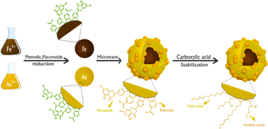
Eco-friendly, Microwave Assisted Green Synthesis of Magnetic Fe@Au (Core-Shell) NPs to Enhance Olive oil Efficiency on Eradication of H. pylori because most of the nanoparticles that to be introduced to the human body acts as a therapeutic agent in (Fig. 1), if its size is less than 50 nm (Jeevanandam et al., 2018; Al-Radadi., 2022).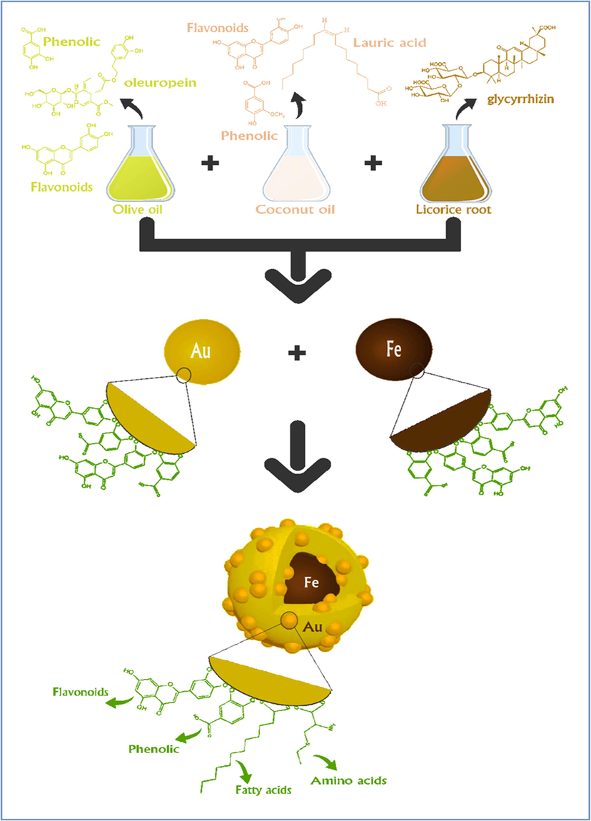
A schematic of the OLC-Fe@AuNPs.
To evaluate the in vivo anti-H. pylori action of Fe@Au Core-Shell NPs with Olive oil, licorice roots and coconut oil (OLC) incorporated in a nanostructured drug delivery system. It is considered as a future treatment strategy with gastroprotective drug delivery systems.
2 Experimental details
2.1 Materials and method
Licorice root was bought from iHerb website, and washed thoroughly with distilled water. About 2 g sun-dried powder of root extract was boiled in 50 ml of sterilized water to get extract. Chloroauric chloride (HAuCl43H2O) was purchased from Sigma–Aldrich. A stock solution contains (1 × 10⁻³ M) auric salt and de-ionized water used for this process. Organic coconut oil was gotten from oil mill. Iron (III) chloride (FeCl3·6H2O) was also purchased from Sigma–Aldrich. A stock solution contains (1 × 10⁻³ M) iron salt and de-ionized water and olive oil was gotten from farm in Aljouf.
2.2 Biosynthesis of gold coated iron nanoparticles (Fe@AuNPs)
To avoid any contamination in the results, before any reaction, all glassware was cleaned and sterilized. The synthesis consists of two main steps.
Synthesis of Fe-NPs by adding 3 ml of (0.001 M) aqueous solution of FeCl3·6H2O to OLC extract (2 ml of olive oil stock, 2 ml of licorice extract, and 1 ml of stock solution of coconut oil), where is natural antioxidants acted as a reducing agent, the process was assisted by microwaves (CEM Discover Microwave™) for 60 min at 50 °C. The color transformation from light yellow to dark brown indicated the reduction of Fe3+ to Fe0 nanoparticles.
for synthesis of Au-NPs, 5 ml of (1 × 10⁻³ M) aqueous HAuCl4·3H2O solution was mixed with natural antioxidants, consist of 2 ml of licorice extract, 2 ml of olive oil and 1 ml of coconut oil. In addition, magnetic bar was put in the mixture and microwave radiations were provided for 60 min at 25 °C. The change in the color of solution from yellow to red, indicated the reduction process of Au3+ to Au0 and formation of the gold nanoparticles. Subsequently, once the gold nanoparticles were formed, Fe-NPs were added to it for core shell nanoparticles and stabilized the mixture under microwave radiation for 10 min. Thus, the color transformation confirmed the synthesis of nanoparticles.
2.3 Characterization of Fe@Au nanoparticles
Due to combination of gold coated iron NPs (Fe@AuNPs), was morphologically characterized by a variety of spectroscopic techniques. The absorbance spectra were recorded using double beam scanning spectrophotometer and quartz cuvettes (Cary 100 UV–Vis Spectrometer from Agilent) at 350–700 nm. Fourier Transform Infrared Spectroscopy (FT-IR) (a Nicolet 6700) was performed in the spectral range of 200–4000 cm−1 and was utilized to determine the possible functional groups of the biomolecules that reside in the plant extract. The X-ray diffraction (XRD) (Shimadzu XRD-6000), were conducted to get data about the crystalline nature of nanoparticles. The internal structure of nanoparticles was examined by using Transmission electron microscope (TEM). Energy dispersive X-ray (EDX) (model-JSM-5610 LV) confirms the presence of gold and iron, demonstrating that the gold coated iron nanoparticles were formed. High Performance Liquid Chromatography (HPLC) analysis was performed to determine the percentage of natural antioxidants.
2.4 DPPH free radical scavenging assay
Protocol of (Choi et al., 2002) with minor modification was used to test the antioxidant potential of (OLC) extract and Fe@AuNPs. DPPH stock was prepared and 1 ml of (0.1 mM) DPPH methanolic solution was pipette out and gently mixed with 1 ml of (OLC) extract and Fe@AuNPs having varying Conc of (7, 31, 62, 125, 250 ,500 and 1000 μg/ml). The reaction mixture was incubated in the dark for half an hour and after incubation was exposed to absorbance at 517 nm against a blank solution. As a control 1 ml of methanol and 1 ml DPPH mixer was used. However, as a standard control butylated hydroxyl toluene (BHT) was used. The following formula was used to calculate the percent inhibition. AC is the absorbance of control while AT is of sample and the values are calculated according to ascorbic acid reference.
2.5 Analysis of cytotoxicity of Fe@AuNPs with extract
A 20% of FBS (WELGEN Inc.), 20 µg/ml of bFGF, 100 units/ml of penicillin and 100 μg/ml of streptomycin supplemented with M199 medium was used to grow the human umbilical vein endothelial cells at 37°and 5% of CO2. MTT assay was used to check the effect of (OLC) extract, and OLC-Fe@AuNPs on the viability of HUVECs. The basics of the MTT assay is the conversion of MTT (4,5‐dimethylthiazol‐2‐yl) (2,5‐diphenyl tetrazolium bromide) to insoluble MTT formazan. It happens by dehydrogenase which cleaved the tetrazolium ring in surviving cells. The process was performed in 24-well culture plates, after overnight incubation the M199 medium was supplemented with 1% of FBS and varying Conc of OLC extract and OLC-Fe@AuNPs solution followed by incubation for 24 h at 37° in humidified 5% carbon dioxide supplemented environment. Next, 5 mg/ml aqueous solution of MTT were added to each well and 0.3 ml additive DMSO was added to dissolve the MTT formazan quickly. The experiment repeated three times and the treated samples were exposed to absorbance at 570 nm to measure the cell viability (Rodríguez-León et al., 2019).
2.6 Anti-ulcer (ulcer-preventive) activity study
To check the anti-ulcer activity of OLC-Fe@AuNPs, (OLC) extract, PBS, (Clarithromycin + Amoxicillin + Omeprazole) and ethanol, a total of 7 groups of Wister rats were enrolled in the study. PBS (10 ml·kg−1) was administered orally to Group 1 for time period of 11 days and label it as control group. Group 2 were administered with PBS (10 ml·kg−1) for 10 days and on the 11th day, absolute ethanol (5 ml·kg−1) weas administered and labeled as ulcer control. Group 3 and 4 were respectively administered with 250 and 500 mg·kg−1 of OLC-Fe@AuNPs, while group 5 with triple regimen consisting of Clarithromycin 500 mg + Amoxicillin 1 g + 20 mg of Omeprazole for 10 days. Group 6 and 7 were respectively administered with 250 and 500 mg·kg−1 of (OLC) extract. After 1 complete day of fast, OLC-Fe@AuNPs or drug was administered at respective doses. After half an hour group 2–7 animals were administered with ethanol (5 ml·kg−1) for induction of ulcer. All the animals were served with an aesthetic ether and scarified. Pylorus ligation method was used to measure gastric volumes. Under dissecting microscope each stomach was examined for gastric erosion. Gastric mucosal ulcer was measured by plane glass square length × width (10 × 10 mm) and the ulcerate area (UA) was calculated. The % of protection (P%) availed to the animals through various treatments was calculated using the formula:
Small piece of stomach was fixed in paraffin wax after measuring the ulcerate area. Using standard technique, 5 µm thick sections were cut in a microtome and kept on a glass slide and the tissues were stained with Hematoxylin-Eosin (H&E) stain and viewed under microscope. As previously mentioned in HPLC and Antioxidants, that the phytochemical studies of the extract of (OLC) proved the existence of flavonoids and phenolics, amino acids, fatty acids, peptides and vitamins.
3 Results and discussion
3.1 High performance liquid chromatography (HPLC)
Licorice root extract was used in combination with olive oil and coconut oil (OLC), the hydroxyl and alcoholic groups of OLC helped in reducing the
and
ions, the OLC extract stabilizes the nanoparticles and helped in formation of bond between oxygen, iron and gold in biosynthesis of nanoparticles, in addition it has been used as an environmentally benign polymer. The three plant extracts (OLC) was considered a strong antibiotic for bacteria because they are rich in natural antioxidants such as amino acids, phenols, flavonoids, fatty acids and minerals such as (Fe, Ca, Na, K, Zn, S, Mg, P and Si) and vitamins such as (E, A, K and D), α-Tocopherol (E) and Retinol, these all acted as an anti-inflammatory agents, that restored damaged tissues to vitality and accelerates the healing of ulcers, and we found that one of the best plant extracts is the virgin olive oil: it is a powerful antibiotic for bacteria, it is considered to strengthen the immune system, protects the body from diseases and infections, prevents the oxidation of fats, prevents the formation of free radicals that cause cancerous tumors, increases gastric secretions and thus facilitates the process of absorption of natural antioxidants, and works to regulate the level of sugar in the blood, it slows the digestive process in the stomach, thereby helping to slow the increase in sugar level, it also contains natural antioxidants and a high amount of polyphenolic and flavonoids, it has more than forty compounds, as shown in Table 1 and Fig. 1S. Olive oil is also rich in saturated fatty acids, shown in Table 2, Figs. 2 and 3, such as palmitic acid, stearic acid and myristic acid, as well as unsaturated fatty acids. In addition, olive oil is very rich in phenols Fig. 2S (a-c) such as oleuropein, and also rich in three powerful natural antioxidants that are hydroxytyrosol, the vanillic acid, the verbascoside, and also contains a number of unsaturated fatty acids such as oleic acid, linoleic acid. The presence of green chlorophyll pigment (pheophytin) Fig. 3S, sterols and squalene Fig. 3S, it has also peptides. Olive oil is considered an antibiotic because it contains a high amount of triglycerides Fig. 3S, containing Retinol which restores damaged tissues. But olive oil lacks amino acids Table 3 and Fig. 4S that helps stabilize synthesized nanoparticles, and to improve the quality of olive oil and increase its efficiency as a plant extract, therefore we combined it with licorice root extract because it is rich in natural antioxidants such as carboxylic acids, and amino acids Table 3 and Fig. 4S, including glycerrhizin Fig. 5S, which has anti-ulcer activity, and its derivatives include carbenoxolone Fig. 5S, which has an effective affect in the treatment of ulcers of the stomach, duodenum and small intestine, anti-inflammatory, rich in phenols, flavonoids and steroids Fig. 6S, containing minerals, rich in aromatic compounds, and rich in proteins. Because of the damage and side effects already mentioned, due to high blood sugar level and fluid retention in the body Table.4, we added a small amount of it (2 ml) and combined it with coconut oil because it is rich in lauric acid and containing vitamin α-Tocopherol (E). It is a fatty acid Table.2 and is considered a strong antiparasitic, and strengthens the body's immunity by protecting it from many diseases, stimulator of beneficial bacteria, rich in vitamin E, which restores the vitality of damaged tissues and contains several vitamins including vitamin A as anti-inflammatory and stomach ulcer preventor and accelerates the healing of ulcers. 0 : undetected.
Phenolic
Concentration in Olive oil
Tyrosol
Caffeic acid
Oleuropein
Protocatechulic
11.04
12.1
39.56
9.55
Retention time
10.5
8.0
14.02
13.5
Flavonoids
Concentration in Olive oil
Luteolin
Rutin
Kampherol
Querestein
15.7
30.12
19.27
9.77
Retention time
13.2
5.4
10.3
15.02
Phenolic
Concentration in Coconut oil
Coumaric
Caffeic acid
Syringic
Vanillic acid
Gallic
8.14
4.01
9.16
29.56
18.12
Retention time
5.0
8.2
9.3
11.6
12.5
Flavonoids
Concentration in Coconut oil
Luteolin
Rutin
Isoquerestin
25.14
15.02
7.44
Retention time
13.2
5.39
8.01
Phenolic
Concentration in licorice root
Sinapic
Ellagic
Protocatechulic
Ferulic
25.14
18.09
9.12
7.04
Retention time
11.02
6.2
13.5
7.5
Flavonoids
Concentration in licorice root
Luteolin
Rutin
Kampherol
50.15
27.1
12.5
Retention time
13.1
5.4
10.3
Glycosides
Concentration in licorice root
linamarin
pinoresinol
laricinesol
50.12
18.30
4.22
Retention time
6.8
9.5
6.8
Organic acids
Concentration in licorice root
Succinic acid
Ascorbic acid
Fumaric acid
Gallic acid
13.41
10.87
2.17
4.56
Retention time
9.1
11.0
10.7
4.21
Triterpenes
Concentration in licorice root
Glycyrrhizin
81.737
Retention time
8.112
Fatty acid
Caprylic (C8)
Capric (C10)
Lauric (C12)
Myristic (C14)
Olive oil content (%)
0.0
0.0
0.13
0.0
Coconut oil content (%)
6.54
9.21
46.75
16.96
Licorice root content (%)
0.0
0.0
0.21
4.08
Retention time(min)
22.3
24.9
26.1
26.8
Fatty acid
Pentadecanoic (C15)
Palmitic (C16)
Palmitoleic (C16:1)
Heptadecanoic (C17)
Olive oil content (%)
0.0
14.25
0.0
2.26
Coconut oil content (%)
0.47
9.61
0.0
0.34
Licorice root content (%)
1.89
25.36
0.27
2.26
Retention time(min)
27.9
30.2
30.9
31.7
Fatty acid
Stearic (C18)
Oleic (C18:1)
Linoleic (C18:2)
Linolenic (C18:3)
Olive oil content (%)
1.61
66.69
16.34
0.56
Coconut oil content (%)
1.29
7.62
0.0
0.0
Licorice root content (%)
7.63
35.72
10.83
3.24
Retention time(min)
33.8
35.7
37.2
40.04
Fatty acid
Arachidic (C20)
Arachidonic (C20:1)
Behenic (C22)
Lignoceric (C24)
Olive oil content (%)
0.0
0.0
0.05
0.04
Coconut oil content (%)
0.23
0.0
0.0
0.0
Licorice root content (%)
8.16
0.35
0.0
0.0
Retention time(min)
39.4
39.8
42.3
43.1
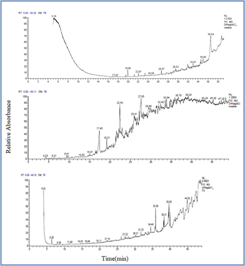
Mass spectrum of the main fatty acid compounds in the Olive oil, Coconut oil and Licorice root.
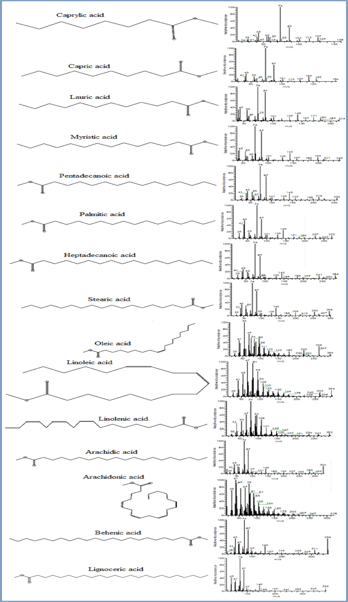
Mass Fragmentation and Chemical structure of the main fatty acid compounds in the Olive oil, Coconut oil and Licorice root.
Coconut oil
Olive oil
Licorice root
RT (min.)
Amino acids
Essential Amino Acids (EAA) (mg/100 g)
0
0
9.66
19.2
L-Threonine
0
0
10.25
24.8
L-Valine
0
0
5.69
27.7
L-IsoLeucine
0
0
12.78
31.9
L-Methionine
0
0
5.26
42.3
L-Phenyl alanine
0
0
2.8
51.3
L-Leucine
0
0
7.23
58.7
L-Lysine
Non-Essential Amino Acids (NAA) (mg/100 g)
0
0
9.24
12.8
Aspartic acid
0
0
4.98
20.6
Arginine
0
0
10.45
28.9
Glutamic acid
0
0
8.82
40.1
Glycine
0
0
5.24
42.2
Alanine
0
0
6.21
47.6
Proline
0
0
–
58.5
Tyrosine
0
0
7.19
52.4
Serine
0
0
3.76
50.2
Histidine
0
0
8.07
59.7
Cysteine
Antioxidant
total phenolic
total flavonoids
DPPH
ABTS
(mg/g)
(mg/g)
(µg/ml)
(µg/ml)
Olive oil
27.4 ± 1.92
9.2 ± 1.48
301.2 ± 34.8
458.4 ± 38.2
Coconut oil
3.25 ± 0.31
5.42 ± 0.64
405.0 ± 30.1
549.3 ± 40.5
Licorice root
15.26 ± 0.72
25.14 ± 1.98
86.9 ± 23.7
63.4 ± 9.3
We carried out HPLC analysis of plant extracts to determine the percentage of natural antioxidants Table. 4 such as phenols, flavonoids, amino acids, fatty acids, vitamins and minerals. In Table. 5 proximate composition and energetic value of olive oil, coconut oil and licorice root has been mentioned. In Table.6 different sugars are listed. The studied amino acids are Glutamic acid, Valine, Alanine, Proline, Phenylalanine, Serine, Threonine, Glycine, Isoleucine, Methionine, Tyrosine, Leucine, Arginine, Lysine, Aspartic acid, Histidine and Cysteine. In OLC extract the oleic acids were found in between 35 and 67% and thus it is categorized the highest fatty acid present in the OLC extract, while Lauric, Linoleic, Myristic acid etc were found in trace amounts. Green synthesis of magnetic bimetallic nanoparticle (Core-Shell NPs) from iron ion solution and gold ion solution with plant extracts olive oil, licorice root and coconut oil, with the help of microwave oven without polymer or organic solvent in the medium interaction. Microwave-assisted synthesis of NPs is electromagnetic and showing spectrum with frequency of (3 0 0) MHz to (3 0 0) GHz (TEM Discover Microwave™), it depends on electromagnetic radiation that relies on heating the solution directly with high efficiency and proves the dissolution of the reactants and the formation and growth of a rapid and homogeneous nucleus in Fe@AuNPs, which improves reaction conditions, green synthesis of NPs occur in one step, fast and short time to reduce the chance of side reactions, giving pure NPs, and producing high degree of dispersion of surface morphology, and stabilizing nanoparticles by prevent the aggregation as shown in Fig. 7. The surface area was large and produced small sized nanoparticles, finally, in an environmental friendly and safe manner that does not result in the generation of toxic or harmful waste. In medical sector gold nanoparticles drawn a significant consideration due to its potential biomedical applications. Fe@AuNPs were biosynthesized in the current study using OLC as a capping and reducing agent assisted by microwave radiation. Furthermore, factors such as metallic salt concentration and microwave radiation that affecting the synthesis of NPs were also studied. Synthesis of nanoparticles were confirmed by the color transformation to dark reddish brown and it was monitored by UV Spectroscopy (El-Naggar et al., 2016; Al-Radadi, 2021b).
Proximate composition
Ash
Moisture
Total Proteins
Total Lipids
Total Carbohydrates
Total Cholesterol
Triglycerides
(g/100 mg dry weight)
(g/100 mg dry weight)
(g/mg dry weight)
(mg/g)
(g/mg dry weight)
(mg/gm)
(mg/gm)
Olive oil content
0.35
4.51
0.19 ± 0.05
989.4 ± 5.7
0.0
0.19 ± 0.05
2.41 ± 0.23
Coconut oil content
0.14
7.2
1.04 ± 0.42
958.2 ± 7.4
0.0
0.52 ± 0.14
0.91 ± 0.15
Licorice root content
17.22
16.4
21.42 ± 0.94
87.1 ± 9.5
24.51 ± 31.3
–
–
Sugars
Sucrose
glucose
Xylose
Arabinose
mannose
Olive oil
0.0
0.0
0.0
0.0
0.0
Coconut oil
0.0
0.0
0.0
0.0
0.0
Licorice root
149.6
57.2
0.0
0.0
0.0
Retention time(min)
8.8
7.9
3.5
5.3
6.2
Sugars
galactose
Lactose
Rhamnose
fructose
Olive oil
0.0
0.0
0.0
0.0
Coconut oil
0.0
0.0
0.0
0.0
Licorice root content
1.3
0.0
0.0
4.1
Retention time (min)
7
9.5
10.5
4.7
![(A) UV–visible spectrum of AuNPs synthesized using various volumes [(1 ml Coconut oil + 2 ml Licorice root) + (0.5–2.0 ml Olive oil)] with 5 ml 1 × 10⁻³ M HAuCl4 solution after 60 min. (B) UV–visible spectrum of AuNPs produced via various volumes (1–5) ml 1 × 10⁻³ M HAuCl4 solution with 5 ml extract after 60 min. (C) UV–visible spectrum of AuNPs as a task of 5 ml 1 × 10⁻³ M HAuCl4 solution and 5 ml of extract after 60 min of addition. (D) UV–Vis spectra as a purpose of effect of different (2–8) pH of 5 ml 1 × 10⁻³ M HAuCl4 solution and 5 ml of extract after 60 min. (E)UV–visible spectrum of AuNPs as a task of 5 ml 1 × 10⁻³ M HAuCl4 solution and 5 ml of extract after 60 min. as a function of temperature (15–30 °C).](/content/184/2022/15/5/img/10.1016_j.arabjc.2022.103685-fig5.png)
(A) UV–visible spectrum of AuNPs synthesized using various volumes [(1 ml Coconut oil + 2 ml Licorice root) + (0.5–2.0 ml Olive oil)] with 5 ml 1 × 10⁻³ M HAuCl4 solution after 60 min. (B) UV–visible spectrum of AuNPs produced via various volumes (1–5) ml 1 × 10⁻³ M HAuCl4 solution with 5 ml extract after 60 min. (C) UV–visible spectrum of AuNPs as a task of 5 ml 1 × 10⁻³ M HAuCl4 solution and 5 ml of extract after 60 min of addition. (D) UV–Vis spectra as a purpose of effect of different (2–8) pH of 5 ml 1 × 10⁻³ M HAuCl4 solution and 5 ml of extract after 60 min. (E)UV–visible spectrum of AuNPs as a task of 5 ml 1 × 10⁻³ M HAuCl4 solution and 5 ml of extract after 60 min. as a function of temperature (15–30 °C).
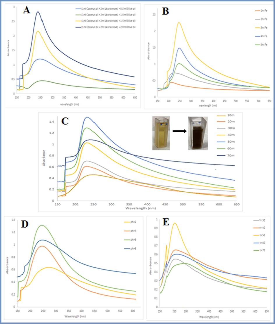
(A) UV–visible spectrum of FeNPs produced via various volumes (1 ml Coconut oil + 2 ml Licorice root) + (0.5–2.0 ml Olive oil) with 3 ml 1 × 10⁻³ FeCl3·6H2O solution after 60 min. (B)UV–visible spectrum of FeNPs produced via various volumes (1–5) ml 1 × 10⁻³ M FeCl3·6H2O solution with 5 ml extract after 60 min. (C) UV–visible spectrum of FeNPs as a task of 3 ml 1 × 10⁻³ M FeCl3·6H2O solution and 5 ml of extract after 60 min. (D) FeNPs UV–Vis spectra as a purpose of effect of different (2,4,6 and 8) pH of 3 ml 1 × 10⁻³ FeCl3·6H2O solution and 5 ml of extract after 60 min. (E) UV–visible spectrum of FeNPs as a task of 3 ml 1 × 10⁻³ FeCl3·6H2O solution and 5 ml of extract. as a function of temperature (30–70 °C).
UV–visible spectrum of Fe@AuNPs with (OLC) extract after 10 min.
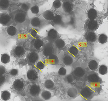
Transmission electron microscopy (TEM) micrograph of iron coated with gold nanoparticle.
3.2 Factors affecting the synthesis of AuNPs
3.2.1 Volume of extract and volume of Au metal
The biosynthesis of gold nanoparticles was confirmed by UV–Vis absorption spectroscopy. Fig. 4A, is the UV spectra of microwave assisted and OLC mediated gold nanoparticles. Olive oil was taken in quantities of 0.5, 1, 1.5 and 2 ml. whereas, licorice root extract and coconut oil was taken in quantities of 2 ml and 1 ml. A gradual increase in the absorbance intensity was observed by increasing the OLC Conc. from 0.5 ml to 2 ml, maximum peaks were observed at 2 ml, some minor peaks were also observed that corresponds to the presence of trace biomolecules in the extract. In the case of concentration, in Fig. 4B it was taken in different quantities of 1, 2, 3, 4 and 5 ml, where the results showed increasing in the intensity of absorbance with increasing in volume, the highest absorption was observed at maximum wavelength of 540 nm at 5 ml of extract. The color change from yellow to red was the primary indication of the biosynthesis of Au-NPs with ideal SPR peaks, at low volumes the broadened peak was observed which confirm the presence of large sized Au-NPs (Al-Radadi, 2022).
3.2.2 The effect of time on AuNPs
In Fig. 4C, the gold nanoparticles gave a characteristic peak at 540 nm which confirmed its synthesis again and proved the OLC extracts are good capping and reducing agents. However when the time exposure of microwave radiation increased from 60 to 70 min, sharpness of peak was observed which indicated the stability and maximum size of the nanoparticles.
3.2.3 The effect of pH on AuNPs stability
The UV–visible spectra of gold nanoparticles displayed in Fig. 4D with different pH factors investigated, it was found that the most preferable pH for extract (OLC) was pH = 6 at = 540 nm, which gave sharper and higher absorbance peak and confirmed by TEM images indicating that at this pH value the gold nanoparticles were more homogeneous shape with smaller size (Al-Radadi & Al-Youbi, 2018a), In contrast to pH values below 6, the particles size was found large due to agglomeration.
3.2.4 The effect of temperature on AuNPs
Temperature is one of the important parameters in the synthesis of nanoparticles. Different peaks were observed at (15, 20, 25 and 30 °C). A narrow UV band at 540 nm was observed at temperature of 25°, while the absorbance intensity became weaken at 30 °C as shown in Fig. 4E. The increase in temperature above than 25°resulted in aggregation of nanoparticles and thus the peaks became weaken. The best synthesis of nanoparticles was observed at 25°.
3.3 Factors affecting on the synthesis of FeNPs
3.3.1 Volume of extract and volume of Fe metal
Fig. 5A shows the UV–visible absorption spectrum of iron nanoparticles at a spectral range of 150–650 nm (Pattanayak & Nayak, 2013). In addition, the absorption spectra of the synthesized Fe0 nanoparticles it varies with the amount of iron from 1 to 5 ml. Where several absorption peaks were observed at 220 nm at 1 ml, 225 nm at 2 ml, 250 nm at 3,4 and 5 ml due to the excitation of surface plasmon vibrations in the FeNPs solution. The highest absorption was observed at 3 ml and is the indication of the small Fe nanoparticles (Carroll et al., 2010; Hosseynizadeh Khezri et al., 2012).
The extract was prepared as previously presented with gold in terms of quantities taken from licorice root extract, olive oil and coconut oil. The UV–visible absorption spectrum of (OLC) extract in Fig. 5B showed suitable (SPR) to synthesize the iron nanoparticles via high UV spectra at 5 ml..
3.3.2 The effect of time on FeNPs
The time factor was studied during preparing iron nanoparticles, where the results in Fig. 5C showed that the best time it took to prepare iron nanoparticles in the microwave was 60 min, where a change in color was observed from yellow to brown, indicated the formation of small Fe nanoparticles. It was also observed that the reactivity of FeNPs was high, which confirms the small size of the Fe0 nanoparticles, because it is linked to their particle size: smaller sizes lead to higher reactivities (Machado et al., 2014). Hence, the nanoparticles was prevented from agglomerating.
3.3.3 The effect of pH on FeNPs stability
The pH was determined by using digital pH meter. The pH of the reduced Fe nanoparticle solution found to be 6. The pH factor of the Fe nanoparticles was studied as shown in Fig. 5D, by adding hydrochloric acid or sodium hydroxide to obtain the best results. It was observed that the natural pH = 6 is the preferable and standard pH for synthesis of FeNPs.
3.3.4 The effect of temperature on FeNPs
A narrow UV band at 250 nm was observed at temperature of 50°, while the absorbance intensity became weaken at 70 °C as shown in Fig. 5E. The increase in temperature above than 50° resulted in the aggregation of nanoparticles, and thus the peaks became weaken. The best synthesis of nanoparticles was observed at 50°. At 70 °C the SPR peak decreased.
3.4 The UV–Vis spectrum of Fe@AuNPs synthesized using microwave radiation
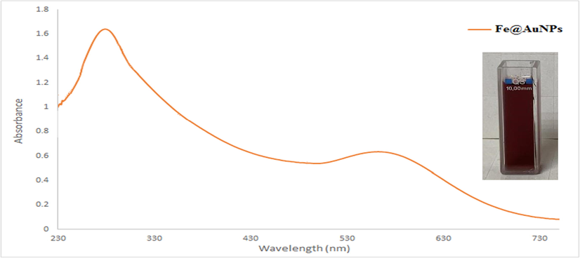
The Fe@AuNPs was first characterized by UV–Vis Spectroscopy as shown in Fig. 6 It is observed that FeNPs showed a prominent absorption peak at λmax = 279 nm, whereas AuNPs displayed an characteristic peak at λmax = 565 nm A red shift was also observed in the peak for Fe@AuNPs as shown in Fig. 6. The bathochromic shift indicated the large sized OLC mediated nanoparticles (Bandyopadhyay et al., 2014)
3.5 Transmission electron microscopic analysis (TEM) and HR-TEM of Fe@AuNPs
This special type of microscopy was used to determine the shape and size of core–shell iron and gold nanoparticles. The TEM image illustrated and confirmed that the core–shell iron and gold nanoparticles were spherical in shape, homogeneous, not aggregated and small in size (Al-Radadi, 2021b). The size of the nanoparticles was 2.9, 3.4, 5.7 and 10 nm as shown in Fig. 7, and the HR-TEM micrograph of spherical Fe@AuNPs is shown in Fig. 8A. The HAADF-STEM images of the Fe@Au nanoparticles and the elemental mapping images is shown in the Fig. 8B that clearly demonstrates the Fe core and Au shell in a bright and dark contrast, which can be further verified by corresponding energy dispersive x-ray spectroscopy (EDX) in Fig. 11B (Wu et al., 2009). By the analysis of size distribution histogram the size of the nanoparticles was determined and shown in Fig. 8C (Chung & Shih, 2014).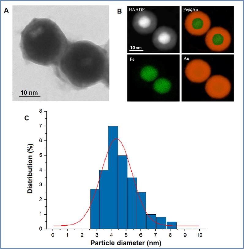
(A) HR-TEM image of spherical Fe@AuNPs, (B) HAADF-STEM images of the Fe@AuNPs and the elemental mapping images and (C) Size distributions of Fe@AuNPs.
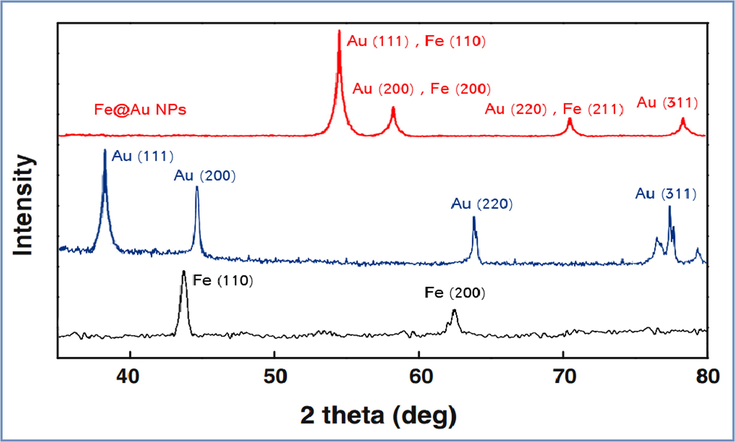
X-ray diffraction (XRD) patterns of green synthesized iron, gold, and Core-Shell Iron@Gold Nanoparticles.
3.6 X-ray diffraction (XRD) of Fe@AuNPs
The X-ray diffraction (XRD) pattern in Fig. 9 illustrated the OLC mediated nanoparticles are crystalline in nature (Al-Radadi & Al-Youbi, 2018b) and showed characteristic peaks at scattering angles of (2θ) values of 38.13°, 44.23°, 64.71°, and 77.49°, which was indexed to (1 1 1), (2 0 0), (2 2 0), (3 1 1) (Singh et al., 2013). In addition, the (XRD) of iron nanoparticle showed characteristic peaks at scattering angles (2θ) values of 43.7°, 62.5°, which was indexed to (1 1 0), (2 0 0). Also, the (XRD) of Fe@AuNPs, due to overlap of diffraction peaks the iron peaks are under the gold at 2θ = 54.37°, 58.12°, 70.31, and 78.13° as shown above (Zhang et al., 2006; Kvitek et al., 2019). The average crystallite size according to Debye–Scherrer equation was calculated and found to be 5.5 nm.
3.7 Fourier transform infrared spectroscopy (FTIR) of Fe@AuNPs
FT-IR analysis were conducted to know about the possible involvement of OLC extract functional groups in the synthesis and reduction of iron and gold nanoparticles, the FT-IR spectra of NPs were compared against spectra of extract. Multiple characteristic peaks were observed for extract between 400 and
4000
as in shown in Fig. 10. The bands shown at 3712.5
was due to the stretching and vibrations of alcohol (O—H) bonds, the peaks at 3472
indicates the existence of primary amino bond, 3267
indicates to aromatic carbon ring, and the (C—H) aliphatic group at 2921.8
supports that fatty acids in olive oil and coconut oil are long-chain carboxylic acids. The strong peak at 1733
corresponds to carbonyl group (C⚌O) bond stretching, disappearance and vibration of flavonoids and polyphenols present in the OLC extract. Due to the coordination and reduction of bio-molecules with Au, a small shift was observed towards lesser wave numbers for the above described peaks (Kumaret al., 2021; Ahmad et al., 2015). The peak at 1025
indicates the existence of (C—N), similar characteristic peaks of FeNPs with extract were also observed. Also, similar peaks at 3587, 3148, and 1337
appeared in the FT-IR spectra of Fe@AuNPs with extract and thus indicating the presence of similar functional groups in the capping of iron nanoparticles.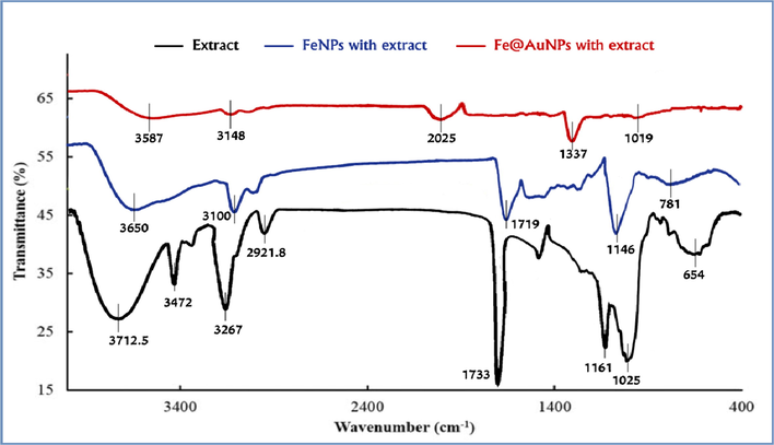
FT-IR spectra of olive oil and coconut oil and Glycyrrhiza root extract, FeNPs with aqueous extract and Fe@AuNPs with aqueous extract.
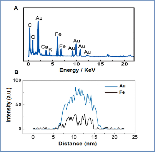
(A) Energy-dispersive X-ray spectroscopy (EDX) spectrum of Fe@AuNPs with aqueous extract and (B) Line scanning profiles of Fe@AuNPs.
3.8 Energy dispersive X-ray spectroscopy (EDX) of Fe@AuNPs
Elemental composition of the nanoparticles was estimated by EDX, confirms the presence of gold nanoparticles were produced by reduction of the ions, in addition, presence of iron nanoparticles by reduction of the ions. The EDX image showed the core–shell structure, the Au is distributed around the whole Fe core, as manifested by the line scanning image in which only Au appears in the external edges, while Fe show up in the middle, as shown in Fig. 11B, Au ligands are stronger than those of Fe, indicating the higher content of Au in the nanoparticles, where the atomic ratio of Au against Fe is roughly 70:30. EDX spectrum Fig. 11A shows the presence of additional carbon, oxygen, calcium and potassium (Ban et al., 2005).
3.9 Magnetic hysteresis loops of Fe@AuNPs
Fig. 12 Illustrated the prepared Fe@AuNPs has magnetic characteristic due to the presence of iron nanoparticles. The magnetic nanoparticles were designed to target affected tissue by applying an magnetic field (Liu et al., 2020), the magnetic properties were confirmed and studied by measuring hysteresis loops at 25 °C. As in Fig. 12A the magnetization vs the applied magnetic field curve, the Fe@AuNPs showed characteristic superparamagnetic behavior and high saturation fields around 36.25 amu/g (Pana et al., 2007). At room and ordinary temperatures, the Fe@Au core–shell nanoparticles are ferromagnetic in nature (Ban et al., 2005). Taking into consideration that ferromagnetic minerals tend to produce narrow loops (Mooney, 2002), and the results showed that Fe@AuNPs are the narrower loop compared to other minerals (Ovejero et al., 2015). The image of the magnetic nanoparticles is shown in Fig. 12B.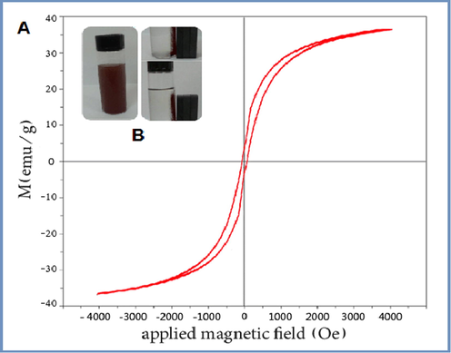
(A) Magnetic hysteresis loops of Fe@AuNPs, (B) Image of the magnetic nanoparticles.
3.10 Biological activity
3.10.1 Antioxidant activity of Fe@AuNPs with extract
It is revealed from different studies that phenolic compounds in the (OLC) extract acts as an antioxidant and reducer of FeCl3·6H2O and HAuCl4 to Fe@AuNPs. Free radicals usually damage the cells and tissues therefore the role of antioxidant is very important. Recently natural plant as a source of antioxidants was widely used to determine the antioxidant potential of nanoparticles usually DPPH was used (Kumar et al., 2018). Upon reduction of DPPH the color of iron transform into brown, and the color of gold changes to red. The color transformation is due to production of H+ ions (Adawiyah et al., 2019). In Fig. 13A, it was found that the DPPH scavenging activity increases with the increase of the concentration of (OLC) and Fe@AuNPs, due to inhibition of the interaction with free radicals. Antioxidant activities of the compounds, present in the extract may depend on structural features, such as the number of phenolic or methoxy groups and flavones hydroxyl. The synthesized Fe@AuNPs showed scavenging of DPPH free radicals (Francis et al., 2018; Vijayan et al., 2018; Abdoli et al., 2021).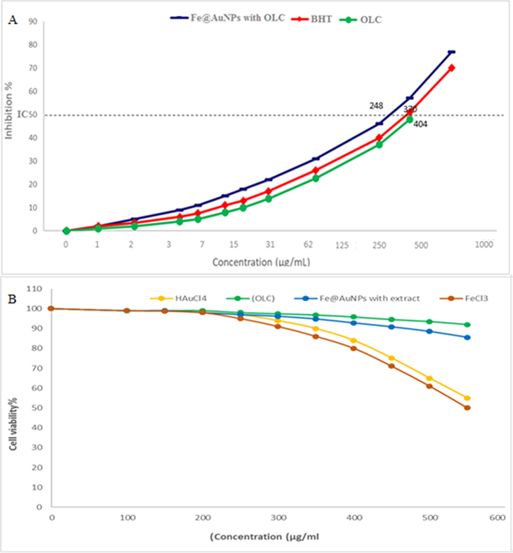
(A) Antioxidant potential of Fe@AuNPs with extract, (OLC), and BHT (Butylated hydroxyltoluene) and (B)Percent viability measured on human umbilical vein endothelial cells after treatment with present Fe@AuNPs with extract, (OLC), HAuCl4 and FeCl3.
Owing its capability to cape free radicals, DPPH antioxidant assay is widely used technique DPPH when encounters antioxidants, it retrieved the hydrogen ions A purple color is produce as a result and can be detected at 517 nm (Shunmugam, et al., 2021). DPPH is more stabilized when it gets more hydrogen ions and upon proper reduction its initial purple color changes from purple to yellow and thus DPPH gets stabilized DPPH free radical scavenging effect of (OLC) and Fe@AuNPs with extract in varying Cons of (0, 1, 3, 7, 15, 31, 62, 125, 250, 500, and 1000 μg/ml) indicated impressive prevention similar to BHT. The IC50 of (OLC), BHT, and Fe@AuNPs with extract was found 404, 370, and 248 μg/ml, respectively (kumar et al., 2021; Al-Radadi, 2021a) as shown in Fig. 13A. In correspondence to our studies, previous studies have reported potential antioxidant activities by using metallic nanoparticles.
3.10.2 Cytotoxicity survey of Fe@AuNPs with extract
Test cells (HUVEC) were treated with varying Cons of (OLC) and OLC-Fe@AuNPs extract and cytotoxicity were determined by MTT assay. After incubation of 48 h the activity were detected at absorbance rate of 570 nm, which showed extra ordinary compatibility even at highest concentration of 1000 μg/ml of (OLC) and Fe@AuNPs as shown in Fig. 13B. previously studies has reported that combination of biological compounds with metals in nanoparticles reduce its cytotoxicity (Kandasamy et al., 2021, Mohammadi et al., 2021).
3.10.3 Antimicrobial activity
Antimicrobial activities of the (OLC) extract and Fe@AuNPs and control was evaluated against bacteria and fungi. Conventional paper disk diffusion method was used to determine the inhibition zones and MIC values. As a positive controls Norfloxacin, and Nystatin were used. The results were represented in Tables 7 and 8. The OLC extract showed IZ near to Norfloxacin, and Nystatin, while, Fe@AuNPs showed potential zones of inhibition against test organisms as compared to drug of reference. OLC extract exhibited moderate to potent anti-bacterial activities with inhibition zone (IZ) ranged between 22 and 30 mm for Bacillus subtilis and Escherichia coli compared with that of norfloxacin that have IZ ranged between 24 and 27 mm. Moreover, Fe@AuNPs showed potent IZ between 28 and 29 mm for Bacillus subtilis and Salmonella typhi. For fungi the (OLC) extract was presented antifungal activity with inhibition zone (IZ) ranging between 18 and 19 mm for Fusarium oxysporum, Aspergillus flavus and Penicillium citrinum. In comparison to nystatin as a therapeutic anti-fungal agent (17–20 mm). Moreover, Fe@AuNPs showed IZ great results more than the standard drug between 21 and 22 mm for Aspergillus niger and Fusarium oxysporum. The minimal inhibition concentration (MIC) for (OLC) extract with antibacterial potential was ranged between 3.9 µg/mL and 15.62 µg/mL against Salmonella typhi and Bacillus subtilis respectively. From the other hand Fe@AuNPs showed promising MIC which ranged between 0.03 µg/mL and 6.56 µg/mL against Salmonella typhi and Bacillus subtilis, respectively. In case of antifungal activity MIC for (OLC) extract the results were ranged between 15.62 µg/mL and 62.5 µg/mL toward Aspergillus niger and Fusarium oxysporum, respectively. At the same time Fe@AuNPs showing 6.56 µg/mL and 13.5 µg/mL against Candida albicans and Aspergillus niger, respectively. This result was the highest than the nystatin (standard drug) which recorded MIC against Candida albicans and Aspergillus niger as 7.81 µg/mL and 20.5 µg/mL, respectively. As previously mentioned in HPLC and antioxidants that the (OLC) extract contained large amounts of natural, phenolic, flavonoids, vitamins, fatty acids, amino acids, peptides and minerals. The presence of multiple phenolic and flavonoids have shown many health-promoting benefits such as anticancer, anti-inflammatory, anti-ulcer, antibacterial and antifungal (Al-Radadi, 2021a; Wang et al., 2015) and that is consider the main reason and interpretation for the antimicrobial activity for (OLC) extract. Against multiple microorganisms the antibacterial potential of OLC were evaluated (Sarwar et al., 2020). Burkholderia cepacia and Klebsiella pneumonia is inhibited by Myricetin, while flavonoids kaempferol and myricetin are also have the potential to cure gastric ulcers. anti-Helicobacter pylori activity is also possessed by these compounds and thus used as a potent agent in therapeutics (Sharifi et al., 2019). Echinacin-permethyl ether and apigenin-5,4-dimethyl ether are the derivatives of echinacin and apigenin-7-O-glucoside per methylate which is formed by their methylation. The above compounds were evaluated for its antifungal activity against conidia's germination of Alternaria tenuissima Wiltshire, that cause pigeon pea leaf blight disease (Cajanuscajan) and these compounds were also used to control the pigeon pea Alternaria blight disease (Akintelu et al., 2021). The hydroxyl groups pattern of flavonoids is (myricetin > luteolin > quercetin > kaempferol) more hydroxyl groups lead to potent antibacterial activity against antibiotic resistant bacteria. The extract of (OLC) is changing the extracellular surface tension and thus leads to the lysis of bacterial cell membrane. Additionally, lipophilic flavonoids could also interrupt microbial membranes. Mechanism of antibacterial and antifungal activity of aqueous and organic extracts of the (OLC) extract and Fe@AuNPs possibly due to their ability to form a complex with bacterial cell walls and extracellular soluble proteins and that is lead to damage of microbial membranes then causing killing microbial cells. Some of the organic complexes of Au (I & III) and Fe (II & III) ions are antibacterial in nature. Fe@AuNPs are antifungal, but with conflicting results on their antibacterial activity (Inbaraj et al., 2020; Sathiyanarayanan et al., 2017). Results are shown in Fig. 14.
Bacterial test strains
Mean diameter of inhibition zone (mm)/minimum inhibitory concentration (MIC) (µg/ml).
(OLC) extract
Fe@AuNPs
Norfloxacin (standard)
Inhibition zone
MIC
Inhibition zone
MIC
Inhibition zone
MIC
Bacillus subtilis
(ATCC 6633)22 ± 0.22
15.62 ± 0.05
28 ± 0.32
6.56 ± 0.26
24 ± 0.56
3.9 ± 0.12
Staphylococcus aureus
(ATCC 29213)
24 ± 0.01
31.25 ± 0.45
29 ± 0.51
3.9 ± 0.55
23 ± 0.5
2.5 ± 0.35
Escherichia coli
(ATCC 25922)
30 ± 0.32
7.51 ± 0.77
32 ± 0.01
1.95 ± 0.46
27 ± 0.98
1.57 ± 0.69
Pseudomonas aeruginosa (ATCC 27853)
25 ± 0.45
15.62 ± 0.02
28 ± 0.12
2.5 ± 0.87
25 ± 0.87
3.9 ± 0.47
Salmonella typhi
(ATCC 6539)27 ± 0.34
3.9 ± 0.08
29 ± 0.52
0.03 ± 0.14
23 ± 0.16
1.57 ± 0.25
Fungal test strains
Mean diameter of inhibition zone (mm)/ minimum inhibitory concentration (MIC) (µg/ml)
(OLC) extract
Fe@AuNPs
Nystatin (standard)
Inhibition zone
MIC
Inhibition zone
MIC
Inhibition zone
MIC
Candida albicans
(ATCC 10231)14 ± 0.34
31.25 ± 0.16
18 ± 0.56
6.56 ± 0.41
15 ± 0.2
7.81 ± 0.16
Aspergillus niger
(RCMB 002007)
16 ± 0.23
15.62 ± 0.33
21 ± 0.61
13.5 ± 0.85
18 ± 0.2
20.5 ± 0.52
Aspergillus flavus
(ATCC 16883)
19 ± 0.52
41.6 ± 0.26
20 ± 0.71
26.75 ± 0.56
17 ± 0.12
31.25 ± 0.45
Fusarium oxysporum
(RCMB 008002)18 ± 0.34
62.5 ± 0.75
22 ± 0.45
31.25 ± 0.55
20 ± 0.32
26.4 ± 0.2
Penicillium citrinum
(RCMB 001011)17 ± 0.16
31.25 ± 0.35
23 ± 0.81
29.75 ± 0.41
20 ± 0.20
31.25 ± 0.15
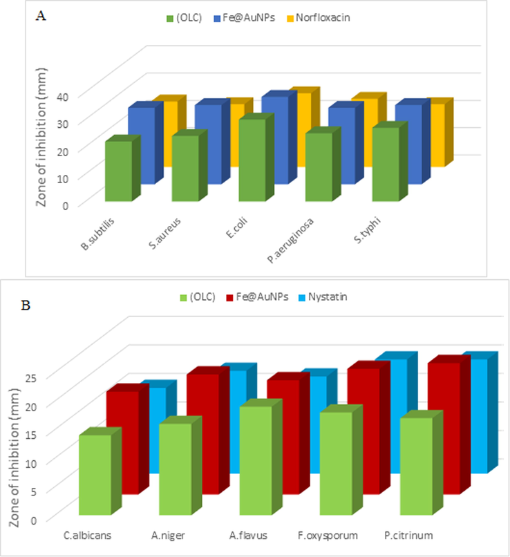
(A) Antibacterial activity of (OLC) extract and Fe@AuNPs and (B)Antifungal activity of (OLC) extract and Fe@AuNPs.
3.10.4 Anti-ulcer (ulcer-preventive) activity of Fe@AuNPs with (OLC) extract
3.10.4.1 Chemicals and reference drug: analytical grade chemicals were used in the current study
The triple regimen consisting of (Clarithromycin + Amoxicillin + Omeprazole) (reference drug) inhibit acid production by inhibiting the enzymes in stomach wall and thus allowing the stomach to heal quickly.
3.10.4.2 Plant material and extract preparation
2 g of licorice root is boiled in de-ionized water and 2 ml was taken from it, 2 ml of stock of olive oil and 1 ml of stock of coconut oil. Extract was taken and dissolved in 10 ml of PBS, 1% tween 80 was also used to make the extract properly dissolved in extract.
3.10.4.3 Animals
91 Wistar rats (180–200 g) and male albino mice (20–25 g) was purchased and kept in ideal environment with supplement of rodent diet. CPCSEA guidelines were followed during experiments and the study was approved by Institutional animal ethical committee.
3.10.4.4 Acute toxicity study
Seven groups of albino mice’s (13 in each group) were exposed to acute toxicity study of OLC extract. All the mice’s were fasted overnight and administered with the OLC-Fe@AuNPs single dose of Conc. of 250, 500, 2000 and 5000 mg·kg−1. Mice’s were served with PBS as a control.
3.10.4.5 Anti-ulcer (ulcer-preventive) activity study
As stated in Table 9, treatment of Wister rats with OLC-Fe@AuNPs protect the stomach mucosal layer from ethanol induced ulcer. OLC-Fe@AuNPs treatment protect the mucosal layer up to 80.37% and 98.36% respectively at 250 and 500 mg·kg−1 doses as compared to control groups. While the reference drug (Clarithromycin + omeprazole + amoxicillin) provided ulcer protection up to 76.31%. Samples without treatment of OLC-Fe@AuNPs showed complete ulceration Fig. 15A. Rats treated with reference drug showed protection as shown in Fig. 15B, 250, 500 mg·kg−1 (OLC) extract Fig. 15C, Fig. 15D and 250, 500 mg·kg−1 OLC-Fe@AuNPs Fig. 15E, Fig. 15F. Rats received almost 250 mg·kg−1 of OLC-Fe@AuNPs and significantly reduced gastric lesion formation and submucosal edema similar to the reference drug treated animals but the higher dose of 500 mg·kg−1 was much better, revealed from histopathological studies (Spósito, et al., 2019). OLC-Fe@AuNPs have various potential biomedical properties such as anti-inflammatory and anticancer. OLC-Fe@AuNPs mediated anti-ulcer activity was reported for the first time in the current study. The significant anti-ulcerative properties as shown in Table 9 revealed that OLC-Fe@AuNPs is gastro protective in nature and it was confirmed by histopathological studies. As antioxidant activities of the compounds, present in the extract may depend on structural features, such as the number of phenolic hydroxyl or methoxy groups and flavones hydroxyl as in Fig. 13A have been identified in the OLC-Fe@AuNPs, the anti-ulcer activity is due to the antioxidant nature of the extract as in Table 10. OLC-Fe@AuNPs is biocompatible as it is confirmed from sub-acute studies. +: Indicates that change was observed; –: Indicates that there was no change.
Treatment
Dose (mg/kg−1)
Ulcer area (mm2)
Protection (%)
Control
NA
0.00 ± 0.0
NA
Ulcer control
0
891.00 ± 4.05
0.0
Drug control (Clarithromycin + Omeprazole + Amoxicillin)
8
211.03 ± 11.60
76.31%
Treatment 1 (OLC) extract
250 Low
596.35 ± 8.07
33.06%
Treatment 2 (OLC) extract
500 High
307.8 ± 6.29
65.45%
Treatment 3 (OLC-Fe@AuNPs)
250 Low
174.9 ± 12.043
80.37%
Treatment 4 (OLC-Fe@AuNPs)
500 High
14.6 ± 9.10
98.36%
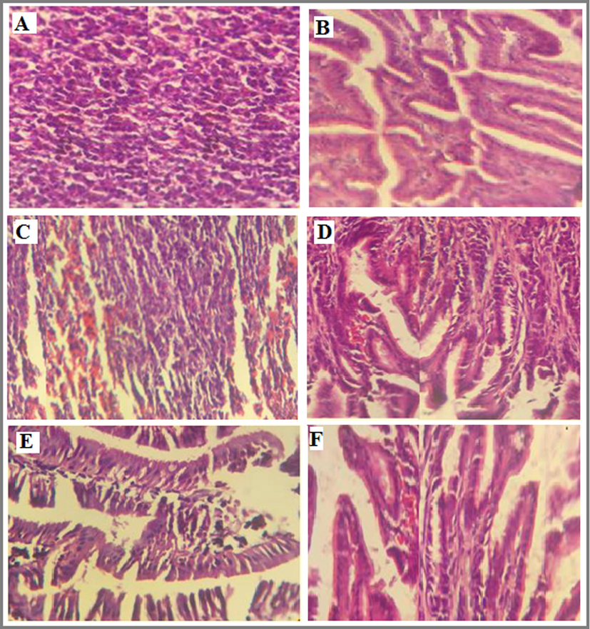
Anti-ulcer activity of: (A) stomach of an ulcer control rat, (B) stomach of a rat treated with the reference drug, (C) stomach of a rat treated with 250 mg kg−1 (OLC) extract, (D) stomach of a rat treated with 500 mg kg−1 (OLC) extract, (E) stomach of a rat treated with 250 mg kg−1 OLC-Fe@AuNPs, (F) stomach of a rat treated with 500 mg kg−1 OLC-Fe@AuNPs.
Time after administration (h)
Gross activity
2
3
5
7
12
24
Respiration
–
–
–
–
–
–
Writhing
–
–
–
–
–
–
Tremors
–
–
–
–
–
–
Convulsion
–
–
–
–
–
–
Salivation
–
–
–
–
–
–
Diarrhea
–
–
–
–
–
–
Mortality
–
–
–
–
–
–
Hind limb paralysis
–
–
–
–
–
–
Sedation
–
–
–
–
–
–
Skin irritation
–
–
–
–
–
–
Eye irritation
–
–
–
–
–
–
CNS Depression
–
–
–
–
–
–
The final part of this study measured direct effects of OLC-Fe@AuNPs, (OLC) extract, olive oil, licorice root extract and coconut oil of levels of anti-H. pylori. Results given in Fig. 16 showed that all samples increased production of specific antibodies, supporting the hypothesis that (OLC) extract has strong anti-inflammatory and anticancer properties. In samples, OLC-Fe@AuNPs and OLC extract are showing the strongest affects followed by olive oil and licorice root extract with a medium difference, while coconut oil was the lowest one among them (Vetvicka et al., 2016).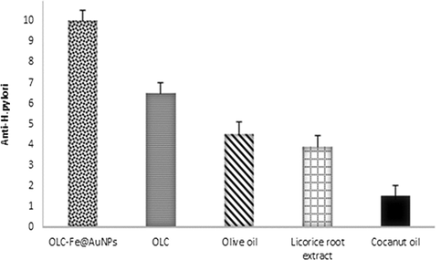
Effects of OLC-Fe@AuNPs, (OLC) extract, olive oil, licorice root extract and coconut oil on anti-H.pylori.
4 Conclusion
It was concluded that using OLC extract as a capping g and reducing agent is a good choice for the synthesis of the Fe@AuNPs core–shell nanoparticles. Extracts of olive oil, licorice root extract and coconut oil have potent antimicrobial activities. It was also concluded that microwaves, temperature and pH optimization help to achieve the small sized nanoparticles that to be used in the therapeutics. The biosynthesized nanoparticles were characterized and confirmed by UV–Visible spectroscopy, Energy dispersive X-ray spectroscopy (EDX), X-ray diffraction (XRD), High resolution Transmission electron microscope (HR-TEM), Fourier Transform Infrared Spectroscopy (FT-IR) and high-performance liquid chromatography (HPLC), High angle annular dark-field scanning TEM (HAADF-STEM), Particle-Size Distribution (PSD), Magnetic hysteresis loops. Helicobacter pylori was successfully eradicated by iron coated gold nanoparticles and iron coated gold nanoparticles can be used as a therapeutic in stomach ulcer. The anti H pylori and anti-ulcer potential of biosynthesized nanoparticles were checked in vivo in animal model (Wister rats). The antimicrobial properties revealed that nanoparticles encounter with bacterial cell membrane and leads to its damage and thus proved itself a good option to be considered as an antimicrobial. The OLC mediated Fe@Au nanoparticles were found nontoxic at high concentration but proper cytotoxic studies are recommended to use nanoparticles in vivo.
Acknowledgement
I would like to express my sincere thanks to Saudi Patent Office at (SAIPKSA) for providing adequate information and facilitate the registration of the patent number (7403), titled “Microwave Assisted Green Synthesis of Fe@Au core-shell Nanoparticales Magnetic to Enhance Olive oil Efficiency on Eradication of Helicobacter Pylori” in (24/12/2020).
Declaration of Competing Interest
The authors declare that they have no known competing financial interests or personal relationships that could have appeared to influence the work reported in this paper.
References
- Novel biosynthesis, characterization and bio-catalytic potential of green algae (Spirogyra hyalina) mediated silver nanomaterial’s. Saudi J. Biol. Sci.. 2022;29(1):411-419.
- [CrossRef] [Google Scholar]
- Green synthesis of gold nanoparticles using Centaurea behen leaf aqueous extract and investigating their antioxidant and cytotoxic effects on acute leukemia cancer cell line (THP-1) Inorg. Chem. Commun.. 2021;129:108649.
- [CrossRef] [Google Scholar]
- Plants against Helicobacter pylori to combat resistance: an ethnopharmacological review. Biotechnol. Rep,. 2020;26:e00470.
- [CrossRef] [Google Scholar]
- Elucidation of synergistic effect of eucalyptus globulus honey and Zingiber officinale in the synthesis of colloidal biogenic gold nanoparticles with antioxidant and catalytic properties. Sustain. Chem. Pharm.. 2019;13(June):100156.
- [CrossRef] [Google Scholar]
- Silver and gold nanoparticles from Sargentodoxa cuneata: synthesis, characterization and antileishmanial activity. RSC Adv.. 2015;5(90):73793-73806.
- [Google Scholar]
- Biosynthesis of gold nanoparticles: a green approach. J. Photochem. Photobiol. B: Biol.. 2016;161:141-153.
- [CrossRef] [Google Scholar]
- Green synthesis, characterization, and antibacterial investigation of synthesized gold nanoparticles (AuNPs) from Garcinia kola pulp extract. Plasmonics. 2021;16(1):157-165.
- [CrossRef] [Google Scholar]
- Artichoke (Cynara scolymus L.), mediated rapid analysis of silver nanoparticles and their utilisation on the cancer cell treatments. J. Comput. Theor. Nanosci.. 2018;15(6–7):1818-1829.
- [CrossRef] [Google Scholar]
- Green synthesis of platinum nanoparticles using Saudi’s Dates extract and their usage on the cancer cell treatment. Arab. J. Chem.. 2019;12(3):330-349.
- [CrossRef] [Google Scholar]
- Green biosynthesis of Pt-nanoparticles from Anbara fruits: toxic and protective effects on CCl4 induced hepatotoxicity in Wister rats. Arab. J. Chem.. 2020;13(2):4386-4403.
- [CrossRef] [Google Scholar]
- One-step synthesis of au nano-assemblies and study of their anticancer activities. J. Comput. Theor. Nanosci.. 2018;15(6–7):1861-1870.
- [CrossRef] [Google Scholar]
- Environmentally-safe synthesis of gold and silver nano-particles with AL-Madinah Barni fruit and their applications in the cancer cell treatments. J. Comput. Theor. Nanosci.. 2018;15(6–7):1853-1860.
- [CrossRef] [Google Scholar]
- Facile one-step green synthesis of gold nanoparticles (AuNp) using Licorice root extract: antimicrobial and anticancer study against HepG2 cell line. Arab. J. Chem.. 2021;14
- [CrossRef] [Google Scholar]
- Green biosynthesis of flaxseed gold nanoparticles (Au-NPs) as potent anticancer agent against breast cancer cells. J. Saudi. Chem. Soc.. 2021;25(6)
- [CrossRef] [Google Scholar]
- Biogenic proficient synthesis of (Au-NPs) via aqueous extract of red dragon pulp and seed oil: characterization, antioxidant, cytotoxic properties, anti-diabetic antiinflammatory, anti-alzheimer and their anti -proliferative potential against cancer cell lines. Saudi J. Biol. Sci. 2022
- [CrossRef] [Google Scholar]
- Green-nanochemistry for safe environment: bio-friendly synthesis of fluorescent monometallic (Ag and Au) and bimetallic (Ag/Au alloy) nanoparticles having pesticide sensing activity. J. Nanostruct. Chem.. 2016;6(4):373-395.
- [CrossRef] [Google Scholar]
- Helicobacter pylori infection among patients presenting with dyspepsia at a primary care setting in Cameroon: seroprevalence, five-year trend and predictors. BMC Infect. Dis.. 2019;19(30):1-9.
- [CrossRef] [Google Scholar]
- Green synthesized gold nanoparticle dispersed porous carbon composites for electrochemical energy storage. Mater. Sci. Technol.. 2019;2(3):389-395.
- [CrossRef] [Google Scholar]
- Effect of propolis in gastric disorders: inhibition studies on the growth of Helicobacter pylori and production of its urease. J. Enzyme Inhib. Med. Chem.. 2016;31:46-50.
- [CrossRef] [Google Scholar]
- The synthesis of core-shell iron@gold nanoparticles and their characterization. J. Mater. Chem.. 2005;15(43):4660-4662.
- [CrossRef] [Google Scholar]
- Applied Surface Science Synthesis and in vitro cellular interactions of superparamagnetic iron nanoparticles with a crystalline gold shell. Appl. Surf. Sci.. 2014;316:171-178.
- [CrossRef] [Google Scholar]
- In vitro inhibition of helicobacter pylori growth by redox cycling phenylaminojuglones. Oxid. Med. Cell. Longevity. 2018;1–9
- [CrossRef] [Google Scholar]
- Inhibition of Helicobacter pylori growth in vitro by Bulgarian propolis: preliminary report. J. Med. Microbiol.. 2003;52:417-419.
- [CrossRef] [Google Scholar]
- One-pot aqueous synthesis of Fe and Ag core/shell nanoparticles. Chem. Mater.. 2010;22(23):6291-6296.
- [CrossRef] [Google Scholar]
- Green approach for synthesis of gold nanoparticles from Nigella arvensis leaf extract and evaluation of their antibacterial, antioxidant, cytotoxicity and catalytic activities. Artif. Cells Nanomed. Biotechnol.. 2018;46(3):579-588.
- [CrossRef] [Google Scholar]
- Creating functional water by treating excited gold nanoparticles for the applications of green chemistry, energy and medicine: a review. J. Ind. Eng. Chem.. 2018;60:9-18.
- [CrossRef] [Google Scholar]
- Green synthesis, characterization, cytotoxicity, antioxidant, and anti-human ovarian cancer activities of Curcumae kwangsiensis leaf aqueous extract green-synthesized gold nanoparticles. Arab. J. Chem.. 2021;14(3):103000.
- [Google Scholar]
- Helicobacter pylori infection associated with an increased risk of colorectal adenomatous polyps in the Chinese population. BMC Gastroenterol.. 2019;19(14):1-6.
- [CrossRef] [Google Scholar]
- A novel electrochemical immunosensor for highly sensitive detection of prostate-specific antigen using 3D open-structured PtCu nanoframes for signal amplification. Biosens. Bioelectron.. 2019;126(October 2018):187-192.
- [CrossRef] [Google Scholar]
- Antioxidant activity and free radical scavenging capacity between Korean medicinal plants and flavonoids by assay-guided comparison. Plant Sci.. 2002;163(6):1161-1168.
- [CrossRef] [Google Scholar]
- Preparation of multifunctional Fe@ Au core-shell nanoparticles with surface grafting as a potential treatment for magnetic hyperthermia. Materials. 2014;7(2):653-661.
- [CrossRef] [Google Scholar]
- Omeprazole absorption and fasting Gastrinemia after Roux-en-Y gastric bypass. Obes. Surg.. 2017;27:2303-2307.
- [CrossRef] [Google Scholar]
- The properties of lauric acid and their significance in coconut oil. J. Am. Oil Chem. Soc.. 2014;92(1):1-15.
- [CrossRef] [Google Scholar]
- A review on metallic gold and silver nanoparticles. Res. J. Pharm. Technol.. 2019;12(2):935-943.
- [CrossRef] [Google Scholar]
- Comparative antibacterial effect of gallic acid and catechin against Helicobacter pylori. LWT – Food Sci. Technol.. 2013;54:331e335.
- [CrossRef] [Google Scholar]
- Impact of a two-bacterial-strain formula, containing Bifidobacterium animalis lactis BB-12 and Enterococcus faecium L3, administered before and after therapy for Helicobacter pylori eradication. Minerva Gastroenterol. Dietol.. 2020;66(2):100156.
- [Google Scholar]
- Eco-friendly microwave-assisted green and rapid synthesis of well-stabilized gold and core-shell silver-gold nanoparticles. Carbohydr. Polym.. 2016;136:1128-1136.
- [CrossRef] [Google Scholar]
- Syzygium aromaticum L.: traditional herbal medicine against cagA and vacA toxin genes-producing drug resistant Helicobacter pylori. J. Tradit. Complement. Med.. 2020;10(4):366-377.
- [CrossRef] [Google Scholar]
- Can the treatment duration be shortened in bismuth-containing therapies for Helicobacter pylori eradication? Turkish J. Gastroenterol.. 2019;30(8):667-672.
- [CrossRef] [Google Scholar]
- Potential transmission sources of Helicobacter pylori infection: detection of H. pylori in various environmental samples. J. Environ. Health Sci. Eng.. 2019;17(1):129-134.
- [CrossRef] [Google Scholar]
- Use of nanoparticles in tissue engineering and regenerative medicine. Front. Bioeng. Biotechnol.. 2019;7(May):1-22.
- [CrossRef] [Google Scholar]
- Curcuma longa mediated synthesis of copper oxide, nickel oxide and Cu-Ni bimetallic hybrid nanoparticles: characterization and evaluation for antimicrobial, anti-parasitic and cytotoxic potentials. Coatings. 2021;11:1-23.
- [CrossRef] [Google Scholar]
- Helicobacter pylori: molecular basis for colonization and survival in gastric environment and resistance to antibiotics. A short review. Infectious Diseases. 2019;51(6):399-408.
- [CrossRef] [Google Scholar]
- Olive oil consumption and human health: a narrative review. Maturitas. 2018;118(October):60-66.
- [CrossRef] [Google Scholar]
- Green synthesis of Stereospermum suaveolens capped silver and gold nanoparticles and assessment of their innate antioxidant, antimicrobial and antiproliferative activities. Bioprocess Biosyst. Eng. 2018
- [CrossRef] [Google Scholar]
- Core – shell nanoparticles: synthesis and applications in catalysis and electrocatalysis. Chem. Soc. Rev.. 2015;44:7540-7590.
- [CrossRef] [Google Scholar]
- Physicochemical properties, antioxidant capacities, and metal contents of virgin coconut oil produced by wet and dry processes. Food Sci. Nutrit.. 2018;6(5):1298-1306.
- [CrossRef] [Google Scholar]
- Perspectives for nano-biotechnology enabled protection and nutrition of plants. Biotechnol. Adv.. 2011;29(6):792-803.
- [CrossRef] [Google Scholar]
- Potential health benefits of olive oil and plant polyphenols. Int. J. Mol. Sci.. 2018;19(3)
- [CrossRef] [Google Scholar]
- Evaluation of clarithromycin and metronidazole resistance of helicobacter pylori infection in symptomatic Iranian children. Int. J. Pediatrics-Mashhad. 2019;7(2):8925-8933.
- [CrossRef] [Google Scholar]
- Instantaneous phytosynthesis of gold nanoparticles via Persicaria salicifolia leaf extract, and their medical applications. Adv. Powder Technol.. 2021;32(8):2891-2904.
- [CrossRef] [Google Scholar]
- Synthesis of Gold Nanoparticles Using Mimosa tenuiflora Extract, Assessments of Cytotoxicity, Cellular Uptake, and Catalysis. Rodríguez-León et al. Nanoscale Research Letters. 2019;14:334.
- [CrossRef] [Google Scholar]
- To evaluate of the effect of adding licorice to the standard treatment regimen of Helicobacter pylori. Brazil. J. Infect. Dis.. 2016;20(6):534-538.
- [CrossRef] [Google Scholar]
- Biosynthesis of iron oxide nanoparticles using Escherichia coli. Iraqi J. Sci.. 2019;60(3):453-459.
- [CrossRef] [Google Scholar]
- Application of citrate-stabilized gold-coated ferric oxide composite nanoparticles for biological separations. J. Magn. Magn. Mater.. 2008;320(15):2049-2055.
- [CrossRef] [Google Scholar]
- Pure iron nanoparticles prepared by electric arc discharge method in ethylene glycol. EPJ Appl. Phys.. 2012;59(3)
- [CrossRef] [Google Scholar]
- Green synthesis, characterization and evaluation of catalytic and antibacterial activities of chitosan, glycol chitosan and poly(γ-glutamic acid) capped gold nanoparticles. Int. J. Biol. Macromol.. 2020;161:1484-1495.
- [CrossRef] [Google Scholar]
- Nanotechnology and global security. Connect.: Quart. J.. 2016;15(2):31-47.
- [CrossRef] [Google Scholar]
- Molecular methods for the detection of Helicobacter pylori. Minerva Biotecnologica. 2020;32(4):182-187.
- [CrossRef] [Google Scholar]
- Review on nanoparticles and nanostructured materials: history, sources, toxicity and regulations. Beilstein J. Nanotechnol.. 2018;9(1):1050-1074.
- [CrossRef] [Google Scholar]
- Phytochemical composition, health effects, and crop management of liquorice (Glycyrrhiza glabra L.): a medicinal plant. Food Rev. Int.. 2018;34(2):182-203.
- [CrossRef] [Google Scholar]
- Green synthesized gold nanoparticles decorated graphene oxide for sensitive determination of chloramphenicol in milk, powdered milk, honey and eye drops. J. Colloid Interface Sci.. 2016;475:46-56.
- [CrossRef] [Google Scholar]
- Nucleolin targeted delivery of aptamer tagged Trichoderma derived crude protein coated gold nanoparticles for improved cytotoxicity in cancer cells. Process Biochem. 2021:325-332.
- [CrossRef] [Google Scholar]
- Nanoparticles: properties, applications and toxicities. Arab. J. Chem.. 2019;12(7):908-931.
- [CrossRef] [Google Scholar]
- Rapid green synthesis and characterization of silver nanoparticles arbitrated by curcumin in an alkaline medium. Molecules. 2019;24(4)
- [CrossRef] [Google Scholar]
- Biomedical applications of green synthesized Nobel metal nanoparticles. J. Photochem. Photobiol., B. 2017;173(May):150-164.
- [CrossRef] [Google Scholar]
- Helicobacter pylori eradication in dyspepsia: new evidence for symptomatic benefit. Best Pract. Res. Clin. Gastroenterol.. 2019;40–41:101637.
- [CrossRef] [Google Scholar]
- Environmental Nanotechnology, Monitoring & Management Utilization of Persea americana (Avocado) oil for the synthesis of gold nanoparticles in sunlight and evaluation of antioxidant and photocatalytic activities. Environ. Nanotechnol. Monit. Manage.. 2018;10(July):231-237.
- [CrossRef] [Google Scholar]
- Spectroscopic and morphological characterization of Nephelium lappaceum peel extract synthesized gold nanoflowers and its catalytic activity. Inorgan. Chem. Commun.. 2021;133
- [CrossRef] [Google Scholar]
- Anxiolytic activities of Matcha tea powder, extracts, and fractions in mice: contribution of dopamine D1 receptor- and serotonin 5-HT1A receptor-mediated mechanisms. J. Funct. Foods. 2019;59:301-308.
- [CrossRef] [Google Scholar]
- Nano-based smart pesticide formulations: emerging opportunities for agriculture. J. Controll. Release. 2019;294:131-153.
- [CrossRef] [Google Scholar]
- Immobilization of Fe @ Au superparamagnetic nanoparticles on polyethylene. Eur. Polym. J.. 2019;110(July 2018):56-62.
- [CrossRef] [Google Scholar]
- Epidemiology of Helicobacter pylori infection. Helicobacter. 2016;21:3-7.
- [CrossRef] [Google Scholar]
- Nanoscience and nanotechnology research at Peking University. ACS Nano. 2018;12(5):4075-4076.
- [CrossRef] [Google Scholar]
- Targeted destruction of cancer stem cells using multifunctional magnetic nanoparticles that enable combined hyperthermia and chemotherapy. Theranostics. 2020;10(3):1181-1196.
- [CrossRef] [Google Scholar]
- Smart nanoparticles for drug delivery application: development of versatile nanocarrier platforms in biotechnology and nanomedicine. J. Nanomater.. 2019;2019
- [CrossRef] [Google Scholar]
- Utilization of food industry wastes for the production of zero-valent iron nanoparticles. Sci. Total Environ.. 2014;496:233-240.
- [CrossRef] [Google Scholar]
- Virgin coconut oil: emerging functional food oil. Trends Food Sci. Technol.. 2009;20(10):481-487.
- [CrossRef] [Google Scholar]
- Low-frequency ultrasound can drive the transport of nanoparticles and molecules in polymer gels for biotechnology applications. EuroBiotech J.. 2019;3(1):1-9.
- [CrossRef] [Google Scholar]
- Organometallic ruthenium nanoparticles: synthesis, surface chemistry, and insights into ligand coordination. Acc. Chem. Res.. 2018;51(2):376-384.
- [CrossRef] [Google Scholar]
- Novel green synthesis of zinc oxide nanoparticles using orange waste and its thermal and antibacterial activity. J. Inorg. Organomet. Polym Mater.. 2021;31:4250-4259.
- [CrossRef] [Google Scholar]
- Study of the mechanism of anti-ulcer effects of virgin coconut oil on gastric ulcer-induced rat model. Sustain. Chem. Pharm.. 2019;15(5):1329-1335.
- [CrossRef] [Google Scholar]
- Helicobacter pylori infections in Ethiopia; prevalence and associated factors: a systematic review and meta-analysis. BMC Gastroenterol.. 2019;19(8):1-15.
- [CrossRef] [Google Scholar]
- An overview on the potential of natural products as ureases inhibitors: a review. J. Adv. Res.. 2015;6:35-44.
- [CrossRef] [Google Scholar]
- Synthesis and characterization of magnetite nanoparticles by co-precipitation method coated with biocompatible compounds and evaluation of in-vitro cytotoxicity. Toxicol. Rep.. 2021;8:331-336.
- [CrossRef] [Google Scholar]
- The use of mineral magnetic parameters to characterize archaeological ochres. J. Archaeol. Sci. 2002
- [CrossRef] [Google Scholar]
- ECurrent knowledge on alleviating Helicobacter pylori infections through the use of some commonly known natural products: bench to bedside. Integ. Med. Res.. 2014;3(3):111-118.
- [CrossRef] [Google Scholar]
- Green synthesis of gold nanoparticles using Curcuma pseudomontana isolated curcumin: its characterization, antimicrobial, antioxidant and anti- inflammatory activities. Environ. Chem. Ecotoxicol.. 2021;3:117-124.
- [CrossRef] [Google Scholar]
- Nafisi, S., and Maibach, H.I., 2017. Nanotechnology in cosmetics. In: Cosmetic Science and Technology: Theoretical Principles and Applications. https://doi.org/10.1016/B978-0-12-802005-0.00022-7.
- Metal nanoparticles as green catalysts. Mater. IEEE Trans. Magn.. 2019;12(21):1-12.
- [CrossRef] [Google Scholar]
- Inhibitory effect of medicinal plants from Cameroon on the growth and adhesion of Helicobacter pylori. Eur. J. Integ. Med.. 2019;30:100957.
- [CrossRef] [Google Scholar]
- Magnetic properties of magnetic nanoparticles for efficient hyperthermia. Nanomaterials. 2014;5(1):63-89.
- [CrossRef] [Google Scholar]
- Helicobacter pylori infection, virulence genes’ distribution and accompanying clinical outcomes: the West Africa Situation. Biomed. Res. Int.. 2019;2019
- [CrossRef] [Google Scholar]
- Plasmid Analysis and Curing of Multidrug Resistant of Helicobacter pylori Isolated from Ulcer Patients in Ondo State, Nigeria. J. Adv. Microbiol.. 2019;16(1):1-8.
- [CrossRef] [Google Scholar]
- Electrochemical synthesis of core-shell magnetic nanowires. J. Magn. Magn. Mater.. 2015;389:144-147.
- [CrossRef] [Google Scholar]
- Colloidal gold: a novel nanoparticle vector for tumor directed drug delivery. Drug Deliv.: J. Deliv. Target. Therap. Agents. 2004;11(3):169-183.
- [CrossRef] [Google Scholar]
- Synthesis & characterization of fluorescent silver nanoparticles stabilized by Tinospora Cordifolia leaf extract-A green procedure. Int. J. Eng. Res. Appl.. 2014;4(9):100-107.
- [Google Scholar]
- Structure, morphology and magnetic properties of Fe-Au core-shell nanoparticles. Surf. Sci.. 2007;601(18):4352-4357.
- [CrossRef] [Google Scholar]
- A review on constituents, pharmacological activities and medicinal uses of Glycyrrhiza Glabra. Univ. J. Pharm. Res.. 2017;2(2):6-11.
- [CrossRef] [Google Scholar]
- Green synthesis and characterization of zero valent iron nanoparticles from the leaf extract of Azadirachta indica (Neem) World J. Nano Sci. Technol.. 2013;2(1):6-09.
- [CrossRef] [Google Scholar]
- Progress in applications of Prussian blue nanoparticles in biomedicine. Adv. Healthcare Mater.. 2018;7(20):1-13.
- [CrossRef] [Google Scholar]
- Aegle marmelos leaf extract and plant surfactants mediated green synthesis of Au and Ag nanoparticles by optimizing process parameters using taguchi method. ACS Sustain. Chem. Eng.. 2015;3(3):483-491.
- [CrossRef] [Google Scholar]
- Applications of nanoparticle systems in drug delivery technology. Saudi Pharmac. J.. 2018;26(1):64-70.
- [CrossRef] [Google Scholar]
- In Vitro Activity of Olive Oil Polyphenols against Helicobacter pylori. J. Agric. Food Chem.. 2007;55(3):680-686.
- [Google Scholar]
- Magnetic properties of transition metal nanoparticles enclosed in carbon nanocages. J. Magn. Magn. Mater.. 2019;472:34-39.
- [CrossRef] [Google Scholar]
- Potential applications of engineered nanoparticles in medicine and biology: an update. J. Biol. Inorg. Chem.. 2018;23(8):1185-1204.
- [CrossRef] [Google Scholar]
- Magnetic nanoparticles in magnetic resonance imaging and diagnostics. Pharm. Res.. 2012;29(5):1165-1179.
- [CrossRef] [Google Scholar]
- Comparative study of Nigella Sativa and triple therapy in eradication of Helicobacter Pylori in patients with non-ulcer dyspepsia. Sustain. Chem. Pharm.. 2010;16(3):207-214.
- [CrossRef] [Google Scholar]
- Sarwar, S., Hanif, M.A., Ayub, M.A., Boakye, Y. D., Agyare, C., 2020. Fenugreek. In: Medicinal Plants of South Asia, pp. 257–271. https://doi.org/10.1016/B978-0-08-102659-5.00020-3.
- Efficacy and tolerability of two quadruple regimens: bismuth, omeprazole, metronidazole with amoxicillin or tetracycline as first-line treatment for eradication of helicobacter pylori in patients with duodenal ulcer: a randomized clinical trial. PLoS ONE. 2018;13(6):e0197096.
- [CrossRef] [Google Scholar]
- Microbial exopolysaccharide-mediated synthesis and stabilization of metal nanoparticles. Crit. Rev. Microbiol.. 2017;43(6):731-752.
- [CrossRef] [Google Scholar]
- Serhan, M., Sprowls, M., Jackemeyer, D., Long, M., Perez, I.D., Maret, W., Tao, N., Forzani, E., 2019. Total iron measurement in human serum with a smartphone. In: AIChE Annual Meeting, Conference Proceedings, 2019-Novem. https://doi.org/10.1039/x0xx00000x.
- Biosynthesis of gold nanoparticles using marine microbe (Vibrio alginolyticus) and its anticancer and antioxidant analysis. J. King Saud Univ. – Sci.. 2021;33(1):101260.
- [CrossRef] [Google Scholar]
- Green synthesis of nanoparticles and its key applications in various sectors. Mater. Today:. Proc. 2021
- [CrossRef] [Google Scholar]
- In vitro anti-Helicobacter pylori activity of aqueous extract from Persian Oak testa. Chin. Herbal Med.. 2019;11(4):394-399.
- [CrossRef] [Google Scholar]
- Nanoscale drug-delivery systems: in vitro and in vivo characterization. Nanocarriers Drug Deliv. 2019:395-419.
- [CrossRef] [Google Scholar]
- Facile green synthesis of variable metallic gold nanoparticle using Padina gymnospora, a brown marine macroalga. Appl. Nanosci. (Switzerland). 2013;3(2):145-151.
- [CrossRef] [Google Scholar]
- In vitro and in vivo anti-Helicobacter pylori activity of Casearia sylvestris leaf derivatives. J. Ethnopharmacol.. 2019;233(April):1-12.
- [CrossRef] [Google Scholar]
- Green Tea and Protection against Helicobacter Infection. Tea Health Dis. Prevent. 2013:593-601. (Chapter 49)
- [CrossRef] [Google Scholar]
- EGaIn-assisted room-temperature sintering of silver nanoparticles for stretchable, inkjet-printed, thin-film electronics. Adv. Mater.. 2018;30(29):1-7.
- [CrossRef] [Google Scholar]
- Pharmacological perspective of Glycyrrhiza glabraLinn.: a mini-review. J. Analyt. Pharmac. Res.. 2017;5(5)
- [CrossRef] [Google Scholar]
- Biological synthesis of metallic nanoparticles. Nanomed. Nanotechnol. Biol. Med.. 2010;6(2):257-262.
- [CrossRef] [Google Scholar]
- Plant polyphenols inhibit VacA, a toxin secreted by the gastric pathogen Helicobacter pylori. FEBS Lett.. 2003;543(1–3):184-189.
- [CrossRef] [Google Scholar]
- Helicobacter pylori: past, current and future treatment strategies with gastroretentive drug delivery systems. J. Drug Target.. 2016;24(10):897-915.
- [CrossRef] [Google Scholar]
- Effects of curcumin on Helicobacter pylori infection. Annal. Transl. Med.. 2016;4(24):1-7.
- [CrossRef] [Google Scholar]
- Indigofera tinctoria leaf extract mediated green synthesis of silver and gold nanoparticles and assessment of their anticancer, antimicrobial, antioxidant and catalytic properties. Artif. Cells Nanomed. Biotechnol.. 2018;46(4):861-871.
- [CrossRef] [Google Scholar]
- Daphnetin: a novel Anti-Helicobacter pylori agent. Int. J. Mol. Sci.. 2019;20(4)
- [CrossRef] [Google Scholar]
- A new label-free electrochemical immunosensor based on dendritic core-shell AuPd@Au nanocrystals for highly sensitive detection of prostate specific antigen. Biosens. Bioelectron.. 2018;99(July 2017):458-463.
- [CrossRef] [Google Scholar]
- The antiviral and antimicrobial activities of licorice, a widely-used Chinese herb. Acta Pharmaceutica Sinica B. 2015;5(4):310-315.
- [CrossRef] [Google Scholar]
- The antiviral and antimicrobial activities of licorice, a widely-used Chinese herb. Acta Pharmaceutica Sinica B. 2015;5(4):310-315.
- [CrossRef] [Google Scholar]
- Helicobacter pylori' infection: a review of current scientific research on the efficacy or potential of herbal medicine for the treatment of 'H. Pylori' infection of the gastric mucosa. Aust. J. Med. Herbal.. 2011;23(3):139-149.
- [Google Scholar]
- Aqueous extracts and polysaccharides from Liquorice roots (Glycyrrhiza glabra L.) inhibit adhesion of Helicobacter pylori to human gastric mucosa. J. Ethnopharmacol.. 2009;125(2):218-223.
- [CrossRef] [Google Scholar]
- The shape evolution of gold seeds and gold@silver core-shell nanostructures. Nanotechnology. 2009;20(30)
- [CrossRef] [Google Scholar]
- A combination effect of epigallocatechin gallate, a major compound of green tea catechins, with antibiotics on helicobacter pylori growth in vitro. Sustain. Chem. Pharm.. 2003;47:0244-0249.
- [CrossRef] [Google Scholar]
- EAnti-Helicobacter pylori activity of Chinese tea: in vitro study. Aliment. Pharmacol. Ther.. 2001;14(5):635-638.
- [CrossRef] [Google Scholar]
- Noble metal-based bimetallic nanoparticles: the effect of the structure on the optical, catalytic and photocatalytic properties. Adv. Colloid Interface Sci.. 2016;229:80-107.
- [CrossRef] [Google Scholar]
- Influences of nanoparticles aggregation/agglomeration on the interfacial/interphase and tensile properties of nanocomposites. Compos. B Eng.. 2017;122:41-46.
- [CrossRef] [Google Scholar]
- Systematic review with meta-analysis: the worldwide prevalence of Helicobacter pylori infection. Aliment. Pharmacol. Ther.. 2018;47(7):868-876.
- [CrossRef] [Google Scholar]
- Laser-assisted synthesis of superparamagnetic Fe@Au core−shell nanoparticles. J. Phys. Chem. B. 2006;110(14):7122-7128.
- [CrossRef] [Google Scholar]
- Anti-osteoclastogenic effect of epigallocatechin gallate-functionalized gold nanoparticles in vitro and in vivo. Int. J. Nanomed.. 2019;14:5017-5032.
- [CrossRef] [Google Scholar]
Appendix A
Supplementary material
Supplementary data to this article can be found online at https://doi.org/10.1016/j.arabjc.2022.103685.
Appendix A
Supplementary material
The following are the Supplementary data to this article:Supplementary data 1
Supplementary data 1


