

Translate this page into:
Oxidative coupling for the spectrophotometric determination of certain cephalosporins and acetaminophen in drug formulations
*Corresponding author zaothman@ksu.edu.sa (Zeid A. ALOthman)
-
Received: ,
Accepted: ,
This article was originally published by Elsevier and was migrated to Scientific Scholar after the change of Publisher.
Peer review under responsibility of King Saud University.

Available online 22 September 2010
Abstract
A spectrophotometric method for the determination of some cephalosporins and acetaminophen is described. The method is based on the hydrolysis of the cephalosporin in sodium hydroxide solution to produce the sulphide ion and the conversion of the sulphide with the p-phenylenediamine to form a violet colour. Acetaminophen is hydrolysed in sulphuric solution and the resulting p-aminophenol is oxidized with sulphide ion in the presence of iron(III) to form a red product. The method has been successfully applied to the assay of some cephalosporins and acetaminophen in drug formulations.
Keywords
Spectrophotometry
Cephalosporins determinations
Oxidative coupling

1 Introduction
Cephalosporins are a group of β-lactam antibiotics and are extensively employed as bacteriostatic antibiotic drugs. Various methods for their determination have been reviewed. These methods include the biological methods (Fogg and Abdalla, 1985) which are very elaborate and not suitable for routine analysis. Other methods include spectrophotometry (Abdalla et al., 1982a,b; Fogg and Abdalla, 1982; Abdalla, 1991; Issopoulos, 1989; Bundgaard, 1977), fluorimetry, (Barbahaiya and Tuner, 1976, 1977; Tusji et al., 1978) electrochemistry (Squella et al., 1981; Fayed, 1979) and titrimetry (Alicino, 1976; Pharmacopoeia, 1980; Kramer and Tolentino, 1999; Mohamed, 1997). The majority of these methods suffer from lengthy treatment and lack of suitability for routine analysis.
Oxidative coupling of p-phenylenediamine with phenols for the determination of either the diamine or the phenols has been described in the literature. Kramer and Tolentino (Kramer and Tolentino, 1999) have described a method for the determination of phenol, 2,6-xylenol using N,N-dimethyl-p-phenylenediamine and a mixture of potassium dichromate and potassium ferricyanide. The use of N,N-diethyl-p-phenylene diamine was found to give better result than the N,N-dimethyl derivatives.
The present work is based on the oxidative coupling of p-phenylenediamine with the hydrogen sulphide that is reproducibly obtained from the alkaline degradation of cephalosporin antibiotics and iron(III). All of the cephalosporins studied gave a reproducible yield of sulphide when they degraded in a 0.5 M sodium hydroxide solution in boiling water bath. The hydrogen sulphide formed can be utilized to produce the violet colour dye in the degraded solution and this procedure has been made as the basis of a spectrophotometric method of determining cephalosporins.
On the other hand drugs such as acetaminophen yield p-amino phenol when they are hydrolysed in 5 M sulphuric acid solution in boiling water-bath. The p-amino phenol formed can be utilized to produce the red colour dye in the hydrolysed solution and the method is being used for the determination of p-aminophenol and p-amino phenol in pharmaceutical formulations.
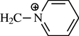
The reaction product could be isolated as picrate and perchlorate salts and subjected to microanalysis, UV-visible and IR identification tests. A λmax of 505 nm was obtained. The IR spectrum of both picrate and perchlorate salts of the isolated reaction product of p-aminophenol with the sulphide ion shows a significant difference from that of the pure compound in region of 3500–3100 cm−1. The disappearance of the doublet peaks that are characteristics of a primary amino group indicates that the nitrogen is a possible reaction site. In addition, analogous structures have been reported before for methylene blue and methylene blue like structure (Abdalla et al., 1982a). From the microanalysis data and the IR spectra a mechanism for the reaction could be suggested as follows: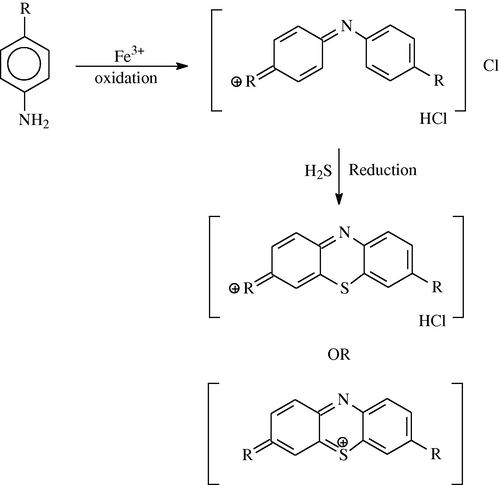
2 Experimental
2.1 Apparatus
A Perkin–Elmer model 330 Spectrophotometer with matching 1.00 cm cell was used for the absorbance measurement.
2.2 Reagents
2.2.1 p-phenylene diamine sulphate solution 0.01 M
Dissolve 0.1 g of p-phenylenediamine sulphate in 1 M sulphuric acid and dilute to 100 ml with 1 M sulphuric acid.
2.2.2 Acetaminophen solution
Dissolve 0.1 g of acetaminophen powder in 20 ml of 5 M sulphuric acid.
2.2.3 Ammonium iron(III) sulphate. 0.25 M
Dissolve 60.8 g of ammonium iron(III) sulphate dodecahydrate in 0.5 M sulphuric acid.
2.2.4 Sodium sulphide solution
Dissolve 1 g of sodium sulphide in 100 ml of water.
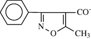
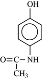
Structure of the cephalosporins and acetaminophen studied are given in Table 1.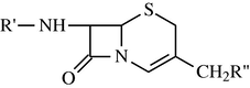
Compound
R’
R”
Cephalexin
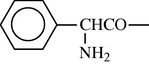
–H
Cephalothin
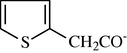
–OCOCH3
Cephaloridine

Cephaxazole
–CH2OCOCH
Acetaminophen
2.3 General procedure
Standard cephalosporin solution, 0.1% w/v. Dissolve 0.1 g (accurately weighed) of cephalosporin in water and dilute to 100 ml in a calibrated flask. Transfer, by means of a pipette, aliquots (0–10 ml) of the solutions into a 100 ml calibrated flask and dilute to volume with 0.5 M sodium hydroxide solution. Heat the flask in a boiling bath for the length of time appropriate for the particular cephalosporin to produce maximum yield of hydrogen sulphide and then cool. Recommended hydrolysis time of heating can be selected for each cephalosporin from Table 2.
Cephalexin
30
Cephalothin
60
Cephaloridine
60
Cephaxazole
60
Add, by pipette, 5 ml of each solution to a 50 ml calibrated flask and add 5 ml of the p-phenylenediamine solution and 2 ml of ammonium iron(III) sulphate and quickly stopper the flask. Shake the flask for 30 s, dilute to mark, mix and read the absorbance at 595 nm against a blank treated similarly.
2.4 For capsules and tablets
Transfer accurately weighed mixed contents of one capsule or one powdered tablet equivalent to 500 mg of the drug into 50 ml calibrated flask, add 30 ml of methanol, shake for 10 min and then make up to volume with methanol. Filter, then transfer 50 ml of the filtrate into 50 ml conical flask and evaporate to dryness. To the residue add 5 ml of water dissolve and then transfer the contents into 50 ml calibrated flask and dilute with 0.5 M sodium hydroxide and then complete the assay as described above.
For the determination of the acetaminophen in pure form or in pharmaceutical formulations. Transfer an accurately weighed mixed content of tablets or its pure powdered form equivalent to 0.1 g of the drug into a 50 ml calibrated flask and add 20 ml of 5 M sulphuric acid. Heat the flask in a boiling water bath for one hour. Cool, then dilute with water. Add by pipette, 5 ml of each solution to 50 ml calibrated flask and add 5 ml of sodium sulphide and 5 ml of ammonium iron(III) sulphate. Shake the flask for 30 s, allow the flask to stand for 5 min. Dilute to mark, mix and read absorbance at 525 nm against a blank treated similarly.
3 Results and discussion
The proposed methods have been used for the quantitative determination of various cephalosporins and acetaminophen in pure form and in real sample.
In the first method, the sulphide that resulted from the alkaline degradation of the cephalosporins studied has been reacted with p-phenylenediamine and ammonium iron(III) sulphate as an oxidizing agent to form the violet dye. The absorbance of the violet dye formed, measured at 595 nm , is a measure of the cephalosporin concentration. In the second method, p-aminophenol formed as a result of acid hydrolysis of acetaminophen has been reacted with sodium sulphide and iron(III) as oxidizing agent to form the red dye and the absorbance of the red colour dye formed, measured at 530 nm is a measure of the acetaminophen concentration.
The optimum conditions were studied by variation of the acidity of the reagent, the concentration of the added reagents such as ammonium iron(III) sulphate in the first method and the ammonium iron(III) sulphate and the sulphide in the second method. In both methods the colour of the dye formed was dependent on the concentration of sulphuric acid and that has reached a maximum with 1 M acid in the case of the first method and 2 M acid in the case of the second method. Higher acidity does not result in greater absorbance in either method. It is also observed that at higher acidity there is a small shift in the wavelength of maximum absorbance. Increase in reagent concentration above 10 ml of either the p-phenylenediamine or the iron(III) in the first method or the iron(III) and the sulphide reagents tends to effect the wavelength of the maximum absorption the same way as with the use of higher acidity. The absorbance of the violet dye at 423 and the repetitive scan show that the reaction is completed in 6 min without mixing. With shaking the colour is developed in 1 min.
3.1 Spectral data
Beer’s law was found to hold over the range 0.1–50 μg/ml for all the five cephalosporins examined and over the range 10–200 μg/ml for the acetaminophen. Table 3 gives data for calibration graphs for the determination of five cephalosporins and the acetaminophen. The calibration graphs show good rectilinearity and low coefficient of variation.
Compound
Concentration range (μg/ml)
Coefficient variation (%)
Cephalexin
5–50
0.76
Cephradine
5–50
0.84
Cephalothin
10–50
0.85
Cephaloridine
10–50
0.77
Cephoxazole
20–100
0.80
Acetaminophen
20–100
0.78
4 Application
The two methods were then applied to the determination of some cephalosporins and acetaminophen in drug formulations (capsules and tablets). Typical results are given in Table 4. The results obtained show good agreement with those expected.
Drug
Nominal composition (mg)
Found (mg)
R.S.D. (%)
Velosef (Squibb)
500
504.1
0.72
250
252.1
0.82
Ultrasporin (Arab phama Ind)
500
499.2
0.91
250
251.3
0.83
Keflodin (Lilly)
500
502.4
0.77
250
251.7
0.75
Prontopyrin (Tablets)
500
503.0
0.78
Saridon (Tablets)
500
502.1
0.67
Calpol (Suspension)
500
502.4
0.71
The results of the two methods were then compared with the results obtained by the official method for the cephalosporins and acetaminophen drug as shown in Tables 5 and 6.The t-test values at 95% confidence limit indicate no significant differences between these methods. The two methods for the determination of the cephalosporins and the acetaminophen have the advantage of using the visible region rather than the ultra-violet, where many degradation products and excipients are usually present in dosage forms, such as starch and lactose.
Compound
Recovery (%)a
tb
Proposed method (μg)
Official method (μg)
Cephalexin
100.3±0.3
101.4±0.3
0.84
Cephradine
101.2±0.2
100.2±0.3
0.88
Cephlothin
99.3±0.2
101.2±0.3
0.85
Cephaloridine
99.7±0.3
102.3±0.2
0.88
Cephoxazole
100.2±0.3
101.2±0.3
0.79
Sample or drug
Recovery (%)a
tb
Proposed method (μg)
Official method (μg)
Saridon
101.82
102.1
1.2
Prontopyrin
100.62
100.2
1.3
Calpol
98.88
99.1
1.4
The second method that is used for the determination of acetaminophen is selective and can be used for the determination of acetaminophen in the presence of ortho- and meta-aminophenol (see Table 7). The method also has the advantage of being a stability indicating method.
Drug
Present (μg)
Found
SDa
Recovery
Saridonc
10
10.22
0.18
102.2
Saridon
10
10.21
0.20
102.1
Prontopyrinb
10
10.31
0.14
103.1
Prontopyrinc
10
10.24
0.17
102.4
The first method, however, has the disadvantages that it is unlikely to be a stability indicating method and will not distinguish between cephalosporins and other sulphide producing degradation products.
References
- Anal. Lett.. 1991;24(1):55.
- Analyst. 1982;107:213.
- Analyst. 1982;107:53.
- J. Pharm. Sci.. 1976;65:300.
- Arch. Pharm. Chem. Sci.. 1977;28:149.
- J. Pharm. Pharmacol.. 1976;28:791.
- Chin. Acta. 1977;77:373.
- British Pharmacopoeia, 1980, H.M. Stationaly, Office, London, 1980.
- Fayed, N.M., Ph.D. Thesis, Loughboroughh University of Technology, 1979.
- Analyst. 1982;107:449.
- J. Pharm. Biomed. Anal.. 1985;3(4):315.
- Analyst. 1989;114:237.
- Anal. Chem.. 1999;43:834.
- Talanta. 1997;44:61.
- Talanta. 1981;28:855.
- Pharm. Pharmacol.. 1978;30:811.


