

Translate this page into:
Saussurea costus for sustainable and eco-friendly synthesis of palladium nanoparticles and their biological activities
-
Received: ,
Accepted: ,
This article was originally published by Elsevier and was migrated to Scientific Scholar after the change of Publisher.
Peer review under responsibility of King Saud University.
Abstract
Abstract
Saussurea costus biomolecules were used to synthesize Palladium nanoparticles by green route. The biosynthesized Palladium nanoparticles were physiohemically characterized by advance spectroscopic techniques. The biosynthesized Palladium nanoparticles showed potent anticancer, antibacterial, anti-alziehmer, and anti- inflammatory properties. The Palladium nanoparticles were found effective to inhibit HepG2, MCF-7, and HCT-116 cells and therefore can be used as targeted anticancerous drug.
Abstract
Palladium nanoparticles have been evaluated as a viable candidate in the realm of biological applications due to their unique features. Saussurea costus extract was used as a stabilizing and reducing agent for the synthesis of palladium nanoparticles with average grain size of 17.6 ± 1.2 nm. The synthesized PdNPs were evaluated for their antioxidant activity, anti Alzheimer's activity, antibacterial and anticancer activities. The nanocharacterization was carried out using different spectroscopic techniques, including UV–visible spectroscopy, Transmission Electron Microscopy, Fourier Transformed Infrared spectroscopy, X-ray Diffraction analysis, X-ray Photoelectron spectroscopy, Energy Dispersive X-ray Spectroscopy, Size distribution, and Zeta potential. The characterization data explained the PdNPs mediated by S.costus extract have spherical form and are disseminated without agglomeration. FTIR and XPS supported the hypotheisis that the biomolecules of S.costus are activing as a reducing and stabilizing agents. The antioxidant activity of PdNPs was assessed using a free radical scavenging assay (DPPH) which exhibited similar results to the ABTS assay i.e. 90 μg/mL IC50 value. Moreover Alzheimer's disease can easily be inhibited by S.costus@PdNPs at 400 mg/mL, with 79.23 ± 1.11 % of inhibition rate against AChE and 76.13 ± 0.43 % towards BChE. S.costus@PdNPs showed comparatively greater antibacterial activity against all four Staphylococcus aureus, Bacillus subtilis Escherichia coli and Pseudomonas aeruginosa microorganisms. Supplementary research carried out on the anti-tumor effects of the generated PdNPs using the colon cancer (HCT-116), hepatocellular carcinoma (HepG2), and breast adenocarcinoma (MCF-7) cell lines. PdNPs showed potent anticancerous activity against all the cell lines. Thus we recommend S.costus@PdNPs as a thearapeutic agent after successful clinical trails in future.
Keywords
Saussurea costus
Antioxidant
Anticancer
Anti-bacterial
Anti-inflammatory
Anti- Alzheimer

1 Introduction
Nanoscience is a new interdisciplinary science that has risen as one of the fastest growing field of science (Al-Radadi, 2022). Since nanoparticles successfully revolutionalized life sciences material sciences and human health, nanotechnology is currently gaining a lot of attention from scientists and researchers all over the world (Abd El-Aziz et al., 2021; Al-Radadi, 2021a). Nanoparticels bear a unique properties related to nanoscaled size and large surface area (Ng and Lim, 2022; Gioria et al., 2019; Fan et al., 2022), it can controll the average grain size and morphology of nanoparticles including their physiochemical properties (Dash et al., 2022; Srinoi et al., 2018). Due to development in nanotechnology there is an intense need of efficient and cost effective, eco-friendrly metallic nanoparticles (Al-Radadi and Al-Youbi, 2018a). Metallic nanoparticles has been synthesizd with ease and efficiency by green routes previously by many researchers (Abdullah et al., 2022; Faisal et al., 2021; Deokar and Ingale., 2017). Due to stability and cost effectiviness the metallic nanoparticles biosynthesis is an emerging field in green chemistry (Hano and Abbasi, 2022; Tran et al., 2022; Deokar and Ingale.,2018). Metallic nanoparticles have efficiency in targeting the site of infection and therefore widely used for drug delivery (Bendre et al., 2022; Chandrakala et al., 2022). As far as toxicity concerns NPs exhibit less toxicity because of targeted action at infection site. The delivery method is also very simplest like by orally, by injecting, and by inhalation (Yaqoob et al., 2020)resulting in a a versatile medical applications (Al-Radadi, 2021b). Out of the metal nanoparticles, nano-configured noble metals have sparked interest and have treamendous potential applications in different fields includingbiochemistry, chemistry, physics, biology, and engineering (Al-Radadi and Al-Youbi, 2018b; Deokar and Ingale., 2016). Amongst noble metals PdNPs gained remarkable attention for biological applications (Zhang et al., 2022) due to their efficient surface energy and area. These characteristics making them inexhaustible in therapeutic sectors. Palladium nanoparticles are also utilised to improve biomedical diagnosis because they are active in the in the infrared range for boimaging (Sonbol et al., 2021). PdNPs are also active catalyst for numerous chemical reactions (Gálvez-Martínez et al., 2021). NPs tends to aggregate and is inherently unstable (Anand et al., 2022; Veisi et al., 2021). Therefore Plant extracts can also be used to make nanoparticles without requiring sophisticated physical or chemical conditions (Al-Radadi, 2022a). Plant biomolecules are regarded as the best reducing agent of Pd to PdNPs (Iftikhar et al., 2020; Sarkar et al., 2022), because they allow eco-friendly and green synthesis (Al-Radadi et al., 2022). Additionally, it is more successful and efficient in regulating the size, shape, and dispersion of nanoparticles (Al-Radadi, 2019) and it contains biologically active compounds. It also works as a capping agent, and coating nanoparticles to reduce their size (El Marghani et al., 2021; Info, 2019). The interaction of NPs at cellular and sub-cellular level make them prominent agent to be used in the prevention from chronic diseases (Al-Radadi and Adam, 2020). Saussurea costus, a synonym of (saussurea lappa), is a potent antibiotic and a plant of the asteraceae family that is extensively dispersed around the world (Park, 2021), particularly in India and the Himalayas (Abdullah et al., 2021). Because of its broad use, it is the most commonly used medicinal plant in ancient medicine (Binti Mohamad and Ahmad, 2021; Shahista, 2019). It is well-known in Islamic medicine and the hadiths of the Prophet Muhammad (may God bless him and grant him peace) (Chloride-, 2022). It is one of the most extensively utilized plants in medicinal systems because of the variety of therapeutic components it contains, which makes it chemically free (Sohrab et al., 2021). A lot of compounds have been discovered in the extract of S. costus including alkaloids, flavonoids, sesquiterpenes, terpenes, anthraquiones, and tannins, as well as components such as lactone, saussureamine etc (Sunil Kumar et al., 2021; Vijayalakshmi et al., 2022. S.costus has anticancer, anti-diabetic, anti- fungal, anti-inflammatory, anti-ulcer, anti-microbial, and antiseptic properties, and it also heals the thyroid gland and covid 19 (Tousson et al., 2020; Deabes et al., 2021; Al-obaidy and Esmaeel, 2021). S.costus contains esquiterpene lactones, such as dehydrocostus lactone and costulonide and alantolactone, cynaropicrin, and saussu-reamine, which contribute to future medication research (Ali and Venkatesalu, 2022; Ellam, 2012; Nadda et al., 2020). For example, isodihydrocostunolide, cynaropicrin, costunolide and dehydrocostus lactone, and lappadilactone, these allcompounds can be utilized to treat cancer, one of the most important health issues in the world (El-Bolkiny et al., 2021). Because it contains d-galactose, S.costus has shown to be useful in reducing inflammation, oxidation, and Alzheimer's disease by formaing tangles of the neurofibrillary (Mohamed et al., 2022). Since it contains, costunolide, dehydrocostuslactone, and dihydrocostunolide, s.costus is also regarded antibacterial, anti-inflammatory, and anticancer (Rathore et al., 2021; Saif-Al-Islam, 2020). Healthcare systems in both developed and under developed countries are impacted by multidrug resistance pathogens, which has been linked to the development of deadly pathogens and chronic illnesses, in order to tackle the emerging diseases there is an intense need of novel drugs, the nanomaterials have been widely used in this aspect and found efficient (Al-Radadi, 2022b; Al Jahdaly et al., 2021). As a result, this work demonstrated a perfect results for the green, eco-friendly synthesis of S.costus-@PdNPs, which are nonaggreagated, spherical, homogeneous, nano sized, and active against the Gram’s positive and negative bacteria, also active against antioxidents and Alzeimer’s disease and active against HCT-116, HepG-2 and MCF-7 cell lines, and whether it is a workable cancer therapeutic approach.
2 Experimental details
2.1 S.costus preparation and S.costus-mediated S.costus-@PdNPs biosynthesis
Saussurea costus of good quality was purchased from a local spice shop, while PdCl2 was purchased from Sigma Aldrich. Saussurea costus extract was prepared by heating Saussurea costusin in 50 mL of distilled water for 5 min at 25 °C. For the preparation of S.costus-@PdNPs 40 mL of plant extract 60 mL of PdCl2 (2 × 10⁻³ M) PdCl2 at 100 °C in reflux condenser for 2 h. The color chage was observed for synthesis of Pd0.
2.2 Characterization of Pd nanoparticles
The physio chemical properties of the bisoynthesized S.costus-@PdNPs were investigated utilizing various advanced spectroscopic procedures and instruments including, UV–vis Spectrometer between wavelengths 800–200 nm using quartz cuvettes later transmission electron microscopy was used to identify the average grain diameter and properties of S.costus-@PdNPs. FTIR spectroscopy in the spectral ange of 400–4000 cm−1 was used for the detection of phytochemicals and the active groups associated with S.costus-@PdNPs. The X-ray diffraction study was used to determine the crystalline structutre of PdNPs. The X-ray photoelectron spectroscopy was used to determine and investigate the surface components of synthesized nanoparticles. EDX was used to further characterise the elements of PdNPs, size distribution and zeta potential was also determined.
2.3 Anti-bacterial assay
Four previously identified bacterial strains Escherichia coli, Staphylococcus aureus, Bacillus subtilis, and Pseudomonas aeruginosa were taken from the Regional Center for Mycology and Biotechnology (RCMB), Al- Azhar University, Saudi Arabia. The strains were cultivated and used for antibacterial activity against S.costus extract and S.costus-@PdNPs. The antibacterial assay was performed according to Agar well diffusion method. Five millimeters wells were bored in the culture media and plant extract, PdNPs were applied against the bacterial strains in a concentration of 15 μg, 25 μg, 50 μg and 100 μg for overnight and the inhibition zones were calculated accordingly. The assay was repeated three times.
2.4 Anti-inflammatory assay
For anti-inflammatory assay murine macrophages RAW264.7 cells ( ) were cultured in Dulbecco's Modified Eagle's Medium (DMEM, Corning, USA) supplemented with 10 % FBS, 100 U/mL of Penicillin, 100 µg/mL of streptomycin sulphate, and 2 mM of l-glutamine in a humidified 5 % incubator. The cells were rinsed in phosphate buffered saline and scraped off the flasks with sterile scrappers for passaging and treatment (SPL, Spain). RAW 264.7 cell stock ( cells/mL) was planted into 96-well micro well plates overnight. The non-induced triplicate wells received media containing the nanoparticle sample the next day. The inducer of inflammation [lipopolysaccharide (LPS) at 100 ng/mL in full culture media] was given to the inflammatory group of triplicate wells. In Triplicate wells, the increasing volumes of the extract (6.25–100 µg/ml) was dissolved in culture media and diluted into culture media containing LPS. The anti-inflammatory positive control was caffeic acid phenacyl ester (CAPE, 5 µM). Griess test was used to measure NO in all wells after 24 h of incubation. To make the colored diazonium salt, equal amounts of culture supernatants and Griess reagent were combined and incubated at 25℃ for 10 min, absorbance were recorded at 540 nm by a Tecan Sunrise™ microplate reader (Austria). The Inhibition percent of the sample was computed in comparison to the LPS-induced inflammation group, and was normalized to cell viability as determined by the Alamar Blue™ decrease assay.
2.5 Anti-cancer assay
The cell lines MCF-7, HCT-116, and HepG-2 were collected from VACSERA Tissue Culture Unit. HCT-116, HepG-2, and MCF-7 cells were cultured in McCoy's 5A medium, Eagle's inimum essential medium, and low glucose Dulbecco's modified Eagle medium respectively. All the culture medias were supplemented by Penicillin, Streptomycin, and 10 % foetal bovine serum.
For MTT assay the HCT-116, HepG-2, and MCF-7 cells were introduced to 12-well plates at seeding density of per well, followed by incubation at 37 ℃ for a complete day in a humidified 5 % incubator. To determine the with reference to standard 0–200 µM of DMSO, 0–400 µg/ml S.costus extract, and S.costus-@PdNPs of 0– 400 µg/ml were applied in to specified wells followed by incubation for 24 h. Just after incubation MTT reagent of 1.25 mg/ml were added in to the wells followed by incubation for two hours. The activity was monitered by microplate reader at 570 nm and was analyzed by using GraphPad Prism longevity software (San Diego, CA, USA).
2.6 Anti-Alzheimer assay
Acetylcholinesterase (Sigma ‘‘101292679″) and Butyrylcholinesterase (Sigma ‘‘101292679″) enzymes were purchased and used in th assay. These enzymes are the key contributers to Alzheimer diseases and the suppression of these enzymes by compound is regarded as their anti-alzheimer activity. By using Elman’s technique nanoparticles of different concentrations from 25 mg/mL to 400 mg/mL were appied against 0.03U/mL AChE and 0.01U/mL of BChE. The methanolic galantamine hydrobromide (Sigma; GI660) were used as a positive control and waited for a color change.
2.7 Antioxidants assay
The free radical and antioxidant activity of S.costus-@PdNPs and S.costus extract was examined by using the DPPH and ABTS assays. DPPH was dissolved in methanol and different concentrations of plant exatract and PdNPs were applied against the free radicals of DPPH in a 96-well microtiter plate followed by incubation for 30 min at room temperature in dark. Ascobic acid was used as positive aand reference control. The reaction was exposed to absorbance of 517 nm and the antioxidant activity was calculated.
The value was determined as the least amount of antioxidant required to scavenge 50 % of the DPPH radicals.
2.8 Analysis of cytotoxicity of S.costus-@PdNPs with extract
The cancer cells were grown cultured in M199 medium supplemented with 20 % FBS, 100 U/mL of Penicillin, 100 µg/mL of Streptomycin Sulphate, and 20 mg/ml bFGF in a humidified 5 %
incubator. at 37 °C and varying concentration of S.costus extract and PdNPs in a 24 well microplate. After incubation 5 mg/ml of aqueos MTT were added to each well to asses the cell viability by MTT assay. The dehydrogenase enzyme of surviving cells convert (4,5-dimethylthiazol-2-yl) (2,5-diphenyl tetrazolium bromide) to insoluble MTT formazan. To dissolve the insoulibe formazan 0.3 mL DMSO were added to wells. The assay was performed three times and exposed to 570 nm to check the cell viability.Fig. 1.
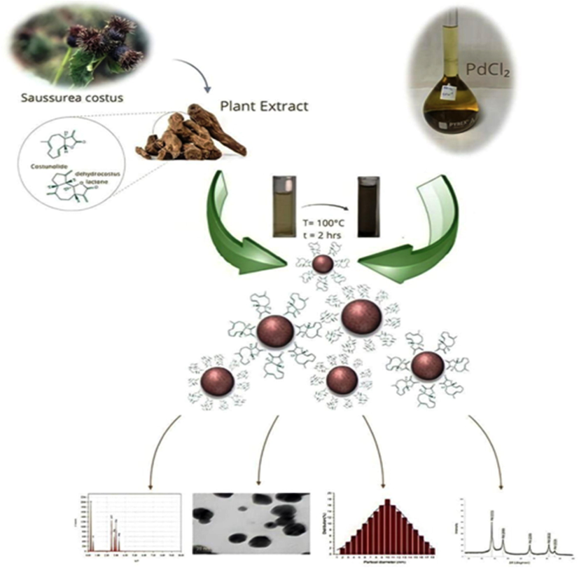
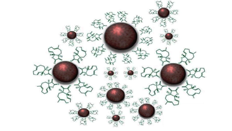
Schematic diagram for synthesis of S.costus-@PdNPs using S.costus extract. S.costus extract was combined with palladium chloride and the nanoparticles were synthesized. The obtained nanoparticles were characterized by advance spectroscopic techniques.
3 Results and discussion
3.1 UV–Visible analysis
The purpose of this analysis was to observe the formation of PdNPs. At 100 °C for 2 h, reactions were carried out with 40 mL of costus root extract and 60 mL of 2
M
. The surface plasmon resonance (SPR) band of PdNPs (Osonga et al., 2020) was characterised by a colour change in reaction solution from pale yellow to dark brown (Kadam et al., 2020). Fig. 2 shows the UV– visible spectra of PdNPs, which showed absorption peaks at
= 350 nm, indicating that
has been reduced to
(Wong et al., 2019).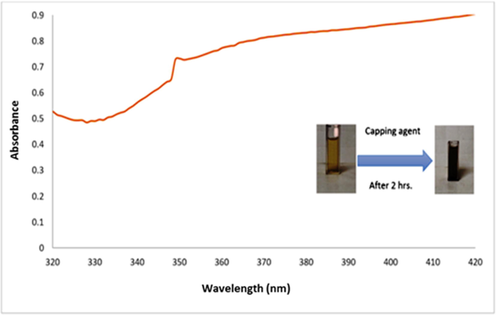
UV–vis. spectra for S.costus-@PdNPs at 100 °C, after 2hrs with 40 mL of S.costus root extract and 60 mL (
) (2
M).
3.2 TEM and HR-TEM and Zeta-potential analysis
Fig. 3 A shows an HR-TEM image of PdNPs, Palladium nanoparticles have a spherical form and are homogeneous, highly distributed and non-aggregated, ultrafine (Lu et al., 2020; Shokouhimehr et al., 2019; Olajire and Mohammed., 2019), with a size range of 1.9 nm to 17.6 ± 1.2 nm on the 10 and 20 nm scale. The reason for uniformity in size was the maintainnace of specific reaction conditions such as pH, temperature, time, salt and extract concentration. Fig. 3 B shows spherical nanoparticles in a TEM picture at 100 nm. Fig. 3 C showing the average particle size distribution histogram investigation, and our results are according with previously published study (Rashidi et al., 2019). The zeta-potential analysis of Pd NPs was found to be (-10.6) mV in Fig. 3 D, describes that the PdNPs found more stable at 25 °C and the negative surface charge of the particle prevented aggregation (Fahmy et al., 2021). Previous report said that 2.5 to 5 nm sized Pd nanoparticles synthesized Anacardium occidentale leaf extract with FCC crystallinity (Sheny et al., (2012).. Another group of researcher reported the Chlorella species of algae has potential to reduce PdCl2. where they reported that amide and polyol group contributed spherical nanoparticle synthesis having 5–20 nm size and FCC structure (Arsiya et al., 2017).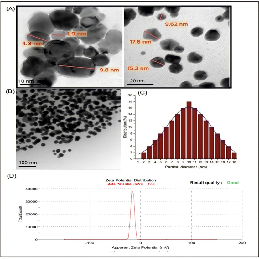
(A) HRTEM image, (B) TEM image of S.costus-@PdNPs at S.costus root extract 40 mL with 60 mL (PdCl₂)(2 ×
M) at 100 °C until 2 hrs. (C)corresponding size distribution graph (D) and Zeta potential of Scostus-@PdNPs.
Recently S. Vinodhini et. al. (Vinodhini et al., 2022) reported the palladium nanoparticles from A.fistulosum, B. alba and T divaricate aqueous extract. Through SEM size calculated was observed 500 nm, 2 μm and 2 μm respectively.
3.3 FTIR analysis
FTIR spectroscopy of S.costus extract and S.costus@PdNPs was used to find out specific functional group associated with PdNPs. The FTIR spectrac oof S.costus revealed absorption peaks at 3408, 2359, 1602, and 896
. The broad peak at 3408
stands for O
H of phenolic or alcoholic functional groups, the peak at 2359
occurs due to the presence of C
C, the peak at 1602
occurs (C⚌O)NH, and several sharp suggestive peaks were found at 896
aromatic stretching. Fig. 4 showed FTIR peaks at 3442.27
, 2083.84
, and 1635.26
respectively, that corresponds to (O
H), C
C, and (C⚌O) NH stretching frequencies of the S.costus bio-molecules, and a sharp peak at 554.30
corresponds to a standard peak of (PdO) due to to the stretching frequency of metal binding to oxygen. The literature (Lee, 2018; Al-Saggaf et al., 2020; Kolahalam et al., 2021; Narasaiah and Mandal, 2020) reported similar findings. Another researcher reported that amide, hydroxyl and glycoside were responsible for the reduction of Pd(II) (Sheny et al., (2012). Recent study on PdNp synthesis by brown alga Padina boryana suggested that the polyols including saponins, tannins and terpenoids are responsible for reduction (Sonbol et al., 2021).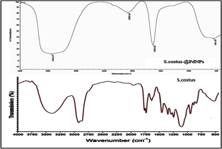
FTIR spectrum of S.costus-@PdNPs and S.costus.
3.4 X-ray diffraction analysis of PdNPs
Purity and crystallinity of S.costus-@PdNPs were determined by XRD. In Fig. 5 The five peaks at diffraction angles of 39.79°, 46°, 67.5°, 81°, 86° corresponds to the interlayer planes of (1 1 1), (2 0 0), (2 2 0), (3 1 1), (2 2 2). The planes were found consistent with the face-centered cubic crystallinity of PdNPs according to reference JCPDS file no. 87–0641 (Gholipour et al., 2022; Sriramulu and Sumathi, 2018; Jeevanantham et al., 2022). PdNPs average crystalline size was calculated using the Debye-Scherrer equation
and was found 2.88 nm.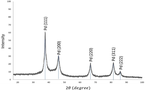
X-ray diffraction pattern of S.costus-@PdNPs.
3.5 X-ray photoelectron spectroscopy (XPS) analysis
The X-ray Photon Spectroscopy were performed to know about the topology of PdNPs. Fig. 6 A demonstrates XPS peaks at C1s, O1s, and N1s and vanish the peaks of the Pd. The Fig. 6 B describes the signals of Pd
at 340.89 eV and Pd
at 335.63 eV with a band gap of 5.26 eV that corresponds to these energies refer to
reduced to
However, the
peaks at 532.54 eV and 532.85 eV in Fig. 6 C indicate the
in the sample. Fig. 6 D revealed the additional peaks of
at 284.92 eV, 286.69 eV, and 288.84 eV corresponding to
, and in Fig. 6 E by the N1s peak at 399.99 eV, which corresponds to (
) (Veisi et al., 2019a; Mallikarjuna et al., 2021; Miao et al., 2022; Bolhasani et al., 2022; Liang et al., 2022; Hemmati et al., 2018). FTIR analysis reveled the bond of Pd with carbon and oxygen is clearly visible in the XPS scan, indicating that the PdNPs has biomolecules on the surface.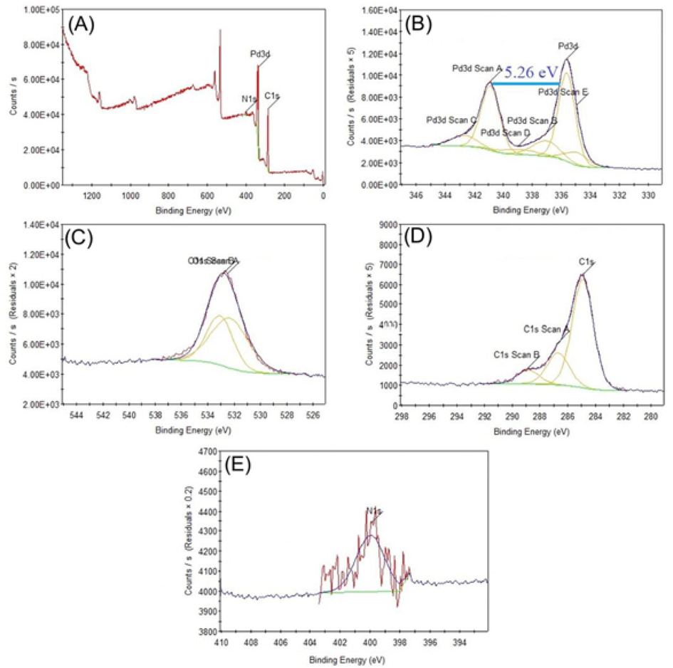
XPS analysis showing survey scan (a), (b) Pd, (c) oxygen and carbon (d),nitrogen (e).
3.6 Energy dispersive X-ray analysis of PdNPs
EDX was used to further characterize the elemental composition of PdNPs. Fig. 7 shows the S.costus- @PdNPs sample used in the EDX analysis. The EDX spectrum peaks at 2.83 keV, indicating that PdNPs have been formed. Metallic nanoparticles, as well as signals of the surface Plasmon resonance (SPR) band, are commonly detected at 3KeV. The presence of carbon, nitrogen, and oxygen were also confirmed using EDX analysis, suggesting the occurrence of S.costus extract biomolecules on the surface of palladium nanoparticles nanoparticles (Veisi et al., 2019b; Sriramulu and Sumathi, 2018; Jeevanantham et al., 2022).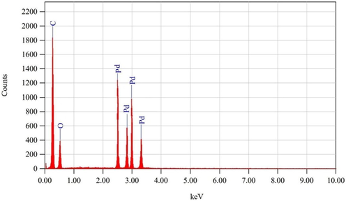
EDX image of S.costus root extract mediated PdNPs synthesized.
3.7 Anti-bacterial activity
Antibacterial activity of PdNPs against bacterial strains were determined by agar well diffusion assay. The largest inhibition zone was obtained in S.costus-@PdNPs with a maximum inhibition at 100 g/mL, 23 ± 0.1 mm against Bacillus subtilis in Fig. 8 A and 22 ± 0.5 mm against Staphylococcus aureus in Fig. 8 B and 22 ± 0.3 mm against Escherichia coli in Fig. 8 C and 19 ± 0.4 mm against Pseudomonas aeruginosa in Fig. 8 D,Whereas S.costus extract showed 18 ± 0.0 mm against Bacillus subtilis in as shown in Fig. 8 A, and 16 ± 0.1 mm of inhibition zone against Staphylococcus aureus as shown in Fig. 8 B. exact 17 ± 0.0 mm against Escherichia coli in Fig. 8 C and 15 ± 0.0 mm against Pseudomonas aeruginosa in Fig. 8 D (see Table 1). The nanoparticles can swiftly breach the cell membrane and enter the cell, bacterial cells may easily assimilate them, and thus PdNPs had greater antibacterial activity and can be very effective in other biological assays also (Abdelsalam and Samer, 2019; Vinodhini et al., 2022; Anjana et al., 2019; Aljohny et al., 2021; Seku, 2022) reported carboxymethylated mediated palladim nanoparticles with potent antibacterial and dye degradation activities. Ghraphene oxide coated palladium nanopartoicles were found effective against E. coli and other bacterial strains, thus these study endorsed our findings (Mallikarjuna et al., 2021; Govindasamy, 2017) Recently S. Vinodhini et. al. reported the antibactierial activity of the leaf extract of leaf extracts of A fistulosum, B alba and T. divaricate Pd nanoparticles for S.aurius showed 11, 12, 9 mm zone respectively, while for B. subtilis 14, 17, 18 mm and E.coli 18 16, 13 mm zone respectively. Here comparatively the S.costus-@PdNPs have superior antibacterial activity while antioxidant activity is less than that of S. Vinodhini et. al.work. (Vinodhini et al., 2022). It is believed that PdNPs can easily target the bacterial cell membrance and produce ROS to damage the bacterial cells, nanoparticles such as Zinc oxide penetrate deep in to the membrane of bacterial cells and the production of free radicals can harm the nucleic acid and contents in cell membrane which leads to bacterial death.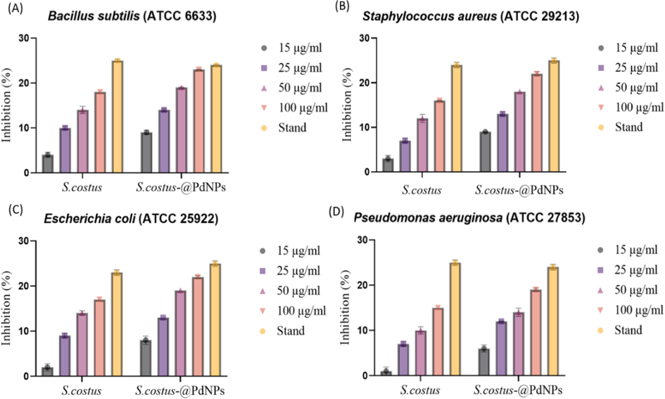
Anti-bacterial potential of S.costus-@PdNP.
Microorganisms
Concentration μg/mL
S.costus
S.costus-@PdNPs
Staphylococcus aureus
15 μg/mL
3 ± 0.7
9 ± 0.1
25 μg/mL
7 ± 0.6
13 ± 0.5
50 μg/mL
12 ± 0.9
18 ± 0.0
100 μg/mL
16 ± 0.1
22 ± 0.5
Standard
24 ± 0.6
25 ± 0.6
Control
0.0 ± 0.0
0.0 ± 0.0
Bacillus subtilis
15 μg/mL
4 ± 0.6
9 ± 0.5
25 μg/mL
10 ± 0.4
14 ± 0.3
50 μg/mL
14 ± 0.8
19 ± 0.1
100 μg/mL
18 ± 0.0
23 ± 0.1
Standard
25 ± 0.3
24 ± 0.2
Control
0.0 ± 0.0
0.0 ± 0.0
Escherichia coli
15 μg/mL
2 ± 0.8
8 ± 0.9
25 μg/mL
9 ± 0.6
13 ± 0.0
50 μg/mL
14 ± 0.5
19 ± 0.0
100 μg/mL
17 ± 0.0
22 ± 0.3
Standard
23 ± 0.6
25 ± 0.6
Control
0.0 ± 0.0
0.0 ± 0.0
Pseudomonas aeruginosa
15 μg/mL
1 ± 0.9
6 ± 0.8
25 μg/mL
7 ± 0.6
12 ± 0.1
50 μg/mL
10 ± 0.8
14 ± 0.9
100 μg/mL
15 ± 0.0
19 ± 0.4
Standard
25 ± 0.6
24 ± 0.6
Control
0.0 ± 0.0
0.0 ± 0.0
3.8 Anti-cancer activity
Cancer treatment is regarded as one of the most difficult tasks in medicine. “Cancer is the second biggest cause of mortality globally (Ahmed et al., 2022), according to the World Health Organization.” One possible answer to this problem and to improve the efficiency of chemotherapeutic agents is to use nanotechnology (Alshaman et al., 2022). Nanoparticles have a wide range of applications in cancer treatment that do not require surgery, such as drug administration and release (Al-Radadi, 2022c). The ability of nanoparticles to target cancer cells with great selectivity, sensitivity, and efficiency (Navya et al., 2019) is due to their high surface area, which allows them to easily infiltrate living cells. This qualifies NPs as a therapeutic agent (Hamida et al., 2020). Pd may also be a promising cancer treatment (Nguyen et al., 2019). Previous research suggested that S. costus includes biomaterials that could be used as a therapeutic treatment. Treatment against HCT-116, HepG-2 and MCF-7 (Shati et al., 2020). Overall, our findings give strong support for previous research. On the colon carcinoma HCT-116, and hepatocellular carcinoma HepG-2, breast cancer MCF-7 cell lines, the cell viability of standard, S.costus and S.costus-@PdNPs were examined. IC50 of standard and S.costus and S.costus-@PdNPs were presented in Fig. 9, Fig. 10 and Fig. 11. IC50 of standard is 8.2 µg/mL, 9.8 µg/mL, 15.6 µg/mL against HCT-116, HepG-2 and MCF-7 respectively, as shown in Fig. 9 A, D, G. IC50 of S.costus was 82.5 µg/mL, 91.5 µg/mL, 114.6 µg/mL against HCT-116, HepG-2 and MCF-7 respectively, as shown in Fig. 9 B, E, H. IC50 of S.costus-@PdNPs 7.8 µg/mL, 11.8 µg/mL, 26.7 µg/mL against HCT-116, HepG-2 and MCF-7 respectively, as shown in Fig. 9 C, F, I. According to the results obtained for the IC50 values shown in Fig. 10 indicated the following order of % cell viability for the HCT-116 was S.costus-@PdNPs is greater than Standard is greater than S.costus. While the order ofthe % cell viability obtained for the HepG-2 cell line, was Standard is greater than S.costus-@PdNPs greater than S.costus. The last cell line % viability for MCF-7 was Standard is greater than S.costus-@PdNPs is greater than S.costus. Overall, we obtained better inhibition towered HCT-116 for S.costus-@PdNPs. In contrast to HepG-2 and MCF-7 colon cancer is the third most frequent type of cancer and continuing to be a major cause of morbidity and mortality globally (Zhou et al., 2022). The difference between the IC50 S.costus-@PdNPs and standard was 2 µg/mL, 11.1 µg/mL of HepG-2, MCF-7, respectively. Our present findings and previous literature suggested that the PdNPs are more potent in anticancerous activity compared to other nanoparticles like ZnO, ZnO-NPs which showed distint IC50 of 50.1 μg/ml against HeLa cells line without harming normal fibroblast cells. The cytotoxicity results of Ag/Fe3O4 nanocomposite revealed IC50 of 55.83 μg/mL against HeLa cells. Chemotherapeutic toxicity has substantial toxic side effects on various organ systems, including the cardiac, pulmonary, renal, hepatic, gastrointestinal, bone marrow, and nervous system (Zeien et al., 2022). Palladium nanoparticles from different plant sources have been utilized against cancerous cell previously and the results found in this study are in accordance with it. The palladium nanoparticles have high specific cytoxicity towards the targeted tumour cells (Sonbol et al., 2021; Arsiya et al., 2017). PdNP synthesis by brown alga P. boryana showed 53 % and 38 % reduction of MCF-7 cellular activity by MTT assay at 125 and 62.5 μg/mL respectively. Chemotherapy medications are not specific to cancer cells; they can also destroy healthy, fast-growing normal cells (Yafout et al., 2021) and they fail to reach the target site. Targeted delivery using nanoparticles has the potential to increase drug accumulation at the target site, reduce toxicity to non-cancerous cells, and overcome the drug resistance (Sheikh et al., 2022), resulting in improved drug delivery with much fewer side effects (Al-Radadi., 2022d). As a result, S.costus-@PdNPs could be a promising cancer therapeutic candidate (Shati et al., 2020) after successful clinical trails.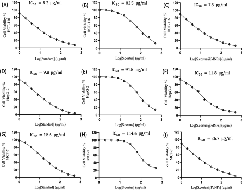
The dose–effect curve for standard µg/mL against (A) HCT-116 (B) HepG-2 (C) MCF-7 and S.costus µg/mL against (D) HCT-116 (E) HepG-2 (F) MCF-7 and S.costus-@PdNPs µg/mL against (G) HCT-11 (H) Hepg-2 (I) MCF-7. The
values were determined using nonlinear regression according to the following equation: log (inhibitor) versus response-variable slope.
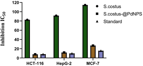
Anti-cancer potential of S.costus and standard and S.costus-@PdNPs.
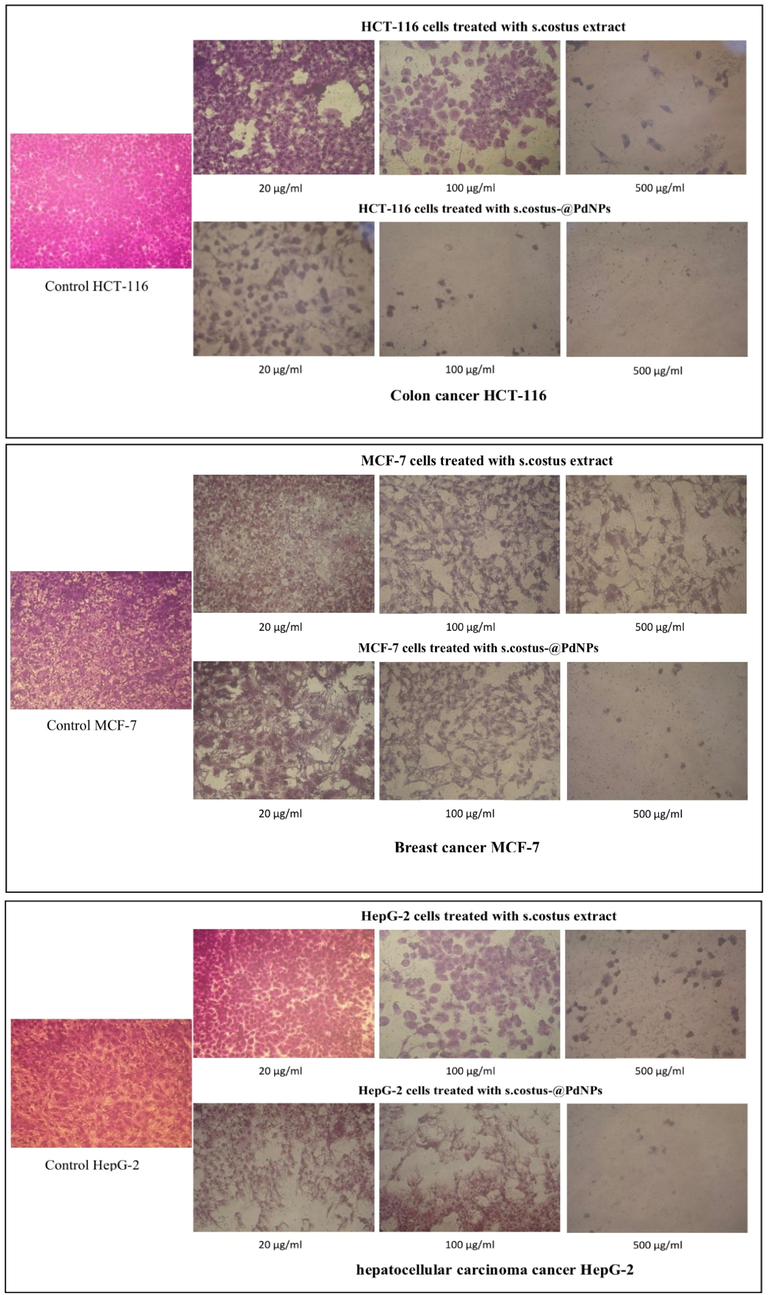
Effect of synthesized. S.costus-@PdNPs on morphological assessment of HCT-116, HepG-2, and MCF-7 cells.
3.9 Anti-Alzheimer activity
The most prevalent form of dementia is Alzheimer's disease. AD is a neurological disease that causes memory loss and cognitive impairment. Alzheimer's disease is expected to double in prevalence every 20 years (Oliver and Reddy, 2019). The physiology of Alzheimer's disease is complicated and involves numerous mechanisms. Both acetylcholine (ACh) and butyrylcholine (BCh) are important for memory learning. The appearance of nicotinic receptors stimulated by ACh is linked to learning ability. When nerve terminals are depolarized, ACh is released from neuron vesicles and binds to the synaptic receptor. This occurs because an AD patient's brain contains a large number of acetylcholinesterase (AChE) and butyrylcholinesterase (BChE) enzymes, which frequently change the neurotransmitters activity and shorten the half-life of ACh and BCh (Gul et al., 2021; Hampel et al., 2019). In this work the production of 5-thio-2-nitrobenzoate and DTNB complex hydrolyzed ATchI to AChE and BTchI to BChE results in the color transformation of the reaction to yellow. The absorbance of the coloured transformed solution was taken at 412 nm and percent enzyme inhibition was calculated which clearly indicated that Alzheimer's disease can easily be inhibited by S.costus@PdNPs at 400 mg/mL, with inhibition rate of 79.23 ± 1.11 % against AChE and 76.13 ± 0.43 % against BChE. As indicated in Fig. 12 (see Table 2), the inhibitory activity was dosage dependent. Nanoparticles assist medications pass the blood–brain barrier (BBB) and allow for targeted administration and regulated release, they offer a new treatment option for Alzheimer's disease (Gong et al., 2022). Though the other complications like in vivo toxicological study needs to be done in both male and female rat.
Enzymes
NPs
25 µg/mL
50 µg/mL
100 µg/mL
200 µg/mL
400 µg/mL
AChE
Pd-NPs
BChE
Pd-NPs
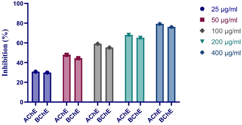
Anti-Alzheimer potential of S.costus-@PdNPs.
3.10 Anti-inflammatory activity
In anti-inflammatory assay 7. 84 %, 3.92 %, 1.96 %, 0.98 %, 0.49 % of low inhibition was showed by PdNPs at a concentration of 100, 50, 25, 12.5, and 6.25 µg/mL respectivily against LPS-induced nitric oxide (NO) shown in Fig. 13. Similar results was also found by researcher in previous studies (Houchi and Messasma, 2022; Singh et al., 2018).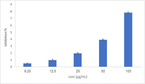
Anti- inflammatory potential of S.costus-@PdNPs.
3.11 Antioxidants DPPH and ABTS activity
DPPH radicals react immediately with antioxidants because antioxidants are stable enough to give an electron to a rogue free radical and tends to neutralize it. It reduces the effects of free radicals. These antioxidants work by scavenging free radicals to postpone or prevent cellular damage (Zangeneh et al., 2019; Kumar et al., 2022). DPPH and ABTS assays were used to assess the antioxidant activity of the S.costus extract and S.costus-@PdNPs. The DPPH, ABTS-scavenging activity was observed to increase with increasing concentration; the DPPH radical scavenging activity data are shown in Fig. 14. The IC50 of DPPH, S.costus-@PdNPs, and S.costus extract were 90 µg /mL, 92 µg /mL respectively. Fig. 15 shows the findings of the ABTS radical scavenging activity. ATBS, S.costus-@PdNPs, and S.costus extract had IC50 of 90, 92 µg /mL respectively. The scavenging activity of DPPH and ABTS radicals was found to be in the following order: ascorbic acid is greater than S.costus extract is greater than S.costus-@PdNPs. The activity of the S.costus-@PdNPs extract was significantly higher than that of the S.costus. The Pd nanoparticles displayed good antioxidant activity and eliminated different free radicals, according to DPPH and ABTS (Mahdavi et al., 2019) found similar results by using Agaricus bisporus for the synthesis of palladium nanoparticles, the synthesized PdNPs scavenged DPPH free radicals up to 77 %. The high phenolic and flavonoid content of S.costus root can be linked to its antioxidant activity(Abdel-Wahhab et al., 2022; Singh and Chahal, 2018).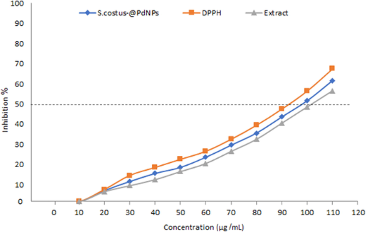
The antioxidant activity of DPPH, S.costus extract, and S.costus-@PdNP.
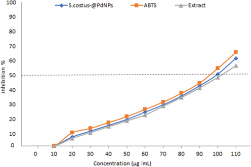
The antioxidant activity of ABTS, S.costus extract, and S.costus-@PdNPs.
3.12 Cytotoxicity of S.costus and S.costus-@PdNPs
Cancer cell lines were used to investigate the cytotoxic effect of S.costus and S.costus-@PdNPs. Surprisingly, S.costus did not show substantial cytotoxicity against a cancer cell line in this study. Fig. 16 shows cell viability was 99, 99, 98, 97, 96.2, 94.8, 92.8, 89.8, 87, and 83 % at concentrations of 100, 150, 200, 250, 300, 350, 400, 450, 500, and 550 µg/mL of PdNPs. The results of the MTT assay and cytotoxicity data demonstrate that PdNPs contact reduces cancer cell viability and is dosage or concentration dependent (BalaKumaran et al., 2020). Metal salts removed their toxicity when they combine with biological molecules (Venil et al., 2021; Zangeneh et al., 2019) As a result, produced PdNPs show an outstanding biocompatibility (Shivakumar et al., 2021). This synthesis approach is therefore a potential procedure for future use (Kiani et al., 2020).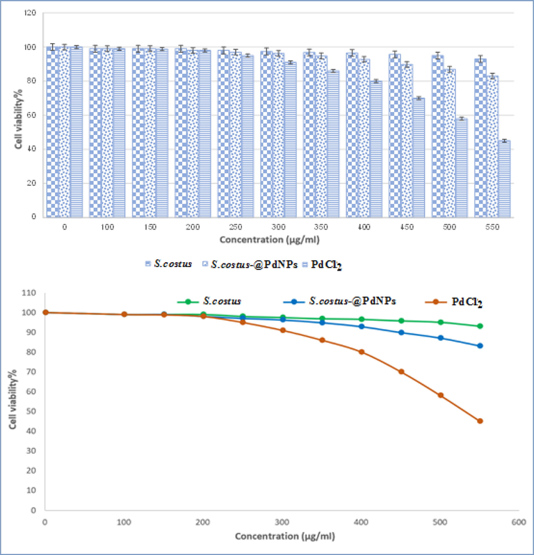
Percent viability measured after treatment with present S.costus, and S.costus- @PdNPs and PdCl2.
4 Conclusion
It is concluded from the study that PdNPs effectively synthesized by utilising S.costus extract using a direct, cost-effective, energy-saving, and environmental friendly method. Herein this study S.costus extract acted as a reducing and capping agent for S.costus-@PdNPs. UV–visible absorption, FTIR, TEM, HR-TEM, zeta-potential, Powder X-ray pattern, XPS, and EDX analytical methods used to analyse and cofirm the formation of spherical, FCC crystalline nanosized particles of palladium synthesized. TEM examination concluded the average grain size of the S.costus-@PdNPs is 1.9 to 17.6 ± 1.2 nm. FTIR and additionally and XPS proved that the nanoparticles od Pd synthesized using bioactive materials in the S.costus extract. The biosynthesizd palladium nanoparticles showed inhibition of MCF-7, HepG-2, HCT-116 cancerous cell lines. In addition as synthesized nanoparticles exhibited good antibacterial, anti-inflamatory, anti-alzeimer and anti-oxidant activity. The S.costus extract in these activites showed less effective comparatively S.costus-@PdNPs Thus we recommend the use of palladium nanoparticles in therapeutic applications only after successful clinical trail in vivo.
5 Future perspective
Metal nanostructures provide tremendous opportunities for targeted drug delivery, detection, diagnosis, and bioimaging, with recent promising outcomes. Nanoparticles are on their way to have a favorable impact on medicine in this case. PdNP-based nanotechnology with significant therapeutic properties can be better used to create a progressive and healthy binding affinity with a variety of biomolecules and targeted medications for cancer, inflammation and other diseases. Nanoparticles have been used against cancer cells MCF-7, HCT- 116, and HepG-2, as well as antibacterial activity against Gram-positive and Gram-negative bacteria (Staphylococcus aureus, Escherichia coli, Pseudomonas aeruginosa, and Bacillus subtilis), as well as inhibition of two Alzheimer's enzymes, AChE and BChE, and inhibition of LPS-induced nitric Pd-based nanopharmaceuticals could soon join the nanomedical repertoire soon.
Declaration of Competing Interest
The authors declare that they have no known competing financial interests or personal relationships that could have appeared to influence the work reported in this paper.
References
- Silver nanoparticles biosynthesis using Saussurea costus root aqueous extract and catalytic degradation efficacy of safranin dye. Saudi J. Biol. Sci.. 2021;28:1093-1099.
- [CrossRef] [Google Scholar]
- Biostimulation of anaerobic digestion using nanomaterials for increasing biogas production. Rev. Environ. Sci. Biotechnol.. 2019;18:525-541.
- [CrossRef] [Google Scholar]
- Effect of oral administration of methanolic root extract of Saussurea costus to rats after propylthiouracil-induced hypothyroid obesity. Comp. Clin. Path. 2022:1-14.
- [Google Scholar]
- Abdullah, Al-Radadi, N.S., Hussain, T., Faisal, S., Ali Raza Shah, S., 2022. Novel biosynthesis, characterization and bio-catalytic potential of green algae (Spirogyra hyalina) mediated silver nanomaterials. Saudi J. Biol. Sci. 29, 411–419. https://doi.org/10.1016/j.sjbs.2021.09.013
- Study the effect of Indian premium (Saussurea costus) extract and sesame oil on the sensitivity of local Acinetobacter baumannii isolates causing infection. J. Biotechnol. Res. Cent.. 2021;15:21-29.
- [CrossRef] [Google Scholar]
- Review of Personalized Cancer Treatment With Machine Learning, in. 2022. p. :44-49.
- Selenium nanoparticles synthesized using an eco-friendly method: dye decolorization from aqueous solutions, cell viability, antioxidant, and antibacterial effectiveness. J. Mater. Res. Technol.. 2021;11:85-97.
- [CrossRef] [Google Scholar]
- Botany, traditional uses, phytochemistry and pharmacological properties of Saussurea costus – An endangered plant from Himalaya- a review. Phytochem. Lett.. 2022;47:140-155.
- [CrossRef] [Google Scholar]
- Antibacterial and catalytic performance of green synthesized silver nanoparticles embedded in crosslinked PVA sheet. J. Polym. Environ.. 2021;29:3252-3262.
- [CrossRef] [Google Scholar]
- Effect of Saussurea costus extracts in the viability of Echinococcus granulosus protoscoleces of sheep origin In vitro. J. Educ. Sci.. 2021;30:73-82.
- [CrossRef] [Google Scholar]
- Green synthesis of platinum nanoparticles using Saudi’s Dates extract and their usage on the cancer cell treatment. Arab. J. Chem.. 2019;12:330-349.
- [CrossRef] [Google Scholar]
- Facile one-step green synthesis of gold nanoparticles (AuNp) using licorice root extract: antimicrobial and anticancer study against HepG2 cell line. Arab. J. Chem.. 2021;14:102956
- [CrossRef] [Google Scholar]
- Green biosynthesis of flaxseed gold nanoparticles (Au-NPs) as potent anti-cancer agent against breast cancer cells. J. Saudi Chem. Soc.. 2021;25:101243
- [CrossRef] [Google Scholar]
- Silver nanoparticles (AgNPs) as a metal nano-therapy : possible mechanisms of antiviral action against COVID-19. Inorg. Nano-Metal Chem. 2022:1-19.
- [CrossRef] [Google Scholar]
- Laboratory scale medicinal plants mediated green synthesis of biocompatible nanomaterials and their versatile biomedical applications. Saudi J. Biol. Sci.. 2022;29:3848-3870.
- [CrossRef] [Google Scholar]
- Biogenic proficient synthesis of (Au-NPs) via aqueous extract of Red Dragon Pulp and seed oil: characterization, antioxidant, cytotoxic properties, anti-diabetic anti-inflammatory, anti-Alzheimer and their anti-proliferative potential against cancer cell. Saudi J. Biol. Sci.. 2022;29:2836-2855.
- [CrossRef] [Google Scholar]
- Microwave assisted green synthesis of Fe@Au core–shell NPs magnetic to enhance olive oil efficiency on eradication of helicobacter pylori (life preserver) Arab. J. Chem.. 2022;15:103685
- [CrossRef] [Google Scholar]
- Single-step green synthesis of gold conjugated polyphenol nanoparticle using extracts of Saudi’s myrrh: their characterization, molecular docking and essential biological applications. Saudi Pharma. J. 2022
- [CrossRef] [Google Scholar]
- Green biosynthesis of Pt-nanoparticles from Anbara fruits: toxic and protective effects on CCl4 induced hepatotoxicity in Wister rats. Arab. J. Chem.. 2020;13:4386-4403.
- [CrossRef] [Google Scholar]
- Environmentally-safe synthesis of gold and silver nano-particles with AL-madinah Barni fruit and their applications in the cancer cell treatments. J. Comput. Theor. Nanosci.. 2018;15:1853-1860.
- [CrossRef] [Google Scholar]
- One-step synthesis of au nano-assemblies and study of their anticancer activities. J. Comput. Theor. Nanosci.. 2018;15:1861-1870.
- [CrossRef] [Google Scholar]
- Al-Radadi, N.S., Abdullah, Faisal, S., Alotaibi, A., Ullah, R., Hussain, T., Rizwan, M., Gul, S., Zaman, N., Iqbal, M., Iqbal, A., Ali, Z., 2022. Zingiber officinale Driven Bioproduction of ZnO Nanoparticles and its Anti-inflammatory, Anti-diabetic, Anti-Alzheimer, Anti-oxidant, and Anti-microbial Applications. Inorg. Chem. Commun. 140, 109274. https://doi.org/10.1016/j.inoche.2022.109274
- Phytosynthesis of selenium nanoparticles using the costus extract for bactericidal application against foodborne pathogens. Green Process. Synth.. 2020;9:477-487.
- [CrossRef] [Google Scholar]
- Formulation and characterization of doxycycline-loaded polymeric nanoparticles for testing antitumor/antiangiogenic action in experimental colon cancer in mice. Nanomaterials. 2022;12:857.
- [CrossRef] [Google Scholar]
- Safer plant-based nanoparticles for combating antibiotic resistance in bacteria: a comprehensive review on its potential applications, recent advances, and future perspective. Sci. Total Environ.. 2022;821
- [CrossRef] [Google Scholar]
- Antibacterial and electrochemical activities of silver, gold, and palladium nanoparticles dispersed amorphous carbon composites. Appl. Surf. Sci.. 2019;479:96-104.
- [CrossRef] [Google Scholar]
- Green synthesis of palladium nanoparticles using Chlorella vulgari. Mater. Lett.. 2017;186(1):113-115.
- [CrossRef] [Google Scholar]
- Comparative analysis of antifungal, antioxidant and cytotoxic activities of mycosynthesized silver nanoparticles and gold nanoparticles. Mater. Technol.. 2020;00:1-11.
- [CrossRef] [Google Scholar]
- Recent developments in microfluidic technology for synthesis and toxicity-efficiency studies of biomedical nanomaterials. Mater. Today Adv.. 2022;13
- [CrossRef] [Google Scholar]
- Tumbuh-Tumbuhan Ubatan Dalam Hadith Tersenarai Kini Sebagai Terancam: Kajian Intertekstual Klasik-Data Saintifik. J. Hadith Stud.. 2021;6:70-91.
- [CrossRef] [Google Scholar]
- Delving into role of palladium nanoparticles-decorated graphene oxide sheets on photoelectrochemical enhancement of porous silicon. Inorg. Chem. Commun.. 2022;135:109081
- [CrossRef] [Google Scholar]
- Review on metal nanoparticles as nanocarriers: current challenges and perspectives in drug delivery systems. Emergent Mater 2022
- [CrossRef] [Google Scholar]
- Chloride-, A.C.T., 2022. Protective Effects of Costus Roots 307–336.
- Applications of inorganic nanoparticles in food packaging: a comprehensive review. Polymers (Basel). 2022;14:1-17.
- [CrossRef] [Google Scholar]
- Antimicrobial activity of bioactive compounds extract from Saussurea costus against food spoilage microorganisms. Egypt. J. Chem.. 2021;64:2833-2843.
- [CrossRef] [Google Scholar]
- Green synthesis of gold nanoparticles (Elixir of Life) from banana fruit waste extract–an efficient multifunctional agent. RSC Adv.. 2016;6:74620-74629.
- [CrossRef] [Google Scholar]
- Green synthesis and study of crystallinity of AuNps. Acta Crystallogr. Sect. A Found. Adv.. 2017;73:C496-C.
- [CrossRef] [Google Scholar]
- Unveiling an unexpected potential of beetroot waste in green synthesis of single crystalline gold nanoplates: a mechanistic study. Arab. J. Chem.. 2018;11:950-958.
- [CrossRef] [Google Scholar]
- Study of antiphlogistic effect of saussurea lappa roots’ ethanol extract in comparison to paracetamol effect. Malaysian J. Microsc.. 2021;17:98-110.
- [Google Scholar]
- Ameliorating effects of rosemary and costus on blood-associated toxicity in Ehrlich-bearing mice treated with cisplatin. Int. J. Cancer Biomed. Res. 2021
- [CrossRef] [Google Scholar]
- Plant review. Plant Eng.. 2012;56:25.
- Green synthesis of platinum and palladium nanoparticles using Peganum harmala L. Seed alkaloids: biological and computational studies. Nanomaterials. 2021;11:1-15.
- [CrossRef] [Google Scholar]
- Faisal, S., Al-Radadi, N.S., Jan, H., Abdullah, Shah, S.A., Shah, S., Rizwan, M., Afsheen, Z., Hussain, Z., Uddin, M.N., Idrees, M., Bibi, N., 2021. Curcuma longa mediated synthesis of copper oxide, nickel oxide and Cu-Ni bimetallic hybrid nanoparticles: Characterization and evaluation for antimicrobial, anti-parasitic and cytotoxic potentials. Coatings 11, 1–22. https://doi.org/10.3390/coatings11070849.
- Enhanced catalytic performance of palladium nanoparticles in MOFs by channel engineering. Cell Reports Phys. Sci.. 2022;3:100757
- [CrossRef] [Google Scholar]
- Catalytic evaluation of citrate-stabilized palladium nanoparticles in the Sonogashira reaction for the synthesis of 1,4-Bis[(trimethylsilyl)ethynyl]benzene. Catal. Commun.. 2021;153
- [CrossRef] [Google Scholar]
- Single-pot tandem oxidative/C-H modification amidation process using ultrasmall PdNP-encapsulated porous organosilica nanotubes. RSC Adv.. 2022;12:4276-4287.
- [CrossRef] [Google Scholar]
- Microreactors for the continuous and green synthesis of palladium nanoparticles: enhancement of the catalytic properties. J. Environ. Chem. Eng.. 2019;7:103136
- [CrossRef] [Google Scholar]
- The long and the short of current nanomedicines for treating Alzheimer’s disease. J. Transl. Intern. Med.. 2022;1–3
- [CrossRef] [Google Scholar]
- Biogenic using green synthesis, characterization and antibacterial efficacy of palladium nanoparticles synthesized using Filicium decipiens leaf extract. J. Mol. Struct. 2017;1138:35-40.
- [Google Scholar]
- Medicinal plants and biogenic metal oxide nanoparticles: a paradigm shift to treat alzheimer’s disease. Coatings. 2021;11:1-16.
- [CrossRef] [Google Scholar]
- Oxidative stress and apoptotic responses elicited by nostoc-synthesized silver nanoparticles against different cancer cell lines. Cancers (Basel). 2020;12:1-25.
- [CrossRef] [Google Scholar]
- Revisiting the cholinergic hypothesis in Alzheimer’s disease: emerging evidence from translational and clinical research. J. Prev. Alzheimer’s Dis.. 2019;6:2-15.
- [CrossRef] [Google Scholar]
- Plant-based green synthesis of nanoparticles: production, characterization and applications. Biomolecules. 2022;12:1-9.
- [CrossRef] [Google Scholar]
- Green synthesis of Pd nanoparticles supported on reduced graphene oxide, using the extract of: Rosa canina fruit, and their use as recyclable and heterogeneous nanocatalysts for the degradation of dye pollutants in water. RSC Adv.. 2018;8:21020-21028.
- [CrossRef] [Google Scholar]
- Exploring the inhibitory potential of Saussurea costus and Saussurea involucrata phytoconstituents against the Spike glycoprotein receptor binding domain of SARS-CoV-2 Delta (B.1.617.2) variant and the main protease (Mpro) as therapeutic candidates. J. Mol. Struct.. 2022;1263:133032.
- [CrossRef] [Google Scholar]
- Green synthesis of silver nanoparticles using grewia optiva leaf aqueous extract and isolated compounds as reducing agent and their biological activities. J. Nanomater.. 2020;2020
- [CrossRef] [Google Scholar]
- Green formulation of palladium nanoparticles on photocatalytic behaviour of fabric dyes removal and its antibacterial assay. SSRN Electron. J. 2022
- [CrossRef] [Google Scholar]
- Green mediated synthesis of palladium nanoparticles using aqueous leaf extract of Gymnema sylvestre for catalytic reduction of Cr (VI) SN Appl. Sci.. 2020;2:1-13.
- [CrossRef] [Google Scholar]
- High-gravity-assisted green synthesis of palladium nanoparticles: the flowering of nanomedicine. nanomedicine nanotechnology. Biol. Med.. 2020;30:102297
- [CrossRef] [Google Scholar]
- Saussurea lappa plant rhizome extract-based zinc oxide nanoparticles: synthesis, characterization and its antibacterial, antifungal activities and cytotoxic studies against Chinese Hamster Ovary (CHO) cell lines. Heliyon. 2021;7:e07265.
- [Google Scholar]
- Antioxidant and phytonutrient activities of Spirulina Platensis. Energy Nexus. 2022;6:100070
- [CrossRef] [Google Scholar]
- Supplementary materials. Comput. Solid Mech. Oil Well Perforator Des.. 2018;367–368
- [CrossRef] [Google Scholar]
- Encapsulating electron-rich PD NPs with Lewis acidic MOF: reconciling the electron-preference conflict of the catalyst for cascade condensation via nitro reduction. ACS Appl. Mater. Interfaces. 2022;14:7949-7961.
- [CrossRef] [Google Scholar]
- Novel covalent organic nanosheets for the construction of ultrafine and well-dispersed metal nanoparticles. New J. Chem.. 2020;44:15354-15361.
- [CrossRef] [Google Scholar]
- Ziziphora clinopodioides Lam leaves aqueous extract mediated synthesis of zinc nanoparticles and their antibacterial, antifungal, cytotoxicity, antioxidant, and cutaneous wound healing properties under in vitro and in vivo conditions. Appl. Organomet. Chem.. 2019;33:1-16.
- [CrossRef] [Google Scholar]
- Phytogenic synthesis of Pd-Ag/rGO nanostructures using stevia leaf extract for photocatalytic H2 production and antibacterial studies. Biomolecules. 2021;11:1-15.
- [CrossRef] [Google Scholar]
- Miao, X., Tian, F., Bai, M., Zhang, Y., Wang, W., Zhao, Z., Shao, X., 2022. of Formic Acid.
- Ameliorative effect of probiotic-fermented milk and costus extract in Alzheimer’s disease model induced by D-galactose and aluminum chloride. Egypt. J. Chem.. 2022;65:411-421.
- [CrossRef] [Google Scholar]
- Aucklandia costus (Syn. Saussurea costus): ethnopharmacology of an endangered medicinal plant of the himalayan region. J. Ethnopharmacol.. 2020;263:113199
- [CrossRef] [Google Scholar]
- Remediation of azo-dyes based toxicity by agro-waste cotton boll peels mediated palladium nanoparticles. J. Saudi Chem. Soc.. 2020;24:267-281.
- [CrossRef] [Google Scholar]
- Current trends and challenges in cancer management and therapy using designer nanomaterials. Nano Converg.. 2019;6
- [CrossRef] [Google Scholar]
- Complex interplay between colloidal stability, transport, chemical reactivity and magnetic separability of polyelectrolyte-functionalized nanoscale zero-valent iron particles (nZVI) toward their environmental engineering application. Colloids Interface Sci. Commun.. 2022;46:100582
- [CrossRef] [Google Scholar]
- Transferrin-conjugated pH-sensitive platform for effective delivery of porous palladium nanoparticles and paclitaxel in cancer treatment. Colloids Surf. B Biointerfaces. 2019;176:265-275.
- [CrossRef] [Google Scholar]
- Green synthesis of palladium nanoparticles using Ananas comosus leaf extract for solid-phase photocatalytic degradation of low density polyethylene film. J. Environ. Chem. Eng.. 2019;7:103270
- [CrossRef] [Google Scholar]
- Small molecules as therapeutic drugs for Alzheimer’s disease. Mol. Cell. Neurosci.. 2019;96:47-62.
- [Google Scholar]
- Synthesis, characterization and antifungal activities of eco-friendly palladium nanoparticles. RSC Adv.. 2020;10:5894-5904.
- [CrossRef] [Google Scholar]
- Park, N., 2021. Saussurea costus (Falc .) Lipsch MERELY AN ESCAPE OR WILD IN NANDA DEVI Saussurea costus (Falc .) Lipsch MERELY AN ESCAPE OR.
- Green synthesis of Pd nanoparticles supported on modified Nonpareil almond shell using almond hull extract: a beneficial nanocatalyst for convenient reduction of organic dyes. J. Mater. Sci. Mater. Electron.. 2019;30:18111-18122.
- [CrossRef] [Google Scholar]
- Kuth Saussurea costus (Falc.) Lipsch.: a critically endangered medicinal plant from Himalaya. J. Appl. Res. Med. Aromat. Plants. 2021;20:100277
- [CrossRef] [Google Scholar]
- Saussurea costus may help in the treatment of COVID-19. Sohag Med. J. 2020
- [CrossRef] [Google Scholar]
- Glycosmis pentaphylla (Retz.) DC leaf extract mediated synthesis of selenium nanoparticle and investigation of its antibacterial activity against urinary tract pathogens. Bioresour. Technol. Reports. 2022;17:100894
- [CrossRef] [Google Scholar]
- Shahista, S., 2019. Studies on the screening of phytochemical, antioxidant and antibacterial activities of certain medicinal plants of kashmir 9, 1–14. https://doi.org/10.13140/RG.2.2.26683.64803
- An efficient biosynthesis of palladium nanoparticles using Bael gum and evaluation of their catalytic and antibacterial activity. International Journal of Biological Macromolecules. 2022;209:912-922.
- [Google Scholar]
- Secondary metabolites of Saussurea costus leaf extract induce apoptosis in breast, liver, and colon cancer cells by Caspase-3-dependent intrinsic pathway. Biomed Res. Int.. 2020;2020
- [CrossRef] [Google Scholar]
- Recent development of aptamer conjugated chitosan nanoparticles as cancer therapeutics. Int. J. Pharm. 2022:121751.
- [Google Scholar]
- Rapid green synthesis of palladium nanoparticles using the dried leaf of Anacardium occidentale. Spectrochimica Acta Part A Molecular and Biomolecular Spectroscopy.. 2012;91:35-38.
- [CrossRef] [Google Scholar]
- Shivakumar, M., Manjunatha, S., Dharmaprakash, M.S., M, S.B., 2021. Biological activity of PdNPs derived from hemicellulose via microwave assisted green synthesis. Curr. Res. Green Sustain. Chem. 4, 100150. https://doi.org/10.1016/j.crgsc.2021.100150
- Palladium nanocatalysts on hydroxyapatite: Green oxidation of alcohols and reduction of nitroarenes in water. Appl. Sci.. 2019;9
- [CrossRef] [Google Scholar]
- Phytochemical analysis and in vitro antioxidant capacity of different solvent extracts of Saussurea lappa L. roots. J. Pharmacogn. Phytochem.. 2018;7:427-432.
- [Google Scholar]
- Singh, R., Chahal, K.K., Singla, N., 2018. Chemical composition and pharmacological activities of Saussurea lappa : A review 6, 1298–1308
- Phytochemical competence and pharmacological perspectives of an endangered boon—Costus speciosus (Koen.) Sm.: a comprehensive review. Bull. Natl. Res. Cent.. 2021;45
- [CrossRef] [Google Scholar]
- Padina boryana mediated green synthesis of crystalline palladium nanoparticles as potential nanodrug against multidrug resistant bacteria and cancer cells. Sci. Rep.. 2021;11:5444.
- [CrossRef] [Google Scholar]
- Bimetallic nanoparticles: enhanced magnetic and optical properties for emerging biological applications. Appl. Sci. (Switzerland) 2018
- [CrossRef] [Google Scholar]
- Biosynthesis of palladium nanoparticles using Saccharomyces cerevisiae extract and its photocatalytic degradation behaviour. Adv. Nat. Sci. Nanosci. Nanotechnol.. 2018;9
- [CrossRef] [Google Scholar]
- Pharmacological actions of contents of kabasura kudineer-a siddha formulation for fever with respiratory illness. Indian J. Pharm. Educ. Res.. 2021;55:36-55.
- [CrossRef] [Google Scholar]
- Costus root aqueous extract modulates rat liver toxicity, dna damage, injury, proliferation alterations induced by plant growth regulator ethephon. Brazilian J. Pharm. Sci.. 2020;56:1-10.
- [CrossRef] [Google Scholar]
- Green synthesis of ZrO2 nanoparticles and nanocomposites for biomedical and environmental applications: a review. Environ. Chem. Lett.. 2022;20:1309-1331.
- [CrossRef] [Google Scholar]
- Sonochemical in situ immobilization of Pd nanoparticles on green tea extract coated Fe 3 O 4 nanoparticles: an efficient and magnetically recyclable nanocatalyst for synthesis of biphenyl compounds under ultrasound irradiations. Mater. Sci. Eng. C. 2019;98:584-593.
- [CrossRef] [Google Scholar]
- In situ biogenic synthesis of Pd nanoparticles over reduced graphene oxide by using a plant extract (Thymbra spicata) and its catalytic evaluation towards cyanation of aryl halides. Mater. Sci. Eng. C. 2019;104:109919
- [CrossRef] [Google Scholar]
- Bio-inspired synthesis of palladium nanoparticles fabricated magnetic Fe3O4 nanocomposite over Fritillaria imperialis flower extract as an efficient recyclable catalyst for the reduction of nitroarenes. Sci. Rep.. 2021;11:1-15.
- [CrossRef] [Google Scholar]
- Green synthesis of silver nanoparticles using canthaxanthin from Dietzia maris AURCCBT01 and their cytotoxic properties against human keratinocyte cell line. J. Appl. Microbiol.. 2021;130:1730-1744.
- [CrossRef] [Google Scholar]
- Foresight on Phytoconstituents and Associated Pharmacological Activities of Traditional Medicinal Plant: Saussurea costus (Falc.) Lipschitz. Topical Collection on Naturopathy, Nanotechnology, Nutraceuticals, and Immunotherapy in Cancer Research.. 2022;8:281-289.
- [CrossRef] [Google Scholar]
- Green synthesis of silver nanoparticles by employing the Allium fistulosum, Tabernaemontana divaricate and Basella alba leaf extracts for antimicrobial applications. J. King Saud. University–Sci.. 2022;34(4):101939.
- [CrossRef] [Google Scholar]
- Green synthesis of palladium nanoparticles using aqueous plant extracts and its biomedical applications. J. King Saud Univ. - Sci.. 2022;34:102017
- [CrossRef] [Google Scholar]
- In-situ generated palladium seeds lead to single-step bioinspired growth of Au[sbnd]Pd bimetallic nanoparticles with catalytic performance. Mater. Chem. Phys.. 2019;183:356-365.
- [CrossRef] [Google Scholar]
- Gold nanoparticles as a drug delivery system for standard chemotherapeutics: a new lead for targeted pharmacological cancer treatments. Sci. Afr.. 2021;11
- [CrossRef] [Google Scholar]
- Gold, Silver, and Palladium nanoparticles: a chemical tool for biomedical applications. Front. Chem.. 2020;8:1-15.
- [CrossRef] [Google Scholar]
- Preparation, characterization, and evaluation of cytotoxicity, antioxidant, cutaneous wound healing, antibacterial, and antifungal effects of gold nanoparticles using the aqueous extract of Falcaria vulgaris leaves. Appl. Organomet. Chem.. 2019;33
- [CrossRef] [Google Scholar]
- Clinical implications of chemotherapeutic agent organ toxicity on perioperative care. Biomed. Pharmacother.. 2022;146:112503
- [CrossRef] [Google Scholar]
- Polymer-based nanofiber-nanoparticle hybrids and their medical applications. Polymers (Basel). 2022;14
- [CrossRef] [Google Scholar]
- Cross talk between acetylation and methylation regulators reveals histone modifier expression patterns posing prognostic and therapeutic implications on patients with colon cancer. Clin. Epigenet.. 2022;1–19
- [CrossRef] [Google Scholar]


