Translate this page into:
The effects of triblock copolymer nano-vesicles loaded with propolis on heart failure model of rats
⁎Corresponding authors at: No.118, Kangfubei Road, Taohualun Sub-district, Heshan District, Yiyang 413000, China (H. He) No.1558, Sanhuan North Road, Wuxing District, Huzhou 313099, China (L. Huang). l9531482@outlook.com (Lei Huang)
-
Received: ,
Accepted: ,
This article was originally published by Elsevier and was migrated to Scientific Scholar after the change of Publisher.
Abstract
Heart failure (HF) is one of the diseases that lead to high mortality of patients. Finding natural compounds with cardioprotective properties and identifying their action mechanisms is of great importance. In this research, the effect of propolis and triblock copolymer nano-vesicles loaded with propolis were studied in Wistar rats HF model. After preparing propolis and Polylactide-block-poly (ethylene glycol)-block-polylactide (PLA-PEG-PLA) loaded with propolis using thin film hydration method, nanoparticles were identified using transmission electron microcopy (TEM) and Zetasizer techniques. Induction of HF in animals was done with 85 mg/kg isoproterenol for 2 consecutive days and 200 mg/kg/day of both propolis and PLA-PEG-PLA nano-vesicle loaded with propolis were administered to rats by oral gavage one week before HF induction and one week after that. On the last day, echocardiography parameters were measured and then, with blood sampling and plasma preparation, the levels of biochemical parameters such as B-type natriuretic peptide (BNP), Galectin-3, interleukin 1β (IL-1β), interleukin 6 (IL-6), interleukin 10 (IL-10), interleukin (IL-17), reduced AMP-activated protein kinase (AMPK) and silent information regulator (SIRT1) were measured using Enzyme-linked immunosorbent assay (ELISA) technique. Both propolis and PLA-PEG-PLA nano-vesicle loaded with propolis led to improvement of echocardiographic parameters in HF rats. Also, the administration of these two compounds decreased the expression of inflammatory cytokines IL-1, IL-6 and IL-17 and increased the expression of IL-10 in HF rats. The levels of SIRT1 and AMPK in HF rats receiving propolis and PEG-PLA loaded with propolis were upregulated compared to HF control ones. In conclusion, it can be indicated that propolis and PEG-PLA loaded with propolis have cardio protection effects through decreasing inflammation and activation of SIRT1-AMPK axis.
Keywords
Heart failure
Propolis
Cytokine
Copolymer
1 Introduction
Heart failure (HF) is one of the most common cardiovascular disorders, which is known as a chronic, progressive and debilitating disorder (Groenewegen et al., 2020). The prevalence of this disease increases with age and HF can occur as a result of complications of infectious, inflammatory, vascular and valvular heart diseases (Ponikowski et al., 2014). In this disease, the inability of the heart to supply blood leads to symptoms such as shortness of breath, dizziness, angina pectoris, edema and ascites, which affects the quality of life of patients (Ekman et al., 2005). Treatment approaches include invasive methods such as the use of a three-cavity pacemaker, implantable cardiac defibrillator (ICD), stem cell transplantation, artificial heart, and heart transplantation, and pharmacological approaches such as the use of diuretics, angiotensin converting enzyme inhibitors (ACEIs), digoxin, and beta-blockers, which lead to improve the quality of life of HF patients (Burnett et al., 2017; Rossignol et al., 2019). However, these treatment approaches are associated with many side effects in the long term, which necessitate alternative or complementary treatments (Ko et al., 2004).
Some medicinal plants and their natural compounds (NPs) can be used for these purposes. NPs should be safe and with few side effects in order to be used in the treatment of diseases (Vahdat-Lasemi et al., 2021). Propolis is a type of natural resin made by bees to protect the hives. This compound is rich in bioactive compounds such as flavonoids, phenolic acids and esters (Ekman et al., 2005). Propolis has pharmacological properties such as antimicrobial, antioxidant, anticancer, neuroprotection, liver and heart protection effects. The protective effects of propolis have been reported in various studies. For example, Wang et al. (2015) showed that flavonoids in propolis have inhibitory effects on myocardial cell apoptosis in chronic heart failure (Wang et al., 2015). Also, in another study, it was reported that the flavonoid extract of this natural compound has protective effects against cardiac hypertrophy, and the mechanism of its effects is exerted through the phosphoinositide 3-kinase (PI3K/AKT) pathway (Sun et al., 2016). Propolis also showed protective effects in diabetic cardiomyopathy due to its antioxidant properties (Lim et al., 2022). Also, recently, it was reported that the flavonoid extract of this compound has protective effects in myocardial infarction by activating the peroxisome proliferator-activated receptor gamma (PRAP-γ) pathway (Wang et al., 2022).
One of the obstacles to using propolis in the pharmaceutical industry is its low solubility in water, oil and other solvents (Kubiliene et al., 2015). This limits the therapeutic potential of this combination. Methods have been developed to solve these obstacles, which include the use of non-ethanolic solvents along with high temperature (Kubiliene et al., 2015) and the use of polyethylene glycol co-solvent (Kubiliene et al., 2018). Nevertheless, it seems that the application of nanocarriers can be an option. Recently, the use of triblock copolymer nano-vesicles loaded with curcumin was reported, which was introduced as an adjuvant treatment in cerebral ischemia (Li et al., 2021). Therefore, in the present study, we synthesized triblock copolymer nano-vesicles loaded with propolis and then studied its protective effects in the HF model of rats.
2 Materials and methods
2.1 Propolis preparation
In the current study, Chinese propolis was used. To prepare the ethanolic extract, propolis was powdered and 3 g of this compound was combined with 100 ml of n-hexane. Then, the mixture was filtered and the remaining propolis compounds were placed at 25 °C to dry. In the next step, the dried part was dissolved in 95% ethanol [3:10 (w/v)] and shaken in the dark for 72 h at 30 °C. The ethanolic extract of propolis was filtered and concentrated by rotary evaporator. Finally, the prepared extract was stored in the refrigerator.
2.2 Total phenols of propolis
The amount of total phenol compounds was measured based on Folin-Ciocalteu colorimetric method. First, the Gallic acid standards solutions in concentrations of 50, 100, 200, 250 and 300 mg/g were prepared and mixed with 2.5 ml of Folin-Ciocalteu reagent and by adding 2 ml of sodium carbonate 7.5% (weight/volume) their absorption intensity was measured at 760 nm and a standard curve was plotted. In the next step, 0.5 ml of propolis ethanolic extract was mixed with 2 ml of sodium carbonate and 2.5 ml of Folin-Ciocalteu reagent and kept in the dark for 8 min, and then its absorbance was read at a wavelength of 760 nm. The amount of phenolic compounds was reported in terms of mg of Gallic acid per gram of dry extract (Lima et al., 2009).
2.3 Total flavonoids
Measurement of total flavonoids was done based on colorimetric test. At first, 0.5 ml of propolis ethanol extract was added to 1 ml of 2% aluminum chloride solution and after incubation for 15 min in the dark, its absorption intensity was measured at 430 nm wavelength. In this section, quercetin was used as a standard and the results were reported on mg of quercetin per gram of dry extract (Popova et al., 2005).
2.4 PLA-PEG-PLA- propolis preparation
Polylactide-block-poly (ethylene glycol)-block-polylactide (PLA-PEG-PLA) copolymer was purchased from Sigma (Product number: 659630, USA). The method described by Li et al. was used for the preparation of PLA-PEG-PLA nano-vesicle loaded with propolis by thin film hydration method (Li et al., 2021). Briefly, 50 mg/ml PLA-PEG-PLA and 20 mg/ml propolis were dissolved in ethanol and then the ethanol was evaporated using a heat gun. Then, the samples were dried in a vacuum oven (50 °C) to be used for nanoformulation identification studies.
2.5 PLA-PEG-PLA- propolis identification
Transmission electron microcopy (TEM) was used to measure the diameter of nanoformulation. Also, the size and zeta potential were evaluated using Zetasizer Nano ZS (model ZEN 3600; Malvern Instrument, Inc., London, UK) at room temperature.
2.6 Experimental groups
48 male Wistar rats were prepared (mean weight: 250–290 g) and placed in the laboratory for one week to acclimatize (temperature 22 °C, relative humidity 65% and cycle of 12 h light and 12 h dark). Then, HF was induced by subcutaneous injections of 85 mg/kg isoproterenol for 2 consecutive days (Feng and Li, 2010) in the animals and they divided in the following groups:
-
Control (healthy rats: n = 8).
-
Control healthy rats received 200 mg/kg/day propolis for 14 days by oral gavage (n = 8).
-
Control healthy rats received 200 mg/kg/day triblock copolymer nano-vesicles loaded with propolis for 14 days by oral gavage (n = 8).
-
HF (n = 8).
-
HF rats received 200 mg/kg/day propolis 1 week before HF induction and 7 days after that by oral gavage (n = 8).
-
HF rats received 200 mg/kg/day triblock copolymer nano-vesicles loaded with propolis 1 week before HF induction and 7 days after that by oral gavage (n = 8).
All animal procedures were complied with the operating procedures stipulated with the State Committee of Science and Technology of the People’s Republic of China.
2.7 Echocardiography
Rats were anesthetized with isoflurane and oxygen 24 h after receiving the last dose . Then the animals were placed on the pad and electrocardiographic parameters such as left ventricle (LV), left ventricular end-diastolic diameter (LVEDd) and left ventricular end systolic dimension (LVESd) were measured by VEVID 7 echocardiography device. Finally, ejection fraction (EF) and fractional shortening (FS) were calculated.
2.8 Biochemical analysis
For biochemical evaluations, at first, blood was taken from the animals, and then they were centrifuged for 15 min at 1600 g at 4 °C and the supernatant (plasma) was kept at −80 °C. ELISA kit (Mybiosource, Cat# MBS9380631, USA) was used to measure B-type natriuretic peptide (BNP). Plasma level of Galectin-3 was measured by Galectin-3 Rat ELISA kit (ThermoFisher, Cat#ERLGALS3, USA).
The plasma levels of inflammatory cytokines including IL-1β (Cat#ab100768), IL-6 (Cat#ab234570), IL-10 (Cat#ab214566) and IL-17 (Cat#ab214028) were measured by ELISA via relevant kits (abcam, UK).
Reduced AMP-activated protein kinase (AMPK) and silent information regulator (SIRT1) expressions were evaluated by Rat total AMP- activated protein kinase (AMPK) (Mybiosource, Cat# MBS1602983, USA) and Rat SIRT1(Sirtuin 1) ELISA Kit (Elabscience, Cat# E-EL-R1102) kits, respectively.
2.9 Statistical analysis
At first, the normal distribution of the data was checked using the Kolmogorov-Smirnov test, and after confirming it, the data was analyzed using one-way analysis of variance (ANOVA) and Tukey's post hoc test at the probability level of P < 0.05.
3 Results
3.1 Total phenols and flavonoids
The content of total phenols of the ethanolic extract of Chinese propolis used in this study was measured as 86.72 mg/g of Gallic acid, and its total flavonoid content was calculated as 38.55 mg/g of quercetin.
3.2 PLA-PEG-PLA- propolis identification
TEM was used to examine the size of PLA-PEG-PLA nano-vesicle loaded with propolis and the results indicated a size range of 23–53 nm (Fig. 1a). Also, it was spherical in shape and overall, the results indicated that it was the right size. Also, the size of PLA-PEG-PLA- Propolis using the DLS technique showed that it has a suitable size range (Fig. 1b). The zeta potential of this nano-vesicle was estimated to be 26.6 mV, which is within the appropriate range (Fig. 1c).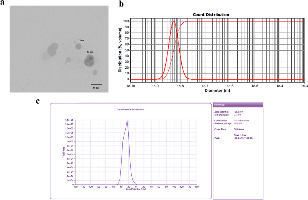
The size of PLA-PEG-PLA nano-vesicle loaded with propolis estimated by transmission electron microcopy (a) and dynamic light scattering (b) as well as its Zeta potential (c).
3.3 Echocardiographic parameters
Induction of HF by subcutaneous injections of 85 mg/kg isoproterenol for 2 consecutive days led to a decrease of 56.88% of EF and 44% of FS in rats. The administration of propolis and PLA-PEG-PLA nano-vesicle loaded with propolis had no significant difference in averages of EF and FS in healthy rats (P > 0.05). However, their administration to HF rats resulted in improvement of echocardiographic parameters. Administration of propolis to HF rats led to an increase of 10.75% and 10.29% in EF and FS parameters, respectively, compared to HF rats, which indicates the improvement of echocardiographic parameters. Also, administration of PLA-PEG-PLA nano-vesicle loaded with propolis led to improvement of EF and FS parameters by 26.75% and 19%, respectively, compared to HF rats (Fig. 2). Therefore, the nanoformulation designed in the present study had a stronger positive effect in improving echocardiographic parameters in HF condition compared to free propolis.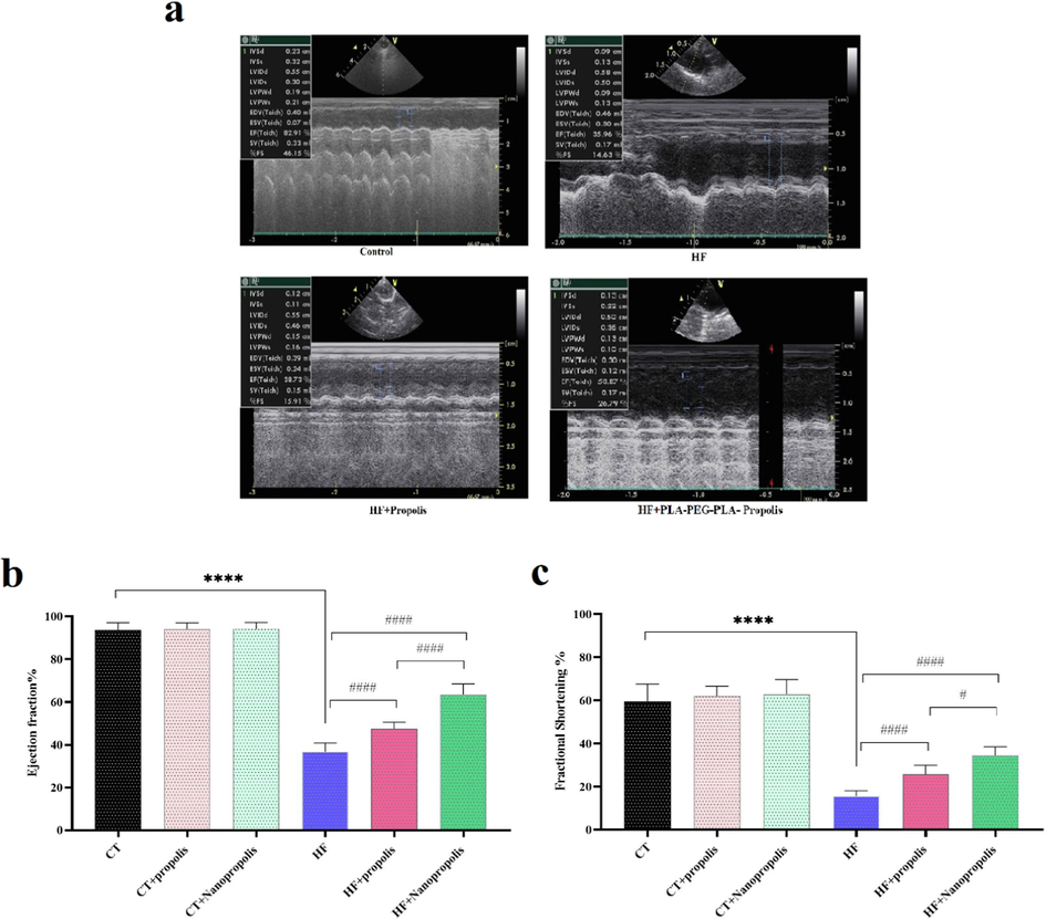
The effects of propolis and PLA-PEG-PLA nano-vesicle loaded with propolis on Echocardiographic parameters (a) ejection fraction (EF, b) and fractional Shortening (FS, c) of healthy and HF rats (n = 8). ****P < 0.0001 VS. Control; #### P < 0.0001 VS. HF; #P < 0.05 VS. HF. 200 mg/kg of propolis and PLA-PEG-PLA nano-vesicle loaded with propolis were given to the rats 1 week before HF induction and 7 days after that and the echocardiographic parameters were measured 24 h after last dosing.
3.4 Biochemical parameters
3.4.1 B-type natriuretic peptide (BNP) and Galectin-3
In the present study, the plasma levels of BNP and Galectin-3 were measured in rats and the results indicated a significant increase of both markers of heart failure in HF rats compared to healthy ones (P < 0.0001). The administration of both propolis and PLA-PEG-PLA nano-vesicle loaded with propolis to HF rats led to a significant decrease in the plasma levels of BNP (Fig. 3a) and Galectin-3 (Fig. 3b) compared to HF rats (P < 0.0001). Comparing the plasma levels of these heart failure markers in HF rats receiving propolis and PLA-PEG-PLA nano-vesicle loaded with propolis indicated a significant decrease in both BNP (P < 0.0001) and Galectin-3 (P = 0.008) parameters (Fig. 3). Therefore, PLA-PEG-PLA nano-vesicle loaded with propolis seems to have the strongest cardioprotective effects compared to free propolis in HF conditions.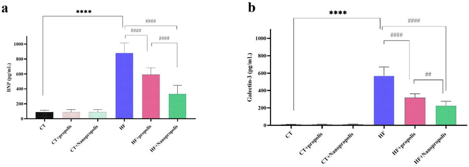
The effects of propolis and PLA-PEG-PLA nano-vesicle loaded with propolis on heart failure biomarkers BNP (a) and Galectin-3 (b) of healthy and HF rats (n = 8). ****P < 0.0001VS. control; ####P < 0.0001VS. HF; #P < 0.05 VS. HF; ##P < 0.01VS. HF. 200 mg/kg of propolis and PLA-PEG-PLA nano-vesicle loaded with propolis were given to the rats 1 week before HF induction and 7 days after that and the plasma levels of BNP and Galectin-3 were measured 24 h after last dosing.
3.4.2 Pro-inflammatory cytokines
Induction of HF in rats resulted in up-regulation of IL-1β (Fig. 4a) and IL-6 (Fig. 4b), down-regulation of IL-10 (Fig. 4c), as well as upregulation of IL-17 (Fig. 4d) cytokines. Nevertheless, a significant decrease in the expression of cytokines IL-1β, IL-6 and IL-17 and a significant increase in the expression level of IL-10 were seen in rats receiving propolis and PLA-PEG-PLA nano-vesicle loaded with propolis compared to HF rats. The expression of IL-1β (P = 0.007) and IL-6 (P = 0.041) in HF rats receiving PLA-PEG-PLA nano-vesicle loaded with propolis was significantly lower than the counterparts receiving free propolis (Fig. 4). Therefore, it seems that PLA-PEG-PLA nano-vesicle loaded with propolis has stronger anti-inflammatory effects compared to free propolis in HF conditions.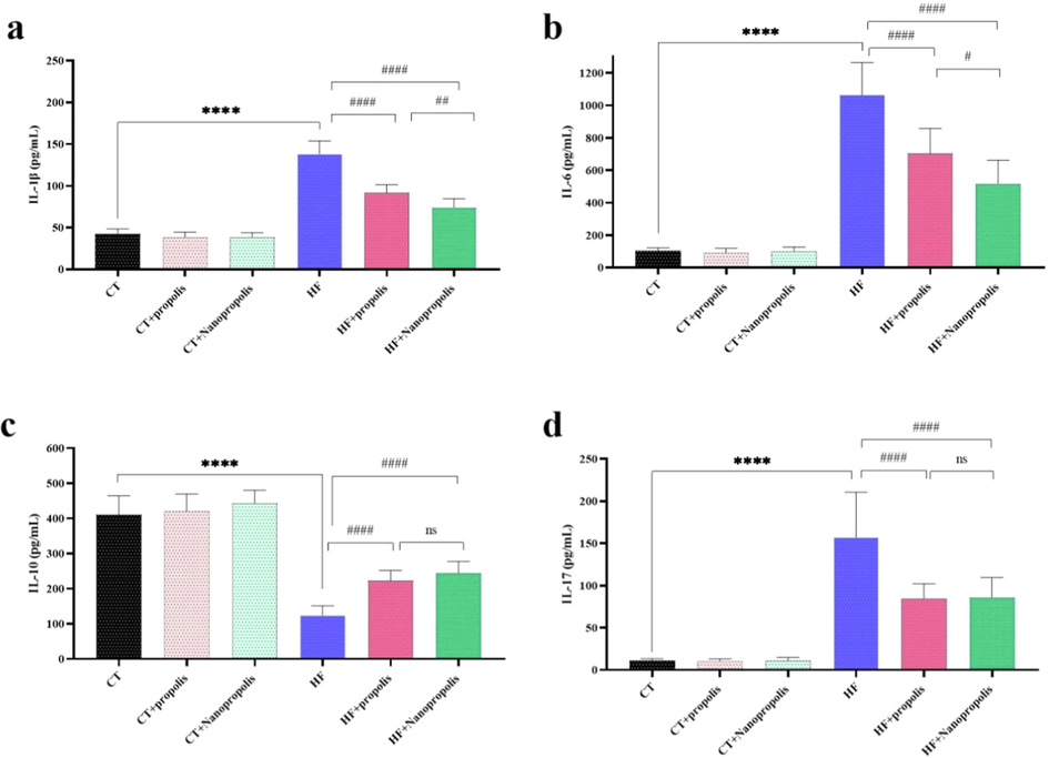
The effects of propolis and PLA-PEG-PLA nano-vesicle loaded with propolis on inflammatory cytokines IL-1β (a), IL-6 (b), IL-10 (c) and IL-17 (d) of healthy and HF rats (n = 8). ****P < 0.0001 VS. control; ####P < 0.0001 VS. HF; #P < 0.05 VS. HF; ##P < 0.01 VS. HF. 200 mg/kg of propolis and PLA-PEG-PLA nano-vesicle loaded with propolis were given to the rats 1 week before HF induction and 7 days after that and the plasma levels of IL-1β, IL-6, IL-10 and IL-17 were measured 24 h after last dosing.
3.4.3 Reduced AMP-activated protein kinase (AMPK) and silent information regulator (SIRT1)
Both the plasma levels of AMPK (Fig. 5a) and SIRT1 (Fig. 5b) were downregulated in HF rats compared to control healthy ones (P < 0.0001). Also, in healthy rats receiving propolis and PLA-PEG-PLA nano-vesicle loaded with propolis SIRT1 levels were increased significantly compared with control (P < 0.0001). However, HF rats receiving both propolis and PLA-PEG-PLA nano-vesicle loaded with propolis showed upregulations of plasma levels of AMPK and SIRT1 compared to HF rats (P < 0.0001). The comparison of AMPK and SIRT1 plasma levels of HF rats receiving PLA-PEG-PLA nano-vesicle loaded with propolis and those receiving free propolis did not show significant change (P > 0.05, Fig. 5).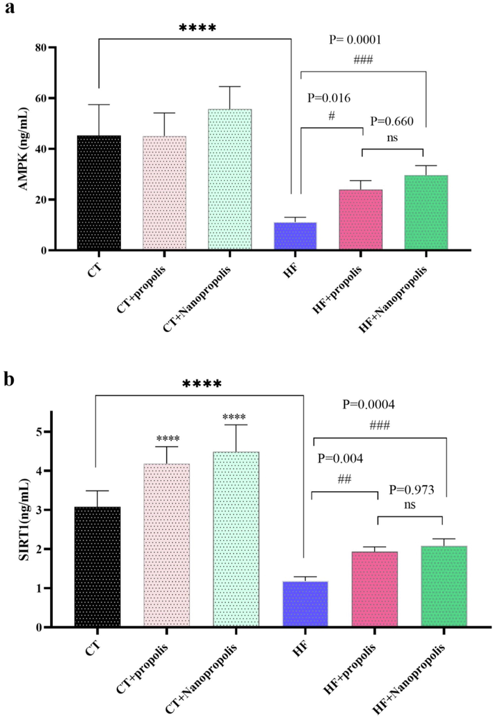
The effects of propolis and PLA-PEG-PLA nano-vesicle loaded with propolis on reduced AMP-activated protein kinase (AMPK, a) and silent information regulator (SIRT1, b) of healthy and HF rats (n = 8). ****P < 0.0001 VS. control; ####P < 0.0001VS. HF; #P < 0.05 VS. HF. 200 mg/kg of propolis and PLA-PEG-PLA nano-vesicle loaded with propolis were given to the rats 1 week before HF induction and 7 days after that and the plasma levels of MAPK and SIRT1 were measured 24 h after last dosing.
4 Discussion
In the present study, administration of propolis and PEG-PLA loaded with propolis led to improvement of echocardiographic parameters and reduction of HF biomarkers (BNP and Galectin-3) in HF model rats. Also, these two compounds decreased pro-inflammatory cytokines (IL-1β, IL-6 and IL-17) and increased the expression of anti-inflammatory cytokine (Il-10) in HF rats. Propolis and PEG-PLA loaded with propolis were able to significantly upregulate the plasma levels of AMPK and SIRT1 in HF rats compared to the positive control (HF). Therefore, the mechanism of cardioprotective effects of propolis seems to be activation of AMPK expression through SIRT1 activation.
Induction of HF in model animals is done through different approaches. However, one of the standard approaches to induce HF is ISO injection, in which conditions similar to HF are created and the vasculature remains intact (Li et al., 2008). Feng and Li (2010) used different doses of ISO to induce HF in rats and concluded that administration of 85 mg ISO for two consecutive days was the most effective in inducing HF (Feng and Li, 2010), so this dose was chosen in the present study.
The present study showed that HF induction in rats leads to an increase in the plasma levels of Galectin-3 in rats. This HF marker has different cellular physiological functions (Ahmed and AlSadek, 2015; de Oliveira et al., 2015) and it has been stated that its levels increase in inflammatory diseases such as CVD and its high expression is directly correlated with stroke (Edsfeldt et al., 2016). The results of the present study showed that the administration of propolis and PEG-PLA loaded with propolis to rats leads to a decrease in the plasma levels of Galectin-3, which can be attributed to their anti-inflammatory effects. In the present study, the anti-inflammatory effects of both propolis and PEG-PLA loaded with propolis were confirmed by reducing the levels of inflammatory cytokines IL-1β, IL-6 and IL-17 and increasing IL-10 expression. Meanwhile, IL-17 plays an important role in inducing inflammation, because increasing its levels causes the release of other inflammatory cytokines such as IL-2, TNF-α, IL-18, IL-8, and IL-6 (Yasumi et al., 2007). So, the reduction of its expression levels by propolis and PEG-PLA loaded with propolis indicates the reduction of inflammation in HF conditions. Therefore, propolis and PEG-PLA loaded with propolis with anti-inflammatory effects can have a protective role for the heart in HF conditions.
Cardioprotective effects of flavonoids in propolis have been reported in many studies. For example, Wang et al. (2015) stated that propolis flavonoids are able to reduce myocardial apoptosis in chronic HF conditions (Wang et al., 2015), and it seems to do this through different signaling pathways. In the condition of cardiac hypertrophy, it was stated that propolis flavonoids are able to show cardioprotective effects through the PI3K/AKT pathway (Sun et al., 2016). It has been stated that propolis flavonoids are able to reduce cardiac fibrosis in myocardial infarction and it does this by activating SIRT1 (Wang et al., 2018), which is in accordance with the findings of the present study. Also, bee propolis combined with metformin reduced diabetic cardiomyopathy (Lim et al., 2022). All these studies indicate the heart protection effects of propolis, and in the current study, its heart protection effects were confirmed in HF conditions.
One of the interesting results of the present study was that the administration of propolis and PEG-PLA loaded with propolis led to inhibition of the development of HF in rats. This can be attributed to their anti-inflammatory effects and especially the upregulations of SIRT1 and AMPK. It seems that the increase in SIRT1 levels can lead to the improvement of the antioxidant status and reduction of apoptosis in myocardial cells (Tanno et al., 2010), and one of the mechanisms of the heart protection effects of propolis and PEG-PLA loaded with propolis is the increase in SIRT1 levels. SIRT1 has been shown to prevent fibrosis of vital organs such as liver, heart, and kidney (Chou et al., 2017; Rizk et al., 2014; Wu et al., 2015). Therefore, the increase of SIRT1 level by propolis and PEG-PLA loaded with propolis may have prevented the development of fibrosis caused by HF. Recently, it was reported that SIRT1 regulates the expression of AMPK (Gu et al., 2014), and it seems that propolis by affecting the expression of SIRT1 led to the improvement of the expression of AMPK and prevented the heart damage caused by HF. The increase in AMPK levels in HF rats treated with propolis and PEG-PLA loaded with propolis can improve energy homeostasis (Gu et al., 2014), which is disturbed in HF conditions.
5 Conclusion
In general, it is concluded that propolis and PEG-PLA loaded with propolis have cardioprotective effects in HF conditions. The mechanism of action of these compounds can be attributed to anti-inflammatory effects and regulations of the SIRT1-AMPK axis. There is a need for clinical studies in this field.
Declaration of Competing Interest
The authors declare that they have no known competing financial interests or personal relationships that could have appeared to influence the work reported in this paper.
References
- Galectin-3 as a potential target to prevent cancer metastasis. Clin. Medicine Insights: Oncol. 2015:9. CMO. S29462
- [Google Scholar]
- Thirty years of evidence on the efficacy of drug treatments for chronic heart failure with reduced ejection fraction: a network meta-analysis. Circulation Heart Failure.. 2017;10(1):e003529.
- [Google Scholar]
- From the Cover: l-Carnitine via PPARγ- and Sirt1-Dependent mechanisms attenuates epithelial-mesenchymal transition and renal fibrosis caused by Perfluorooctanesulfonate. Toxicol. Sci.. 2017;160(2):217-229.
- [Google Scholar]
- Galectin-3 in autoimmunity and autoimmune diseases. Experimental Biol. Med.. 2015;240(8):1019-1028.
- [Google Scholar]
- High plasma levels of galectin-3 are associated with increased risk for stroke after carotid endarterectomy. Cerebrovasc. Dis.. 2016;41(3–4):199-203.
- [Google Scholar]
- The study of ISO induced heart failure rat model. Experimental Mol. Pathol.. 2010;88(2):299-304.
- [Google Scholar]
- Resveratrol, an activator of SIRT1, upregulates AMPK and improves cardiac function in heart failure. Genet Mol Res.. 2014;13(1):323-335.
- [Google Scholar]
- Adverse effects of β-blocker therapy for patients with heart failure: a quantitative overview of randomized trials. Arch. Internal Med.. 2004;164(13):1389-1394.
- [Google Scholar]
- Alternative preparation of propolis extracts: comparison of their composition and biological activities. BMC Complement Altern Med.. 2015;15:156.
- [Google Scholar]
- Comparison of aqueous, polyethylene glycol-aqueous and ethanolic propolis extracts: antioxidant and mitochondria modulating properties. BMC Complementary Alternative Med.. 2018;18(1):165.
- [Google Scholar]
- Triblock Copolymer Nanomicelles Loaded with Curcumin Attenuates Inflammation via Inhibiting the NF-Κb Pathway in the Rat Model of Cerebral Ischemia. Int. J. Nanomed.. 2021;16:3173.
- [Google Scholar]
- Mesenchymal stem cell transplantation attenuates cardiac fibrosis associated with isoproterenol-induced global heart failure. Transplant Int.. 2008;21(12):1181-1189.
- [Google Scholar]
- Stingless bee propolis, metformin, and their combination alleviate diabetic cardiomyopathy. Brazilian J. Pharm. Sci.. 2022;58
- [Google Scholar]
- Nutritional composition, phenolic compounds, nitrate content in eatable vegetables obtained by conventional and certified organic grown culture subject to thermal treatment. Int. J. Food Sci. Technol.. 2009;44(6):1118-1124.
- [Google Scholar]
- Heart failure: preventing disease and death worldwide. ESC Heart Failure. 2014;1(1):4-25.
- [Google Scholar]
- Antibacterial activity of Turkish propolis and its qualitative and quantitative chemical composition. Phytomedicine.. 2005;12(3):221-228.
- [Google Scholar]
- A novel role for SIRT-1 in L-arginine protection against STZ induced myocardial fibrosis in rats. PloS one. 2014;9(12):e114560.
- [Google Scholar]
- Flavonoids extraction from propolis attenuates pathological cardiac hypertrophy through PI3K/AKT signaling pathway. Evidence-Based Complementary Alternative Med. 2016
- [Google Scholar]
- Induction of manganese superoxide dismutase by nuclear translocation and activation of SIRT1 promotes cell survival in chronic heart failure. J. Biol. Chem.. 2010;285(11):8375-8382.
- [Google Scholar]
- Targeting interleukin-β by plant-derived natural products: Implications for the treatment of atherosclerotic cardiovascular disease. Phytother. Res.. 2021;35(10):5596-5622.
- [Google Scholar]
- Flavonoid extract from propolis inhibits cardiac fibrosis triggered by myocardial infarction through upregulation of SIRT1. Evidence-Based Complementary Alternative Med.. 2018;2018
- [Google Scholar]
- Flavonoid extract from propolis provides cardioprotection following myocardial infarction by activating PPAR-γ. Evidence-Based Complementary Alternative Med.. 2022;2022
- [Google Scholar]
- Effects of total flavonoids of propolis on apoptosis of myocardial cells of chronic heart failure and its possible mechanism in rats. Zhongguo Ying Yong Sheng li xue za zhi= Zhongguo Yingyong Shenglixue Zazhi= Chinese J. Appl. Physiol.. 2015;31(3):201-206.
- [Google Scholar]
- Silent information regulator 1 (SIRT1) ameliorates liver fibrosis via promoting activated stellate cell apoptosis and reversion. Toxicol. Appl. Pharmacol.. 2015;289(2):163-176.
- [Google Scholar]
- Interleukin-17 as a new marker of severity of acute hepatic injury. Hepatol. Res.. 2007;37(4):248-254.
- [Google Scholar]


