

Translate this page into:
The protective effects of apple pectin and citrus pectins on post-cerebral I/R depression in mice: The role of NF-κB-p65 and pSTAT3 pathways
⁎Corresponding authors at: NO.76, Yanta West Road, Institute of Neurobiogy, Xi'an Jiaotong University Basic Medical School, Xi'an 710061, China. Liuy5599@mail.xjtu.edu.cn (Yong Liu)
-
Received: ,
Accepted: ,
This article was originally published by Elsevier and was migrated to Scientific Scholar after the change of Publisher.
Peer review under responsibility of King Saud University.
Abstract
One of the serious consequences of brain stroke is depression, which affects a large number of patients. Finding new compounds that have antidepressant properties in these conditions can be very important. Therefore, in the present study, the antidepressant effects of apple pectin (AP) and citrus pectin (CP) in mice after induction of cerebral ischemia were studied. Seven days before transient middle cerebral artery occlusion (tMCAO) and three days thereafter, mice were given orally AP and CP by gavage (50 and 100 mg/kg). Forced swim test (FST), inclined beam-walking test and open field test were used to evaluate the behaviors of mice. Levels of IL-1β, INF-γ, TNF-α, IL-6 NF-κB-p65 and pSTAT3 were studied using enzyme-linked immunosorbent assay (ELISA) and the gene expressions of NF-κB-p65 and pSTAT3 were studied by qRT-PCR. Induction of cerebral I/R injury resulted in depressive-like behaviors in mice, which were confirmed by FST, inclined beam-walking test and OFT. However, beneficial effects of AP and CP were observed in reducing depressive-like behaviors in mice. The downregulation of proinflammatory cytokines and NF-κB-p65 and pSTAT3 proteins were observed as a result of oral AP and CP administration in cerebral I/R mice. AP and CP have antidepressant-like effects on cerebral I/R- induced depression due to reduced inflammation in the brain hippocampus.
Keywords
Post-Stroke Depression
Inflammation
Cytokine
Mice

1 Introduction
Depression is one of the most important psychosocial consequences in patients with stroke. The occurrence of depression is usually common after stroke, so that its prevalence in the first year after stroke is estimated to be more than 30% (Carod-Artal et al., 2009; Kouwenhoven et al., 2011). Post-stroke depression affects a person's cognitive function, health-related quality of life, and performance improvement (Alajbegovic et al., 2014). Depression after stroke is related to the underlying factors and the severity and course of this disorder is associated with other complications of stroke (Sit et al., 2007). Many researchers believe that mood disorders after stroke can also have a significant effect on the cognitive abilities of patients (Mitchell et al., 2017).
One of the important mechanisms of post- stroke depression is inflammatory responses. The role of interleukin-2 (IL-2), tumor-necrosis factor alpha (TNF-α) and interferon alpha (INFα) in the development of depressive symptoms has been demonstrated (Villa et al., 2018). Cytokines such as IL-1β, TNF-α, and IL-8 have also been reported to increase after stroke (Massaro et al., 2018). Therefore, it has been suggested that the production of proinflammatory cytokines in cerebral ischemia in mood-related areas may be associated with post-stroke depression (Spalletta et al., 2006). Also, interleukin-18 (Il-18) has been mentioned as a potential biomarker for the development of post-stroke depression (Yang et al., 2010).
Today, various treatments for this disorder have been proposed and the effectiveness of many antidepressants in reducing the symptoms associated with post-stroke depression has been proven (Olesen et al., 2007). However, chemical drugs are always associated with adverse effects, so finding natural compounds with antidepressant properties that are effective in stroke conditions is important.
Pectins are the natural polymers that make up the cell wall in most plants, about 70% of which is galacturonic acid. Pectins are widely used in the food industry as stabilizers, and the main sources of pectin extraction are citrus peel, apple pulp and sugar beet (Baississe et al., 2010). Although pectins are not digested by the upper gastrointestinal tract, it has many health benefits (Maxwell et al., 2012). Pectin has cholesterol-lowering effects and plays an important role in preventing heart disease(Brouns et al., 2012), high blood pressure and diabetes (Silva et al., 2011). There is also considerable evidence for the role of pectin in the prevention of cancer and metastasis (Fang et al., 2018; do Prado SBR, Ferreira GF, Harazono Y, Shiga TM, Raz A, Carpita NC, , 2017). One study showed that pectins improved memory and antidepressant behavior in mice, which was attributed to upregulation of Il-6 expression and the JAk-STAT signaling pathway (Paderin and Popov, 2018).
Therefore, the aim of the present study was to evaluate the oral administration of apple pectin (AP) and citrus pectin (CP) on depressive-like behaviors of mice after induction of transient middle cerebral artery occlusion (tMCAO). Also, the expression levels of IL-1β, INF- γ, TNF-α and IL-6 cytokines and NF-κB-p65 and pSTAT3 proteins were studied.
2 Materials and methods
2.1 Animals
56 Male BALB/c mice weighing 22–26 g were given two weeks to adapt to the environmental conditions before starting the experiment. The animals were exposed to ambient conditions with a temperature of 24 ± 1.5 °C, 56% humidity and under light conditions with a 12-hour light/dark cycle. They were allowed to free access a diet with 13% protein, 5.5% fat, 55% carbohydrate and 3.9% fiber during the test period. To prevent changes due to the circadian cycle, all tests were performed between 09:00 and 15:00.
Mice were divided into 7 groups as follows:
1. Control group received normal saline solution.
2. Sham group received normal saline solution;
3. Stroke group received normal saline;
4. Stroke group receiving 50 mg/kg AP (CAT Number: 93854, Sigma Aldrich, Germany);
5. Stroke group receiving 100 mg/kg AP.
6. Stroke group receiving 50 mg/kg CP (CAT Number: S419095, Sigma Aldrich, Germany);
7. Stroke group receiving 100 mg/kg CP.
2.2 Administration of pectins
Prior to administration, AP and CP were dissolved in distilled water at specified concentrations and then fed to mice by gavage. Concentrations were determined based on published reports (Paderin and Popov, 2018). Treatments began seven days before cerebral I/R induction and continued for three days thereafter. 96 h after induction of I/R injury, mice were evaluated for neurobehavioral activities and then sacrificed for biochemical evaluations. Brain hippocampal extraction was performed using the method of Mathis et al. (Mathis et al., 2011). For this purpose, after decapitation and making a midline incision along the skull, and its separation, the brain was scooped out and washed with an ice-cooled buffer. The upper part of the hippocampal cortex was separated and after homogenization for 5 min at 4 °C in 0.7 mL of lysis buffer, it was centrifuged at 10,000 g for 10 min. It was then stored at −20 °C for biochemical experiments.
2.3 Induction of tMCAO
Hill and Nemoto (Hill and Nemoto, 2014) methods were used to induce transient middle cerebral artery occlusion (tMCAO) (Hill and Nemoto, 2014). For this purpose, the animals were first anesthetized intraperitoneally (i.p) with ketamine (50 mg/kg) and xylazine (2–8 mg/kg). Then, a shallow incision of 25 mm was made along the midline from the bottom of the mandible to the sternum. The common carotid artery (CCA) was separated from the vagus nerve with complete caution and ligated with 4–0 silk sutures. The CCA was thus occluded without injury. Next, a punctate incision was made in the CCA ventral wall and then the suture was inserted into the ICA until resistance was felt. Under these conditions, suture occluded the middle cerebral artery (MCA) origin. After 90 min, the animals were reanesthetized and the occluding suture was removed and complete restoration of perfusion was performed.
2.4 Neurobehavioral tests
2.4.1 Forced swim test
The forced swimming test was performed according to the method of Wang et al. (Wang et al., 2010). For this purpose, a colorless glass cylinder with a diameter of 10 cm and a height of 25 cm was filled with 23 °C water to a height of 19 cm. The mice were individually placed individually in the cylinder, each given 6 min to swim. The first 2 min were for the animal adaptation with the environment and the duration of immobility was recorded in the last four minutes of the test. Immobility is when each mouse stops trying to swim and stays still in the water without moving, keeping only its head above the water.
2.4.2 Inclined beam-walking test
This test was performed based on the method of Feeney et al. (Feeney et al., 1981) to examine fore and hind limb motor coordination. Initially, the mice were placed individually on a wooden bar at a 60-degree angle to the platform. Each animal was given a score of 0 to 4 based on its motor function, with 0 indicating an animal that could easily traverse the beam. Mice with mild, moderate, or severe defects were given scores of 1, 2, and 3, respectively. A mouse that showed no movement on the beam was given a score of 4. This test was performed four days after induction of I/R cerebral injury.
2.4.3 Open field test
The OFT test is used because of the reduced locomotion and exploration in depression. In the present study, an open field apparatus with a diameter of 60 cm was used, with 15 holes in the bottom (each 1 cm in diameter) with a height of 40 cm. Initially, each mouse was placed separately in the center of the square and its behavior was monitored for 5 min using a camera. The distance traveled, number of squares crossed, and the number of rearing and number of examined holes were measured.
2.4.4 Neurological scores
The method of Garcia et al. (Garcia et al., 1995) was used to calculate neurological scores. In this method, six animal behaviors including spontaneous activity, symmetry in the movement of four limbs, forepaw outstretching, climbing, body proprioception and response to touching vibrissae were evaluated.
Spontaneous activity was assessed for 5 min. A score of 0 was considered when the animal showed no activity. Animals that difficulty moved without rise up were given a score of 1. Animals that moved only to one corner of the cage were given a score of 2, and animals that moved freely in the cage were given a score of 3.
Symmetry in the movement of four limbs by lifting the animal from the tail and observing the movements of all four limbs were done and scores 0 (no movement of limbs), 1 (very little movement in limbs) 2 (mobility of limbs but asymmetry between limbs left and right) and 3 (symmetrical motion of every 4 limbs) were given.
Forepaw outstretching was done by lifting the animal and placing it on the table and observing the movement of the forelimbs, and scores 0 (no movement), 1 (hard movement), 2 (the right forelimb moved more than the left) and 3 (symmetrical movement by both forelimbs) were assigned.
To assess climbing, the animals were placed on the wall of a wire cage and given scores of 1 (without attempting climbing), 2 (asymmetric climbing), and 3 (symmetrical and easy climbing) based on their movement.
Body proprioception was examined by contacting the animals' bodies with a blunt stick and based on how they responded, and scores of 1 (no response to contact), 2 (slow response to contact) and 3 (symmetrical response of both sides to contact) were given.
Response to touching vibrissae was performed by evaluating the response of animals to the stimulus vibrissae on both sides of the body and scores of 1 (no response to the stimulus), 2 (slow response to the stimulus) and 3 (symmetrical response of both sides of the body to the stimulus) were assigned.
2.5 Enzyme-linked immunosorbent assay (ELISA)
Levels of IL-1β, INF- γ, TNF-α and IL-6 cytokines in mice brain hippocampal homogenates were measured using ELISA technique. For this purpose, IL-1β (Cat. No. AB1832P, Meck), INF- γ, (clone 12E7.1, Merck), TNF-α (Cat#430904, BioLegend Inc., USA) and IL-6 (MABF1507, Meck) mouse monoclonal antibodies were used as capture antibodies and Biotin-conjugated IL-1β, IL-18, TNF-α and IL-2 antibodies were used as detection antibodies. Detection was determined using streptavidin-labeled horseradish peroxidase (HRP). The absorbance was read at 492 nm.
ELIZA kits for NF-κB-p65 from Mybiosource (Cat# MBS2508000, San Diego, USA) and pSTAT3 from Abcam (Cat# ab176654, Cambridge, USA) were used to measure the expression levels of NF-κB-p65 and pSTAT3.
2.6 qRT-PCR
To evaluate the expression of NF-κB p65 and STAT3 genes, RNA was extracted using QIAGEN kit (Hilden, Germany) and after determining its quantity and quality with nanodrop and agarose electrophoresis gel, respectively, cDNA synthesis was done using Sigma-Aldrich (USA) kit. In the present study, the glyceraldehyde-3 phosphate dehydrogenase (GAPDH) gene was used as the internal control. The sequences of primers are given in Table 1. The qRT-PCR reaction was then performed using SYBR Green. Relative expression of genes was measured using 2-ΔΔCT method.
Genes
Sequences
STAT3-F
CAGCAATACCATTGACCTGCC
STAT3-R
TTTGGCTGCTTAAGGGGTGG
NF-κB p65-F
CATCTTTCCCTCAGAGCCAGCC
NF-κB p65-R
GTGCTTCTCTCCCCAGGAATAC
GAPDH-F
TGCTGGTGCTGAGTATGTCG
GAPDH-R
TTGA GAGCAATGCCAGCC
2.7 Statistical analysis
After ensuring the normal distribution of the data using the Kolmogorov-Smirnov test, the data were analyzed using one-way analysis of variance (ANOVA). All data were presented as mean ± standard error. Probability level P < 0.05 was considered a significant level. Tukey's multiple range test was used to compare the means. Data analysis was performed using Graphpad Prism 8 software.
3 Results
3.1 Neurobehavioral tests
3.1.1 Forced swim test (FST)
FST results showed that time latency to immobility was significantly reduced in tMCAO-challenged mice compared with control and sham (P < 0.0001). However, pretreatment with AP and CP for 10 days resulted in a dose-dependent increase in latency to immobility after cerebral I/R injury. Both AP and CP at concentrations of 100 mg/kg resulted in increased time latency to immobility in cerebral I/R mice compared with a dose of 50 mg/kg. In I/R mice, the highest latency to immobility was observed in mice receiving 100 mg CP (57.25 s) (Fig. 1a).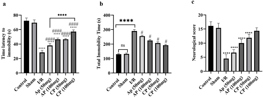
The effects of apple pectin (AP) and citrus pectin (CP) on FST latency (a), FST immobility (b) and neurological score (c) in cerebral I/R mice. ** and **** represent significant differences with control and sham groups at probability levels of P < 0.01 and P < 0.0001, respectively. # and #### Represent significant differences with I/R mice at probability level of P < 0.001 and P < 0.0001, respectively (n = 7).
Total immobility time was measured in the FST test and the results showed a positive effect of AP and CP in reducing total immobility time (Fig. 1b). The results showed a sharp increase in total immobility time in I/R mice compared to sham and control groups; however, when AP or CP were administered to I/R mice, a dose-dependent decrease was observed in total immobility time. Overall, the effect of CP on reducing total immobility time was better than AP, and a concentration of 100 mg/kg resulted in a severe reduction in total immobility time, which may indicate antidepressant-like effects of CP.
3.1.2 Neurological scores
Induction of tMCAO greatly reduced neurological scores in mice (P < 0.0001). However, pretreatment with 100 mg/kg CP significantly increased neurological scores compared to cerebral I/R mice (Fig. 1c).
3.1.3 Open field test (OFT)
The results of the present study indicated the development of a hypoactive phenotype in cerebral I/R mice (Fig. 2). However, AP or CP pretreated I/R mice showed improvement in locomotor activity in all three indicators of square crossings, rearing, and explored holes. The best effect was seen in I/R mice pretreated with 100 mg/kg CP.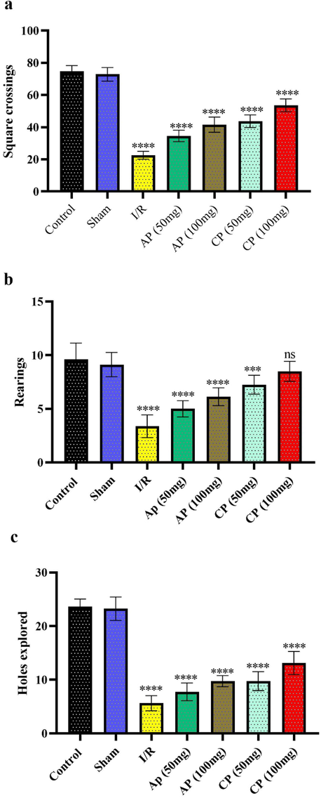
The impacts of apple pectin (AP) and citrus pectin (CP) supplementation for 10 days on the square crossings (a), rearing (b) and hole exploration (c) of mice in the open field test.
3.2 The expression levels of IL-1β, INF- γ, TNF-α and IL-6 cytokines
The results showed an overexpression of IL-1β, INF- γ, TNF-α and IL-6 in cerebral I/R mice hippocampus compared with control and sham groups. IL-1β and INF-γ expression were upregulated in the hippocampal tissue of I/R mice, but 100 mg/kg CP treatment was able to significantly downregulate the expression of this cytokine compared to cerebral I/R mice. 100 mg/kg AP and 50 and 100 mg/kg CP were able to reduce the expression of the proinflammatory cytokine TNF-α and even bring it to a healthy and sham control level. However, a significant decrease in IL-6 expression was observed in cerebral I/R mice when received 50 and 100 mg/kg CP (Fig. 3). Therefore, it seems that 100 mg/kg CP treatment has the greatest effect on the downregulation of the expression of studied proinflammatory cytokines.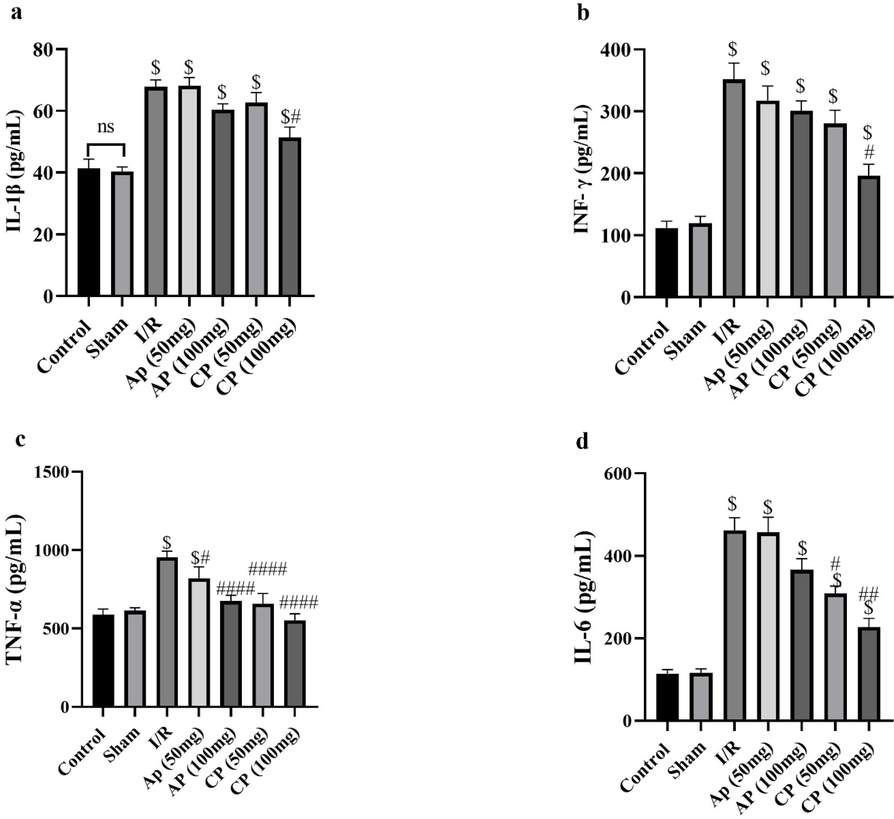
The effects of apple pectin (AP) and citrus pectin (CP) supplementation for 10 days on expression levels of IL-1β (a), INF- γ (b), TNF-α (c) and IL-6 (d) cytokines in mice hippocampi. $ indicates significant differences with control and sham groups and #,## and ####represents significant differences with cerebral I/R groups at probability levels of P < 0.05, P < 0.01 and P < 0.001, respectively (n = 5).
3.3 NF-κB-p65 and pSTAT3 proteins and genes expression levels
The expression levels of NF-κB-p65 and pSTAT3 proteins and genes showed different expression patterns in different groups (Fig. 4,Fig. 5). Induction of cerebral I/R injuries upregulated both NF-κB-p65 and pSTAT3 proteins and genes expression levels. But, AP and CP downregulated NF-κB-p65 and pSTAT3 proteins and genes expression levels which indicated downregulating effects of AP and CP on the expressions of κB-p65 and pSTAT3. 100 mg/kg CP and AP had the strongest downregulating effects on the expressions of NF-κB-p65 and pSTAT3, which may be the reasons for anti-inflammatory effects of AP and CP.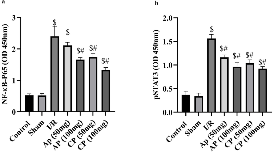
The effects of apple pectin (AP) and citrus pectin supplementation for 10 days on the expression levels of NF-κB (a) and pSTAT3 (b) proteins. $ shows significant differences compared control and sham groups at probability level of P < 0.05 and # represents significant differences compared I/R groups at probability level of P < 0.05 (n = 5).
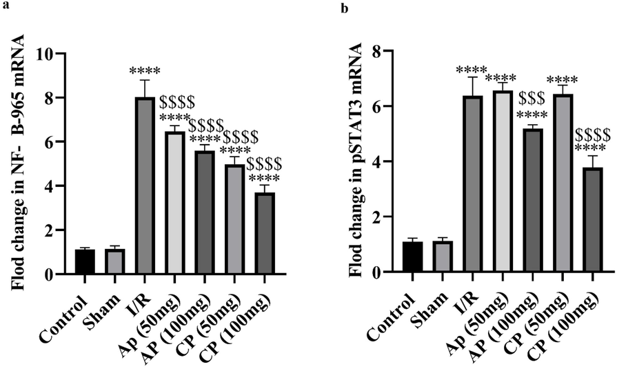
The effects of apple pectin (AP) and citrus pectin supplementation for 10 days on the expression levels of NF-κB (a) and pSTAT3 (b) genes. **** shows significant differences compared control and sham groups at probability level of P < 0.0001 and $$$ and $$$$ represents significant differences compared I/R groups at probability level of P < 0.001 and P < 0.0001 (n = 5).
4 Discussion
At the forefront of psychiatric disorders following brain stroke is depression, which is independently associated with many complications including increased anxiety, disability, and ultimately suicidal ideation (Villa et al., 2018). So far, no suitable treatment has been found for post-stroke depression, and most of the chemical drugs used have been associated with adverse adverse effects. Therefore, it is important to find treatment approaches that improve the symptoms of depression following stroke. The results of the present study showed the antidepressant-like effects of oral administration of AP and CP on cerebral I/R mice and it seems that the concentration of 100 mg/kg led to the maximum antidepressant-like effects in cerebral I/R mice. In the present study, a strong therapeutic effect of CP on depression-like behaviors caused by cerebral I/R injury was observed in comparison with AP. This can be attributed to the strong anti-inflammatory properties of CP compared to AP observed in the current research.
One of the experimental models of cerebral I/R induction is tMCAO, which has been shown to be effective in studies (Liu et al., 2019) and has also been shown to be associated with depressive-like behaviors (Xiong et al., 2020). Therefore, in the current study, inclined beam-walking test, FST and OFT were used to evaluate the depression like behaviors of mice. The results showed that tMCAO and subsequent reperfusion significantly increased neurological scores, confirmed the tMCAO induction of depression-like behaviors. However, oral administration of AP and CP reduced neurological scores, indicating the therapeutic potential of both AP and CP for cerebral I/R condition.
The FST test was used to evaluate the antidepressant-like behavior of AP and CP in cerebral I/R mice in the present study, and the results showed that induction of cerebral I/R significantly increases immobility time, which indicates depressive behavior. This has been shown in other studies (Yan et al., 2007; Yan et al., 2004). However, PA and CP were able to reduce the immobility time in cerebral I/R mice, indicating the antidepressant-like effects of these compounds. OFT results showed that AP and CP had enhancing effects on locomotor and exploratory activities in cerebral I/R mice. The enhancing effects of pectin on locomotor and exploratory activities have been demonstrated in other studies (Viggiano et al., 2006).
Elevated levels of proinflammatory cytokines were observed in the hippocampi of cerebral I/R mice. Also, the results showed that oral administration of AP and CP can prevent the increase of levels of proinflammatory cytokines such as IL-1β, INF-γ, TNF-α and IL-6. Elevated levels of proinflammatory cytokines, such as IL-6 and TNF-α, have been reported in other studies (Fassbender et al., 1994; Zhang et al., 2019), suggesting that stroke induces inflammation and inflammatory responses. However, levels of these cytokines decreased in I/R mice receiving AP and CP, indicating anti-inflammatory effects of the two compounds. An association between IL and 6/TNF-α levels and post-stroke depression has been reported in other studies (Nishuty et al., 2019; Ting et al., 2020; Maes et al., 1995), which is consistent with current research findings. However, there are contradictory results in this regard (Brambilla and Maggioni, 1998; Haack et al., 1999). Studies have shown an association between cytokine secretion and depression (Köhler et al., 2017). Therefore, reducing the expression of proinflammatory cytokines in the hippocampi of mice by oral administration of AP and CP may indicate the antidepressant-like effects of these compounds in cerebral I/R conditions. However, more research is needed to confirm this in clinical settings.
As mentioned, inflammation is involved in the pathophysiology of cerebral I/R, and in this process, the NF-κB transcription factor plays an important regulatory role and, therefore, plays a vital role in inflammatory diseases (Gu et al., 2012). When activated, this transcription factor increases the expression of proinflammatory cytokines such as IL-1, IL-18 and TNF-α (Mitchell and Carmody, 2018), leading to inflammatory reactions and, as noted, there is an association between inflammation and post-stroke depression. In the present study, induction of cerebral I/R injury resulted in increased NF-κB protein expression, which may indicate increased inflammation in these conditions t. However, oral administration of AP and CP reduced the expression of this transcription factor in I/R mice, which may indicate the anti-inflammatory effects of these compounds. Therefore, the antidepressant-like effects observed in the present study as a result of AP and CP administration in cerebral I/R mice can be attributed to the reduction of inflammation in mice brain due to decreased expressions of proinflammatory cytokines as well as decreased NF-κB expression.
The JAK2/STAT pathway is important in diseases such as ischemia and is one of apoptotic signal transduction pathways (Zhang et al., 2015). However, in the present study, decrease of pSTAT3 protein expression was observed in the hippocampus of AP and CP-receiving mice, which may indicate a reduction in apoptosis due to cerebral I/R injury. Downregultion of pSTAT3 has been shown to reduce cellular apoptosis and neurological disorders (Satriotomo et al., 2006).
Expression and secretion of inflammatory cytokines such as TNF, IL-6 and IL-1β is induced by NF-κB, which is one of the main causes of cerebral I/R-induced inflammation (Lan et al., 2013). These inflammatory cytokines, such as IL-6, activate JAK2/STAT3, which leads to an intensification of inflammatory responses (Luo et al., 2016). Thus, decreased expression of inflammatory cytokines, NF-κB and STAT3 in the hippocampal tissue of mice received AP and CP in current study may be responsible for their therapeutic effects in cerebral I/R conditions.
5 Conclusion
In general, it is concluded that AP and CP are effective in reducing depression after cerebral I/R injury by reducing the expression of inflammatory cytokines and NF-κB transcription. Future research in this area in the clinical setting could confirm the beneficial effects of these compounds.
Ethical approval
Animals were handled in accordance with the Regulations of Experimental Animal Administration issued by the State Committee of Science and Technology of the People’s Republic of China.
Declaration of Competing Interest
The authors declare that they have no known competing financial interests or personal relationships that could have appeared to influence the work reported in this paper.
References
- Alajbegovic A, Djelilovic-Vranic J, Alajbegovic S, Nakicevic A, Todorovic L, Tiric-Campara M. Post stroke depression. Medical archives. 2014;68(1):47
- Comparison of structure and emulsifying activity of pectin extracted from apple pomace and apricot pulp. World J. Dairy Food Sci.. 2010;5(1):79-84.
- [Google Scholar]
- Blood levels of cytokines in elderly patients with major depressive disorder. Acta Psychiatr. Scand.. 1998;97(4):309-313.
- [Google Scholar]
- Cholesterol-lowering properties of different pectin types in mildly hyper-cholesterolemic men and women. Eur. J. Clin. Nutr.. 2012;66(5):591-599.
- [Google Scholar]
- Poststroke depression: prevalence and determinants in Brazilian stroke patients. Cerebrovasc. Dis.. 2009;28(2):157-165.
- [Google Scholar]
- do Prado SBR, Ferreira GF, Harazono Y, Shiga TM, Raz A, Carpita NC, et al. Ripening-induced chemical modifications of papaya pectin inhibit cancer cell proliferation. Scientific reports. 2017;7(1):1-17
- Modified citrus pectin inhibited bladder tumor growth through downregulation of galectin-3. Acta Pharmacol. Sin.. 2018;39(12):1885-1893.
- [Google Scholar]
- Proinflammatory cytokines in serum of patients with acute cerebral ischemia: kinetics of secretion and relation to the extent of brain damage and outcome of disease. J. Neurol. Sci.. 1994;122(2):135-139.
- [Google Scholar]
- Responses to cortical injury: I. methodology and local effects of contusions in the rat. Brain Res.. 1981;211(1):67-77.
- [Google Scholar]
- Neurological deficit and extent of neuronal necrosis attributable to middle cerebral artery occlusion in rats: statistical validation. Stroke. 1995;26(4):627-635.
- [Google Scholar]
- Inhibition of NF-κB activation is associated with anti-inflammatory and anti-apoptotic effects of Ginkgolide B in a mouse model of cerebral ischemia/reperfusion injury. Eur. J. Pharm. Sci.. 2012;47(4):652-660.
- [Google Scholar]
- Plasma levels of cytokines and soluble cytokine receptors in psychiatric patients upon hospital admission: effects of confounding factors and diagnosis. J. Psychiatr. Res.. 1999;33(5):407-418.
- [Google Scholar]
- Transient middle cerebral artery occlusion with complete reperfusion in spontaneously hypertensive rats. MethodsX. 2014;1:283-291.
- [Google Scholar]
- Peripheral cytokine and chemokine alterations in depression: a meta-analysis of 82 studies. Acta Psychiatr. Scand.. 2017;135(5):373-387.
- [Google Scholar]
- Depression in acute stroke: prevalence, dominant symptoms and associated factors. a systematic literature review. Disabil. Rehabil.. 2011;33(7):539-556.
- [Google Scholar]
- Electroacupuncture exerts anti-inflammatory effects in cerebral ischemia-reperfusion injured rats via suppression of the TLR4/NF-κB pathway. Int. J. Mol. Med.. 2013;31(1):75-80.
- [Google Scholar]
- Melatonin ameliorates cerebral ischemia/reperfusion injury through SIRT3 activation. Life Sci.. 2019;239:117036
- [Google Scholar]
- Osthole decreases renal ischemia-reperfusion injury by suppressing JAK2/STAT3 signaling activation. Exp. Ther. Med.. 2016;12(4):2009-2014.
- [Google Scholar]
- Increased plasma concentrations of interleukin-6, soluble interleukin-6, soluble interleukin-2 and transferrin receptor in major depression. J. Affect. Disord.. 1995;34(4):301-309.
- [Google Scholar]
- Plasma biomarkers of brain injury in neonatal hypoxic-ischemic encephalopathy. J. Pediatr.. 2018;194(67–75):e1.
- [Google Scholar]
- Preparation of acute hippocampal slices from rats and transgenic mice for the study of synaptic alterations during aging and amyloid pathology. J. Vis. Exp. JoVE. 2011;49
- [Google Scholar]
- Pectin–an emerging new bioactive food polysaccharide. Trends Food Sci. Technol.. 2012;24(2):64-73.
- [Google Scholar]
- NF-κB and the transcriptional control of inflammation. Int. Rev. Cell Mol. Biol.. 2018;335:41-84.
- [Google Scholar]
- Prevalence and predictors of post-stroke mood disorders: a meta-analysis and meta-regression of depression, anxiety and adjustment disorder. Gen. Hosp. Psychiatry. 2017;47:48-60.
- [Google Scholar]
- Evaluation of serum interleukin-6 and C-reactive protein levels in drug-naïve major depressive disorder patients. Cureus. 2019;11(1)
- [Google Scholar]
- The treatment of post stroke-depression and emotionalism in clinical practice. Ugeskrift for laeger. 2007;169(16):1470-1472.
- [Google Scholar]
- The effect of dietary pectins on object recognition memory, depression-like behaviour, and IL-6 in mouse hippocampi. J. Funct. Foods. 2018;43:131-138.
- [Google Scholar]
- JAK2 and STAT3 activation contributes to neuronal damage following transient focal cerebral ischemia. J. Neurochem.. 2006;98(5):1353-1368.
- [Google Scholar]
- Pectin from Passiflora edulis shows anti-inflammatory action as well as hypoglycemic and hypotriglyceridemic properties in diabetic rats. J. Med. Food. 2011;14(10):1118-1126.
- [Google Scholar]
- Associated factors of post-stroke depression among Hong Kong Chinese: a longitudinal study. Psychol. Health Med.. 2007;12(2):117-125.
- [Google Scholar]
- The etiology of poststroke depression: a review of the literature and a new hypothesis involving inflammatory cytokines. Mol. Psychiatry. 2006;11(11):984-991.
- [Google Scholar]
- Ting EY-C, Yang AC, Tsai S-J. Role of interleukin-6 in depressive disorder. International journal of molecular sciences. 2020;21(6):2194
- Annurca apple-rich diet restores long-term potentiation and induces behavioral modifications in aged rats. Exp. Neurol.. 2006;199(2):354-361.
- [Google Scholar]
- Post-stroke depression: mechanisms and pharmacological treatment. Pharmacol. Ther.. 2018;184:131-144.
- [Google Scholar]
- Antidepressant-like effects of the active acidic polysaccharide portion of ginseng in mice. J. Ethnopharmacol.. 2010;132(1):65-69.
- [Google Scholar]
- Neuronal brain injury after cerebral ischemic stroke is ameliorated after subsequent administration of (R)-ketamine, but not (S)-ketamine. Pharmacol. Biochem. Behav.. 2020;191:172904
- [Google Scholar]
- The antidepressant effect of ethanol extract of radix puerariae in mice exposed to cerebral ischemia reperfusion. Pharmacol. Biochem. Behav.. 2004;78(2):319-325.
- [Google Scholar]
- Quetiapine attenuates the depressive and anxiolytic-like behavioural changes induced by global cerebral ischemia in mice. Behav. Brain Res.. 2007;182(1):36-41.
- [Google Scholar]
- The serum interleukin-18 is a potential marker for development of post-stroke depression. Neurol. Res.. 2010;32(4):340-346.
- [Google Scholar]
- Bigelovin inhibits STAT3 signaling by inactivating JAK2 and induces apoptosis in human cancer cells. Acta Pharmacol. Sin.. 2015;36(4):507-516.
- [Google Scholar]
- Interleukin-11 treatment protected against cerebral ischemia/reperfusion injury. Biomed. Pharmacother.. 2019;115:108816
- [Google Scholar]


