

Translate this page into:
6-[3-(4-Fluorophenyl)-1H-pyrazol-4-yl]-3-[(2-naphthyloxy)methyl][1,2,4]triazolo[3,4-b][1,3,4]thiadiazole as a potent antioxidant and an anticancer agent induces growth inhibition followed by apoptosis in HepG2 cells
*Corresponding author. Tel.: +91 824 2474000x3206, mobile: +91 9448523990; fax: +91 824 2474033 isloor@yahoo.com (Arun M. Isloor)
-
Received: ,
Accepted: ,
This article was originally published by Elsevier and was migrated to Scientific Scholar after the change of Publisher.
Available online 9 June 2010
Abstract
In this paper we have investigated the in vitro antioxidant property of two triazolo-thiadiazoles, 6-[3-(4-fluorophenyl)-1H-pyrazol-4-yl]-3-[(2-naphthyloxy)methyl][1,2,4]triazolo[3,4-b][1,3,4]thiadiazole (FPNT) and 6-[3-(4-chlororophenyl)-1H-pyrazol-4-yl]-3-[(phenyloxy)methyl][1,2,4]triazolo[3,4-b][1,3,4]thiadiazole (CPPT) by spectrophotometric DPPH and ABTS radical scavenging methods as well as by lipid peroxide assay. The anticancer activity along with possible mechanism of action of triazolo-thiadiazoles in Hep G2 cells was explored using MTT assay, [3H] thymidine assay, flow cytometry and chromatin condensation studies. Both FPNT and CPPT exhibited a dose dependent cytotoxic effect on hepatocellular carcinoma cell line, HepG2. The IC50 value was very low for both the compounds when compared to standard drug, doxorubicin. Incorporation of [3H] thymidine in conjunction with cell cycle analysis suggested that FPNT inhibited the growth of HepG2 cells. Flow cytometric studies revealed more percentage of cells in sub-G1 phase, indicating apoptosis, which was further confirmed through chromatin condensation studies by Hoechst staining. FPNT was found to be a potent antioxidant when compared to the standard in DPPH, ABTS radical scavenging assays and lipid peroxidation studies.
Keywords
FPNT
CPPT
HepG2 cell lines
Cytotoxicity
Apoptosis
Antioxidant

1 Introduction
Human liver carcinoma is the fifth most common cancer in the world and is accountable for more than 600,000 deaths annually. Most of the patients with hepatocellular carcinoma die within a year after diagnosis. At present, the treatment mainly includes surgery and chemotherapy, but the restorative effects of the existing chemotherapeutic drugs are not good enough and they have many side effects. Therefore, development of highly efficient, selective and less noxious antitumor drug remains a hot research area.
1,2,4-Triazole derivatives are an interesting class of heterocycles which possess a wide range of activity, high oral availability and toxicity. Triazoles and thiadiazoles have been reported to possess analgesic (Mathew et al., 2007), anti-inflammatory (Kamotra et al., 2007), antiviral (Al-Soud et al., 2008), antimicrobial (Demirbas et al., 2004; Swamy et al., 2006), antifungal (Tsukuda et al., 1998; Hirpara et al., 2003; Isloor et al., 2009), antibacterial (Vikrant et al., 2009), antitubercular (Karthikeyan et al., 2007) and antitumor (Shivarama et al., 2002) activities. Reports of compounds that contain two active groups, triazole and thiadiazole in a single molecule have rarely been studied for its anticancer activity. Also, the presence of halogens in biologically active molecules is shown to play a critical role in its pharmacological properties. Moreover, in vitro and in vivo data suggest that certain antioxidants selectively hinder the growth of tumor cells, may cause cellular differentiation, and may change the intracellular redox state, thereby enhancing the effects of cytotoxic therapy (Lamson and Brignall, 1999; Conklin, 2000, 2002). In continuation of our earlier work on the synthesis of triazolo-thiadiazoles (Dhanya et al., 2009), we report in this paper the anticancer properties of two triazolo-thiadiazoles, FPNT and CPPT in HepG2 cells, in vitro. Furthermore, various in vitro assays were also performed to evaluate their antioxidant activities.
2 Materials and methods
2.1 Synthesis
The triazolo-thiadiazoles, FPNT and CPPT were synthesized by cyclization reaction between substituted triazole and substituted pyrazole acid in phosphorous oxychloride medium as per literature methods (Dhanya et al., 2009). Their structures were confirmed by NMR and mass spectra and elemental analysis. The purity was checked using liquid chromatography–mass spectrometric technique. The chemical structures of the two compounds are given in Fig. 1. The stock solution of the compounds were prepared in 10% dimethyl sulphoxide (DMSO) solution and further diluted in phosphate buffer saline (PBS) before use. The final concentration of DMSO in the solution was less than 0.1%.![Chemical structures of FPNT and CPPT. (A) 6-[3-(4-Fluorophenyl)-1H-pyrazol-4-yl]-3-[(2-naphthyloxy)methyl][1,2,4]triazolo[3,4-b][1,3,4]thiadiazole (FPNT). (B) 6-[3-(4-Chlorophenyl)-1H-pyrazol-4-yl]-3-[(phenyloxy)methyl][1,2,4]triazolo[3,4-b][1,3,4]thiadiazole (CPPT).](/content/184/2010/3/4/img/10.1016_j.arabjc.2010.06.002-fig1.png)
Chemical structures of FPNT and CPPT. (A) 6-[3-(4-Fluorophenyl)-1H-pyrazol-4-yl]-3-[(2-naphthyloxy)methyl][1,2,4]triazolo[3,4-b][1,3,4]thiadiazole (FPNT). (B) 6-[3-(4-Chlorophenyl)-1H-pyrazol-4-yl]-3-[(phenyloxy)methyl][1,2,4]triazolo[3,4-b][1,3,4]thiadiazole (CPPT).
2.2 Anticancer studies
2.2.1 Cells and cell culture
Unless otherwise mentioned, all the chemicals used in the present study were from Sigma–Aldrich, USA. The hepatocellular carcinoma cell line, HepG2 was used for every study. HepG2 cell lines were purchased from National Center for Cell Science, Pune, India. The cell line was cultured in DMEM containing 10% fetal bovine serum (FBS) at 37 °C in an atmosphere containing 5% CO2.
2.2.2 MTT assay
Cytotoxic effect of FPNT and CPPT was assessed using 3-(4,5-dimethylthiazol-2-yl)-2,5-diphenyl tetrazolium bromide (MTT) assay (Mosmann, 1983; Kishore et al., 2008). Cells were seeded in duplicates in 96-well plates at 1 × 10⁴ cells/well. After 24 h, the triazolo-thiadiazoles were added at a concentration of 0.0001, 0.001, 0.01 and 0.1 g/L and incubated for another 24 h. 5 × 10⁻³ mol of MTT reagent was added and incubated for additional 4 h. The purple formazan crystals were then dissolved in 100 × 10−6 L of hydrochloric acid (0.4 N):isopropanol (1:24). Cells grown in culture media alone and with appropriate concentration of DMSO were used as control and vehicle control, respectively. Doxorubicin was used as the standard drug. The optical density of each well was measured at 570 nm in an ELISA plate reader. The results are reported in Fig. 2 as percentage survival of the cells when compared to that of the untreated control cells ± standard deviation.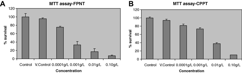
Cytotoxic analysis of FPNT and CPPT in HepG2 cell lines using MTT assay. DMSO acts as vehicle control. In case of control neither the compounds nor DMSO was added. Cells were treated with 0.0001, 0.001, 0.01 and 0.1 g/L of FPNT and CPPT. The percentage viability was calculated considering control wells as 100% and plotted with representation of error bars. (A) FPNT. (B) CPPT.
2.2.3 [3H] thymidine incorporation assay
DNA synthesis was monitored by labeling cells using [3H] thymidine (Kavitha et al., 2009). Around 1 × 10⁹ cells/L were seeded in duplicates, FPNT and CPPT were added at a concentration of 0.001, 0.01 and 0.1 g/L and incubated for 3 h. Cells grown in culture media with appropriate concentration of DMSO were used as control. 18.5 × 10³ Bq of [3H] thymidine was added and incubated for additional 2 h. The cells were collected by Millipore filtration, washed with cold 10% trichloro acetic acid (TCA), methanol and water. Filters were removed into liquid scintillation vials, dried, cocktail added and radioactivity was measured using a liquid scintillation beta counter. Radioactivity is expressed in cpm (counts per minute), which was proportional to the amount of [3H] thymidine incorporated into the DNA of cultured cells ± standard deviation, Fig. 3.![[3H]thymidine incorporation assay to determine effect of FPNT and CPPT on cell proliferation. In case of control DMSO was added. The data presented is result of duplicates and error bars are indicated. (A) FPNT. (B) CPPT.](/content/184/2010/3/4/img/10.1016_j.arabjc.2010.06.002-fig3.png)
[3H]thymidine incorporation assay to determine effect of FPNT and CPPT on cell proliferation. In case of control DMSO was added. The data presented is result of duplicates and error bars are indicated. (A) FPNT. (B) CPPT.
2.2.4 Cell cycle analysis
HepG2 cells were cultured and treated with two concentrations, 0.00003 and 0.00005 g/L of FPNT in duplicates for 24 h to study concentration dependent effect on the cell cycle arrest (Kavitha et al., 2009; Ormerod et al., 1994). Control cells were treated with 0.1% DMSO. Cells were fixed in 2 ml of 70% alcohol, centrifuged at 3000 rpm for 10 min and alcohol was discarded. The cells were washed in 2 ml PBS and to the pellet 15 × 10−6 L of RNase was added, followed by 10 × 10−6 L of propidium iodide. The cells were incubated for 1 h and subjected to flow cytometry. The results were analyzed using cell quest pro software using excitation at 488 nm laser and emission at 560/670 nm. A minimum of 10,000 cells were acquired and histograms analyzed. The total number of cells included in analysis was taken as 100%. The results of flow cytometry studies are represented in Fig. 4. The flow cytometry data is represented in Table 1.
(A–C) Cell cycle analysis histograms. (A) Control (DMSO). (B) 0.00003 g/L of FPNT. (C) 0.00005 g/L of FPNT. (D) Bar diagram showing quantification of cells in each phase of the cell cycle on treatment with FPNT.
Concentration
M1 (% sub-G1-cells)
M2 (% G1 cells)
M3 (% S cells)
M4 (% G2/M cells)
Control
3.57 ± 0.02
50.68 ± 2.42
32.84 ± 1.06
12.92 ± 0.27
0.00003 g/L
4.69 ± 0.07
56.41 ± 1.96
24.48 ± 0.98
14.12 ± 0.63
0.00005 g/L
9.31 ± 0.09
54.20 ± 1.37
27.53 ± 0.67
8.95 ± 0.18
2.2.5 Chromatin condensation studies (Hoechst staining)
Condensation of chromatin is usually the late event in apoptosis. HepG2 cells were grown in 96-well plates in duplicates and subjected to the treatment of FPNT in two different concentrations of 0.01 and 0.1 g /L and incubated for 24 h. Cells grown in culture media with appropriate concentration of DMSO were used as control. About 50 × 10−6 L of the medium was aspirated, the same amount of diluted Hoechst dye was added and incubated for 5 min. 50 × 10−6 L of the medium was removed and viewed under fluorescence microscope. Hoechst dye binds at the adenine–thymine rich regions of DNA and emits blue fluorescence when excited by UV light about 350 nm. The results of chromatin condensation studies are shown in Fig. 5.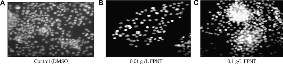
Chromatin condensation studies (Hoechst test). Dose dependent increase in condensed apoptotic chromatin is visible on treatment with FPNT when compared to control. (A) Control (DMSO). (B) 0.01 g /L FPNT. (C) 0.1 g/L FPNT.
2.3 Antioxidant studies
2.3.1 DPPH radical scavenging assay
The DPPH antioxidant assay is based on the ability of DPPH, a stable free radical, to decolorize in the presence of antioxidants. The DPPH radical contains an odd electron, which is responsible for the absorbance at 517 nm and also for visible deep purple color (Sreejayan and Rao, 1996; John and Steven, 1984). When DPPH accepts an electron donated by an antioxidant compound, the DPPH is decolorized which can be quantitatively measured from the changes in absorbance. Hundred microlitre of various concentrations of FPNT was added to respective wells of a 96-well micro plate. Equal amount of DPPH (1,1-diphenyl-2-picryl hydrazide) was also added to each well to make up a final volume of 200 μl. After 20 min incubation in the dark, the ability of FPNT to scavenge the free radical DPPH, was measured by recording the absorbance at 517 nm using an ELISA plate recorder. Experiment was performed in triplicates and average values were considered. An equal amount of methanol and DPPH was added to the control. Ascorbic acid was used as reference standard. Comparison of the antioxidant activity of FPNT and ascorbic acid is shown in Fig. 6.
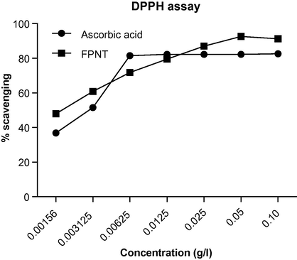
DPPH radical scavenging activity of FPNT added to methanol solution of DPPH. Radical scavenging activity was measured at 517 nm as compared to standard ascorbic acid. Values are the average of triplicate experiments.
2.3.2 ABTS radical scavenging assay
ABTS is chemically 2,2-azino bis 3-ethyl benzo-thiazoline-6-sulphonic acid. The reduction of this radical by FPNT is measured at 690 nm (Vaijanathappa et al., 2008). The electron transfer capability of FPNT was studied using ABTS radical scavenging assay. In a 96-welled microtitre plate, 40 μl of the FPNT/ascorbic acid, 200 μl of methanol and 30 μl of ABTS solution were added. The plate was then incubated at 37 °C for 20 min after which the absorbance was measured at 690 nm using an ELISA plate reader. Sample blank and control were also taken. The experiment was performed in triplicates and average values were considered. The comparative values of the antioxidant activity of FPNT and ascorbic acid is depicted in Fig. 7.
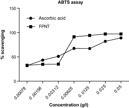
ABTS radical scavenging activity of FPNT measured at 690 nm as compared to standard ascorbic acid. Values are the average of triplicate experiments.
2.3.3 Lipid peroxidation assay
Malonaldehyde, formed from breakdown of polyunsaturated fatty acids, serves as a convenient index for determining the extent of peroxidation reaction (Okhawa et al., 1979). Malonaldehyde reacts with thiobarbituric acid to form TBARS, a red chromogen, which is measured at 535 nm. Albino rats (180–200 g) of either sex were used for the study. After decapitation, the brain was removed carefully. The tissue was immediately weighed and homogenated with cold 1.15% potassium chloride solution to make 10% homogenate. This homogenate was immediately used for in vitro lipid peroxidation study. Lipid peroxidation was initiated by adding 1 ml of 100 μM ferrous sulphate to a 0.5 ml mixture containing rat brain homogenate and 1 ml of different concentrations of FPNT. The resultant solution was incubated for 30 min and the reaction was terminated by adding ice cold TBA–TCA reagent. Further, the mixture was mixed thoroughly and heated for 15 min in boiling water bath. After cooling, the flocculent precipitate was removed by centrifugation at 1000 rpm for 10 min. The absorbance of the supernatant was measured at 535 nm. The experiment was repeated in triplicates and the average values were measured. The same procedure was repeated with curcumin as reference. The results of the in vitro lipid peroxidation assay for FPNT and curcumin is presented in Fig. 8.
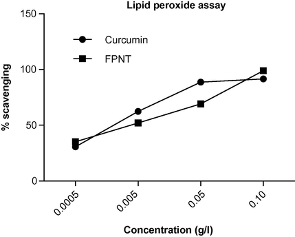
In vitro lipid peroxide assay of FPNT measured at 535 nm as compared to standard curcumin. Values are the average of triplicate experiments.
3 Results
Induction of apoptosis or inhibitions of cell proliferation are important properties associated with chemotherapeutic agents. In our present study, we have used FPNT and CPPT to understand the mechanism of its cytotoxicity. Hepatocellular carcinoma cell lines, HepG2 cells were used exclusively for the entire study.
3.1 Cytotoxic effect of FPNT and CPPT
To investigate the potential effects of FPNT and CPPT, their cytotoxicity was measured after 24 h using MTT assay. The test showed a dose dependent cytotoxic effect of FPNT and CPPT, their IC50 being 0.0005 and 0.006 g/L. These values were found to be significantly low when compared to standard drug, doxorubicin with IC50 0.019 g/L. Since above study suggested that both FPNT and CPPT affects the cell viability, we were interested to know whether they could inhibit cell division.
3.2 FPNT affects cell proliferation
One of the currently employed methods to assess cell proliferation is based on incorporation of radio labeled nucleotides into the DNA of the dividing cells. FPNT treatment showed a dose dependant reduced incorporation of [3H] thymidine drastically, suggesting that it affects the cell viability by inhibiting cell division probably by interfering with DNA replication. However it is also possible that in addition to its effect on cell division, FPNT could also induce apoptosis. The treatment with CPPT did not show a dose dependent reduction in cell proliferation when labeled with tritiated thymidine.
3.3 FPNT affects cell cycle profile upon treatment with appearance of sub-G1 cells
As FPNT induced a reduction in the number of viable cells in MTT assay and also exhibited a reduction in the cell proliferation in tritiated thymidine assay, we were interested in studying the cell cycle distribution by fluorescence-activated cell sorting analysis of PI-labeled cells. The histogram of DMSO treated cells showed a standard cell cycle pattern, which included G1 and G2/M peaks separated by S phase peak. The sub-G1 peak showed very less percentage of dead cells (Fig. 4A), when treated with control, DMSO. Interestingly upon addition of FPNT, a concentration dependant change was observed in the percentage of cells in each phase of the cell cycle (Fig. 4B and C). There was a remarkable dose dependent increase in the percentage of sub ploid cells in the sub-G1 phase. We could also observe more cells in the G1 phase when compared to S and G2/M phase indicating a cell cycle arrest probably in the G1 phase of the cell cycle allowing fewer cells to enter into the S phase confirming the results obtained in the [3H] thymidine incorporation assay. The bar diagram quantifying the percentage of cells in the different phases of the cell cycle in control and treated group is shown in Fig. 4D.
3.4 FPNT induces apoptosis
Apoptosis is known to be a very important mechanism involved in the anticancer effects induced by chemotherapeutic agents. The induction of apoptosis on treatment with FPNT was evident from the concentration dependent increase in the appearance of apoptotic fraction (sub-G1 population) cells. To confirm the action of FPNT through growth inhibition mediated by a DNA replication defect followed by apoptosis, chromatin condensation studies were conducted. DMSO treated wells were used as control (Fig. 5A). The presence of dose dependent increase in condensed apoptotic chromatin in Hoechst staining test confirms the apoptotic action of FPNT (Fig. 5B and C).
3.5 FPNT as a potent antioxidant
Antioxidant capacity is widely used as a parameter for medicinal bioactive components. The antioxidant most made known for its potential benefits to cancer patients is vitamin C (ascorbic acid). Antioxidant supplements may reduce the frequency and severity of toxicity associated with anticancer therapy.
The IC50 values for FPNT and ascorbic acid for scavenging DPPH were found to be 0.0012 and 0.0015 g/L correspondingly. FPNT was found to be more potent than ascorbic acid. FPNT was found to be more active when compared to ascorbic acid in ABTS assay with IC50 values being 0.0021 and 0.0023 g/L, respectively. In vitro lipid peroxidation assay also revealed high antioxidant nature of FPNT compared to reference curcumin with IC50 values 0.0016 and 0.0015 g/L, respectively.
4 Discussion
To explore the antiproliferative property of FPNT and CPPT, initially hepatic cell viability was checked using MTT assay. The results showed that the cell death caused by both the compounds were concentration dependent. Also, the IC50 of both the compounds was markedly lower than the standard drug, doxorubicin which positively correlated to their potency. Reduced incorporation of tritiated thymidine indicated that FPNT could inhibit the proliferation of cells. CPPT did not show any dose dependent reduction in the incorporation of tritium. Results based on cell cycle analysis suggested a G1 arrest in the cell cycle. One of the most interesting observations of cell cycle analysis was the increase in sub-G1 peak in a concentration dependent manner, which suggested the presence of apoptotic cells. Hoechst test conducted confirmed the type of cell death to be apoptotic as dose dependent increase in the condensed apoptotic chromatin was visible under UV light.
The significant antioxidant activity of FPNT with low IC50 values when compared to standard is clearly evident from DPPH, ABTS free radical scavenging and in vitro lipid peroxidation assays. As DPPH scavenging occurred at a lower concentration than ABTS scavenging, it may be presumed that the antioxidant action of FPNT is primarily through the mechanism of hydrogen transfer rather than electron transfer. The in vitro lipid peroxidation assay also proved FPNT to be an excellent antioxidant.
The ability to cross the biomembranes and bioavailability of a compound is dependent on the lipophilic moiety attached. The title compound has naphthyloxy methyl and pyrazole substituents. The fluorine atoms also might have added to the potency of the compound as a fluorine substituent produces simultaneously an increase in lipophilicity, an electron attracting effect and a metabolic obstruction. In FPNT, fluorine atom in the para position could increase the binding affinity of the molecule.
5 Conclusions
The present study reveals FPNT as a potent cytotoxic compound with low IC50 values compared to the standard drug doxorubicin in MTT assay. Accumulation of cells in sub-G1 phase and chromatin condensation studies suggested induction of apoptosis on treatment with FPNT. Also FPNT is found to possess excellent antioxidant properties.
Acknowledgements
The authors are thankful to the Head-SIF, Indian Institute of Science, Bangalore for providing NMR and mass spectral data. A.M.I. is grateful to the Director, National Institute of Technology, Karnataka, India for providing research facilities. The authors express their gratitude to Chairman, RGCB, Thiruvananthapuram, for carrying out the chromatin condensation studies. One of the authors (D.S.) is thankful to Director and Head-Department of Chemistry, Manipal Institute of Technology, Manipal University for rendering us the necessary laboratory facilities.
References
- In vitro anti-HIV and antitumor activity of new 3,6-disubstituted [1,2,4]triazolo[3,4-b][1,3,4]thiadiazoles and thiadiazine analogues. Archiv der Pharmazie. 2008;341:365-369.
- [Google Scholar]
- Dietary antioxidants during cancer chemotherapy, impact on chemotherapeutic effectiveness and development of side effects. Nutrition and Cancer. 2000;37:1-18.
- [Google Scholar]
- Dietary polyunsaturated fatty acids impact on cancer, chemotherapy and radiation. Alternative Medicine Review. 2002;7:4-21.
- [Google Scholar]
- Synthesis and antimicrobial activities of some new 1-(5-phenylamino-[1,3,4]thiadiazol-2-yl)methyl-5-oxo-[1,2,4]triazole and 1-(4-phenyl-5-thioxo-[triazol-3yl)methyl-5-oxo-[1,2,4]triazole derivatives. European Journal of Medicinal Chemistry. 2004;39:793-804.
- [Google Scholar]
- Synthesis, characterization and anticancer activity of 1,2,4-triazolo[3,4-b]-1,3,4-thiadiazoles on HepG2 cell lines. Der Pharma Chemica. 2009;1(2):19-26.
- [Google Scholar]
- Microwave assisted synthesis and biological activities of some bridge-head nitrogen heterocycles. Indian Journal of Chemistry. 2003;42B:1756-1759.
- [Google Scholar]
- Regioselective reaction: synthesis, characterization and pharmacological studies of some new mannich bases derived from 1,2,4-triazoles. European Journal of Medicinal Chemistry. 2009;44(9):3784-3787.
- [Google Scholar]
- Microwave assisted synthesis and biological activity of 3-alkyl/aryl-6-(1-chloro-3,4-dihydronaphth-2-yl)-5,6-dihydro-s-triazolo [3,4-b] [1,3,4]thiadiazoles. Indian Journal of Chemistry. 2007;46B:980-984.
- [Google Scholar]
- Biological studies of some 2,4-dichloro-5-fluorophenyl containing triazolothiadiazoles. Monatshefte fuer Chemie. 2007;138:1309-1316.
- [Google Scholar]
- Novel derivatives of spirohydantoin induce growth inhibition followed by apoptosis in leukemia cells. Biochemical Pharmacology. 2009;77:348-363.
- [Google Scholar]
- Methyl angolensate, a natural tetranortriterpenoid induces intrinsic pathway in leukemic cells. FEBS Letters. 2008;582:4066-4076.
- [Google Scholar]
- Antioxidants in cancer therapy, their actions and interactions with oncologic therapies. Alternative Medicine Review. 1999;4:304-329.
- [Google Scholar]
- Studies on synthesis and pharmacological activities of 3,6-disubstituted-1,2,4-triazolo[3,4-b]-1,3,4-thiadiazoles and their dihydro analogues. European Journal of Medicinal Chemistry. 2007;42:823-840.
- [Google Scholar]
- Rapid colorimetric assay for cellular growth and survival: application to proliferation and cytotoxic assays. Journal of Immunological Methods. 1983;65:55-63.
- [Google Scholar]
- Assay for lipid peroxides in animal tissues by thiobarbituric acid reaction. Analytical Biochemistry. 1979;95:351-355.
- [Google Scholar]
- The role of apoptosis in cell killing by cisplatin: a flow cytometric study. British Journal of Cancer. 1994;69:93-100.
- [Google Scholar]
- New bis-aminomercaptotriazoles and bis-triazolothiadiazoles as possible anticancer agents. European Journal of Medicinal Chemistry. 2002;37:511-517.
- [Google Scholar]
- Synthesis of pharmaceutically important condensed heterocyclic 4,6-disubstituted-1,2,4-triazolo-1,3,4-thiadiazolederivatives as antimicrobials. European Journal of Medicinal Chemistry. 2006;41:531-538.
- [Google Scholar]
- Modeling, synthesis and biological activity of novel antifungal agents. Bioorganic Medicinal Chemistry Letters. 1998;8:1819-1824.
- [Google Scholar]
- In vitro antioxidant activity of Enieostemma axillare. Journal of Health Sciences. 2008;54(5):524-528.
- [Google Scholar]
- Synthesis and antibacterial activity of some novel bis-1,2,4-triazolo[3,4-b]-1,3,4-thiadiazoles and bis-4-thiazolidinone derivatives from terephthalic dihydrazide. European Journal of Medicinal Chemistry. 2009;441:5112-5116.
- [Google Scholar]


