

Translate this page into:
Green synthesis and chemical characterization of a novel anti-human pancreatic cancer supplement by silver nanoparticles containing Zingiber officinale leaf aqueous extract
⁎Corresponding author. carunachalam@ksu.edu.sa (Arunachalam Chinnathambi),
-
Received: ,
Accepted: ,
This article was originally published by Elsevier and was migrated to Scientific Scholar after the change of Publisher.
Abstract
In recent years, silver nanoparticles (AgNPs) have been used as key chemotherapeutic drugs to treat various cancers like prostate, breast, ovarian, and blood cancers. No previous reports demonstrated the in vitro anti-human pancreatic cancer properties of the novel chemotherapeutic drug formulated by silver nanoparticle compounds including Zingiber officinale leaf. To survey the anti-human pancreatic cancer activities of AgNO3, Zingiber officinale leaf aqueous extract, and silver nanoparticles, 3-(4,5-dimethylthiazol-2-yl)-2,5-diphenyl-2H-tetrazolium bromide (MTT) assay was used on common human pancreatic cancer cell lines. According to the Field Emission Scanning Electron Microscopes (FE-SEM) and Transmission electron microscopy (TEM) images, the silver nanoparticles were in an average size of 18.93 nm with a spherical shape. 2,2-diphenyl-1-picrylhydrazyl (DPPH) test revealed similar antioxidant potentials for Zingiber officinale leaf aqueous extract, silver nanoparticles, and butylated hydroxytoluene. Silver nanoparticles inhibited half of the DPPH molecules in the concentration of 172 µg/mL. Silver nanoparticles had very low cell viability and anti-human pancreatic cancer properties dose-dependently against AsPC-1, PANC-1, and MIA PaCa-2. The IC50 values of the silver nanoparticles were 295, 312, and 220 µg/mL against AsPC-1, PANC-1, and MIA PaCa-2 cell lines, respectively. It is thought that the silver nanoparticles obtained can be used as an anticancer drug for the diagnosis of pancreatic cancer in humans after acceptance of the above findings in clinical study trials.
Keywords
Chemotherapeutic drug
Human pancreatic cancer
Chemical characterization
Silver nanoparticles
Zingiber officinale leaf

1 Introduction
Research interest in nanotechnology has increased significantly due to the exponential growth in nanomaterial production and marketing. Metallic nanoparticles can be shaped in different ways; spheres or rods. According to size; Nanomaterials are divided into different groups such as nanoparticles, dendrimers, nanotubes and nanofilms. This further increases the variety of nanoscale materials (Han et al., 2020).
Various approaches and techniques have been developed for the synthesis of nanoparticles to make them one of the most applicable and widely used materials in science. For example, silver nanoparticles are used as biocides and antioxidants and they are utilized either barely or integrated into other structures/substances. In addition, apart from other uses, such as engineering, cosmetics, and agriculture, silver nanoparticles have pharmaceutical applications such as cancer treatment and medical imaging (Arulmozhi et al., 2013). Nano-oncology is a significant field of nanotechnology and has developed as an extension for the use of nanomaterials in the care of many forms of tumors (Hosseinimehr et al., 2011). The role of green silver nanoparticles synthesized by herbs in treating numerous different types of cancer is unique among all nanomaterials (Yang et al., 2017; Judith Vijaya et al., 2017). There is no study however on the pancreatic cancer effects of green-synthesized silver nanoparticles from medicinal herbs.
Many therapeutic supplements and drugs are formulated from traditional medicine for the treatment, control, and prevention of several diseases every year (Velmurugan et al., 2014; Kumar et al., 2012). Researchers have focused on the anticancer properties of traditional medicine for synthesizing and formulating many chemotherapeutic supplements and drugs containing herbs (Wael et al., 2019; Hagh-Nazari et al., 2013). Based on our knowledge, comparative research on anti-pancreatic cancer properties of AgNO3, and AgNPs synthesized by Zingiber officinale leaf against, pancreatic cancer cell lines in cellular models has not been done so far. The purpose of this study was to determine the structures of AgNO3, Zingiber officinale leaf, and AgNPs against pancreatic (PANC-1, AsPC-1, and MIA PaCa-2) cancer cell lines.
2 Material and methods
2.1 Material
Dimethyl sulfoxide (DMSO), Antimycotic antibiotic solution, hydrolysate, decamplmaneh fetal bovine serum, Ehrlich solution, 4-(Dimethylamino) benzaldehyde, DPPH, carbazole reagent, borax-sulphuric acid mixture, Dulbecco's Modified Eagle Medium (DMED), and phosphate buffer solution (PBS) were purchased from Sigma-Aldrich (USA).
2.2 Preparation of Zingiber officinale leaf aqueous extract
At the beginning of the aqueous extracting, the fresh and healthy parts of Zingiber officinale leaf were collected. After shade drying in a mixer, 50 g of powdered plant sample was extracted with distilled water with the increase of polarity at a ratio of 1:15 (v/v). In the end, for concentrating, the rotary evaporator was used (Ghashghaii et al., 2017).
2.3 Synthesis of AgNPs
Green synthesis of silver nanoparticles was started with such a process combination of 100 mL of AgNO3·H2O at concentrations of 1 mM and 10 mL of Zingiber officinal leaf aqueous isolate (20 μg / mL) in a cylindrical flask.
The reaction mixture was kept under magnetic stirring for 12 h at room temperature. At the end of the reaction time, the black colored colloidal solution of Ag was formed. The solution was centrifuged at 10,000 rpm for 15 min. The precipitate was sprayed with water and then resuspended (Ahmeda et al., 2020).
2.4 The characterization of AgNPs
Field Emission Scanning Electron Microscopes (FE-SEM), Transmission electron microscopy (TEM), Fourier-transform infrared spectroscopy (FTIR), and Ultraviolet–visible (UV–Vis) spectrophotometry were employed to characterize the composition, structure, and morphology of the silver nanoparticles. Silver nanoparticles have been verified using UV–Vis spectroscopy nm (Jasco V670 Spectrophotometer) at a scanning scale of 350–650 nm wavelength. The FT-IR spectrophotometer (Shimadzu IR Affinity.1) has been used to track the organic compounds engaged in removing silver nanoparticles. With “FE-SEM (Fe-SEM ZEISS EVO18)” analysis and “TEM (TEM FEI-TECNAI G2-20 TWIN)”, the morphological properties of silver nanoparticles were examined in terms of form and thickness.
2.5 Determination of the antioxidant property of AgNPs
The organic chemical compound DPPH stands for 2,2-diphenyl-1-picrylhydrazyl as an abbreviation. This is a crystalline powder of dark colors, composed of stable free-radical molecules. DPPH has two main laboratory research applications: one of the chemicals monitors the radicals, particularly the common analysis of antioxidants, and the next of the paramagnetic electron resonance signal position and strength (Zangeneh et al., 2020).
The above DPPH was added to the various concentrations of AgNO3, Zingiber officinale leaf aqueous extract, and silver nanoparticles and all samples were transferred to an incubator at the temperature of 37 °C. After 30 min incubating, the absorbance’s were measured at 517 nm. Acceding to the following formula, the antioxidant properties of AgNO3, Zingiber officinale leaf aqueous extract, and silver nanoparticles were determined in detail (Zangeneh et al., 2020):
2.6 Determination of anti-pancreatic cancer effects of silver nanoparticles
The following cell lines have been utilized in this research to precisely invest the cytotoxicity and anti-pancreatic of the AgNO3, Zingiber officinale leaf aqueous extract, and silver nanoparticles using an 3-(4,5-dimethylthiazol-2-yl)-2,5-diphenyl-2H-tetrazolium bromide (MTT) assay:
-
Normal cell line
-
–
HUVEC.
-
-
Pancreatic cancer cell lines
-
–
PANC-1
-
–
AsPC-1
-
–
MIA PaCa-2
-
For culturing the above cells streptomycin, penicillin, and Dulbecco’s modified Eagle’s medium (DMEM) were used. Cell density in 96-well plates was 10,000 cells/right. Both samples were then moved at a temperature of 37 °C to a humidified incubator of %5 CO2. After 24 h incubating, all cells were treated with several concentrations of AgNO3, Zingiber officinale leaf aqueous extract, and silver nanoparticles, then incubated for 24 h. AgNO3, Zingiber officinale leaf aqueous extract, and silver nanoparticles were sterilized using the radiation of UV for 2 h.
2.7 Qualitative measurement
The findings reported were loaded into the “SPSS-22” program and evaluated by “one-way ANOVA”, accompanied by a “Duncan post-hoc” check (p ≤ 0.01).
3 Results and discussion
3.1 The characterization of silver nanoparticles
3.1.1 UV–Vis analysis
Fig. 1 shows a 414 nm absorption band linked to the AgNPs surface plasmon resonance (SPR). We may also note that the SPR band decreases, by growing the volume of aqueous isolate liquid Zingiber officinale strip. It indicates that utilizing a higher concentration of Zingiber officinale extract aqueous extract reduces the average size of AgNPs, and raises the density of AgNPs.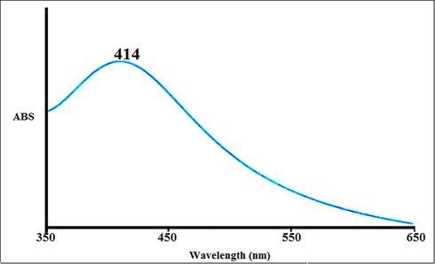
The UV–Vis spectrum of silver nanoparticles green-synthesized using Zingiber officinale leaf. ABS. Absorbance.
Shasha Han et al. reported Gundelia tournefortii L. aqueous extract synthesized AgNPs peaked at 440 nm in the UV–Vis spectrum (Han et al., 2020). Mohammadi et al. observed the peak of silver nanoparticles containing Phoenix dactylifera seed ethanolic extract at the wavelength of 438 nm (Mohammadi et al., 2020). Han et al. recorded 430 nm of the absorption coefficient for silver nanoparticles utilizing the polyol process (Han et al., 2020). Zangeneh et al. revealed the absorbance at 462 nm for silver nanoparticles synthesized by Spinacia oleracea L. (Zangeneh, 2019a). Ahmeda et al. studied Melissa officinalis leaf aqueous extract mediated synthesis of AgNPs. Absorption of the continuum was detected at 462 nm (Ahmeda et al., 2020). Zangeneh et al. recorded Stachys lavandulifolia leaf extract induced AgNP as well as absorption maximum was detected at 440 nm (Ahmeda et al., 2020).
3.1.2 FE-SEM and TEM analysis
The FE-SEM image of Zingiber officinale leaf aqueous extract mediated silver nanoparticles is shown in Fig. 2. As understood from Fig. 2, AgNPs show an agglomerated structure and indicates particulate sizes from 15 to 31 nm for biosynthesized AgNPs. Silver nanoparticles can usually be agglomerated and the hydroxyl groups present in the Zingiber officinale leaf aqueous extract are thought to be responsible for the observed agglomeration (Zangeneh, 2019b; Zangeneh, 2020).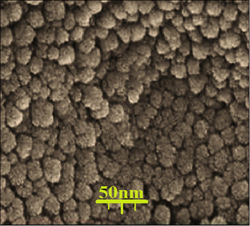
FE-SEM image of silver nanoparticles green-synthesized using Zingiber officinale leaf.
TEM analysis is preferred to detect detailed grain size, size distribution, and morphology of nanoparticles (Katata-Seru et al., 2018). TEM image shows that biosynthesized silver nanoparticles have a particle size distribution between 12 and 30 nm (Fig. 3). Thus, the size of the nanoparticles we obtained was smaller than the literature (Katata-Seru et al., 2018).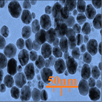
TEM image of silver nanoparticles green-synthesized using Zingiber officinale leaf.
3.1.3 FT-IR analysis
The FT-IR analysis was performed to identify the antioxidant compounds which may be responsible for reducing the Ag+ ions in the Zingiber officinale extract, as well as those that may be concerned with stabilizing the synthesis of AgNPs. The solution was centrifuged at 10,000 rpm for 15 min. The precipitate was sprayed with water and then resuspended (Yang et al., 2017; Judith Vijaya et al., 2017). The composition of the AgNPs suggests that the presence of a maximum at 484 cm−1 is part of the Ag-O tension change in the current case. Thus, the IR spectroscopic approach has been used as a suitable method to identify bioactive components in the field of natural products.
Also, this technique is a valuable tool for detecting the existence of secondary metabolites over the AgNPs in plants. As per the findings, the occurrence of common-IR bars was linked to the nature of different organic compounds in an aqueous extract of Zingiber officinale extract. In addition, a band at 2071 cm−1 referred to aliphatic “C—H” stretching; peaks at 3416 cm−1 linked to “O—H” stretching; peaks at 1079, 1204 and 1287 cm−1 may be compared to “—C—O” stretching and peaks at 1391 and 1612 cm−1 refer to “C⚌C” stretching and “C⚌O” stretching occurring in phenolic and flavonoid compounds (Fig. 4). These peaks may be known for the existence of numerous substances at the Zingiber officinale such as flavonoid, phenolic, and carboxylic substances previously recorded.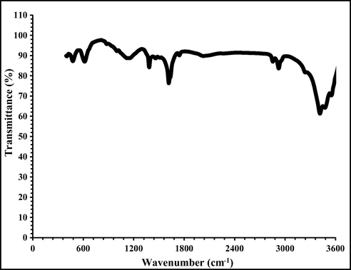
FT-IR spectra of silver nanoparticles green-synthesized using Zingiber officinale leaf.
3.2 Antioxidant potential of silver nanoparticles green-synthesized by Zingiber officinale leaf aqueous extract
Plants have impressive antioxidant capabilities. One alternative to increase the plants' antioxidant ability is to combine them with metallic salts. Recent experiments have shown that their antioxidant function improves significantly as the leaves are mixed with zinc, iron, silver, gold, copper and titanium (Taghavi Fardood and Ramazani, 2016). Several studies have indicated that the antioxidant properties of silver nanoparticles green-synthesized by medicinal plants are the most among all metallic nanoparticles.
In our study, a significant concentration-dependent DPPH radical scavenging effect was demonstrated by the Zingiber officinale leaf aqueous isolate and silver nanoparticles close to BHT. The connection with both the Zingiber officinale leaf aqueous isolate and silver nanoparticles and DPPH may have happened from the conversion of electrons and hydrogen ions into 2,2-diphenyl-1-picrylhydrazyl radical, forming a controlled DPPH complex [24–26]. The IC50 values of Zingiber officinale leaf aqueous extract, BHT, and silver nanoparticles were 275, 203, and 172 μg/mL, respectively (Fig. 5, Table 1).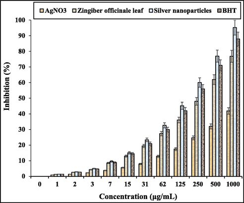
The antioxidant properties of AgNO3, Zingiber officinale leaf, silver nanoparticles, and BHT against DPPH.
AgNO3 (µg/mL)
Zingiber officinale (µg/mL)
Silver nanoparticles (µg/mL)
BHT (µg/mL)
IC50 against DPPH
–
275
172
203
The oxidative efficacy of Zingiber officinale leaf aqueous isolate can be due to the presence of various phytochemicals, which are thought to work dynamically and synergistically to neutralize the “reactive oxygen species (ROS)” and the “reactive nitrogen species (RNS)” (Rajesh et al., 2018; Kaur et al.,2016; Reuter et al., 2010). In an earlier analysis, compounds such as ethylate, bis (2- ethylhexyl) phthalate, cinnamic acid, nepitrine, gallic acid, oleylic alcohol, isorhamnetine-3-O-rutinoside, acteoside and quercetine were found in Zingiber officinale analysis (Demirci Gültekin et al., 2016). These biologically active substances were shown to sustain redox homeostasis through multi-step antioxidant reactions, including initiation, aggregation, splitting, and free radicals (Rehana et al., 2017).
3.3 Cytotoxicity anti-pancreatic cancer potentials of silver nanoparticles synthesized in green with Zingiber officinale leaf aqueous extract
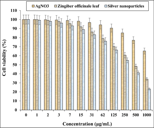
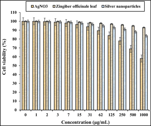
Treated cells with specific concentrations of the present AgNO3, Zingiber officinale leaf aqueous extract, and silver nanoparticles were tested by 48 h MTT study for cytotoxicity, regular anti-pancreatic (HUVEC) and pancreatic (PANC-1, AsPC-1, and MIA PaCa-2 cell lines of cancer (Figs. 6–9, Table 2). The absorbance limit of AgNO3, Zingiber officinale leaf aqueous extract, and silver nanoparticles was measured at 570 nm, indicating exceptional viability even up to 1000 μg/mL on a standard cell line (HUVEC).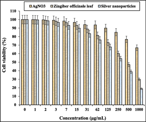
The anti-human pancreatic cancer properties of AgNO3, Zingiber officinale leaf, and silver nanoparticles against PANC-1 cell line.
The anti-human pancreatic cancer properties of AgNO3, Zingiber officinale leaf, and silver nanoparticles against AsPC-1 cell line.
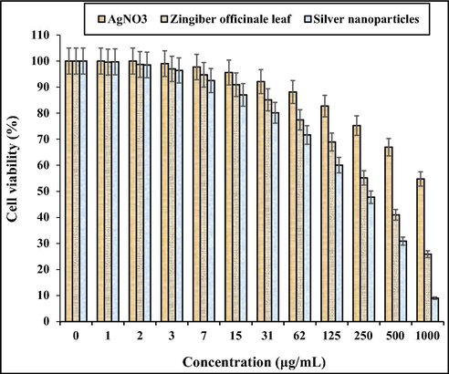
The anti-human pancreatic cancer properties of AgNO3, Zingiber officinale leaf, and silver nanoparticles against MIA PaCa-2 cell line.
The cytotoxicity properties of AgNO3, Zingiber officinale leaf, and silver nanoparticles against HUVEC cell line.
IC50 against “..”
AgNO3 (µg/mL)
Zingiber officinale (µg/mL)
Silver nanoparticles (µg/mL)
HUVEC
–
–
–
PANC-1
–
443
295
AsPC-1
–
479
312
MIA PaCa-2
–
359
220
In typical human pancreatic, their cell viability decreased dose-dependently with various concentrations (1–1000 μg/mL) of AgNO3, Zingiber officinale leaf aqueous extract, and silver nanoparticles current.
There were 443 and 295 μg/mL for specific human pancreatic cancer cell lines, 479 and 312 μg/mL for Zingiber officinale leaf aqueous extract and 312 μg/mL for silver nanoparticles against PANC-1 cell lines, and 359 and 220 μg/mL for MIA PaCa-2 cell lines, respectively. The strongest findings of the cytotoxicity and anti-human pancreatic cancer ability of silver nanoparticles against the above cell lines were shown in the PANC-1 cell line, and BHT against DPPH.
The chemotherapeutic results of metallic nanoparticles, in particular iron, zinc and silver nanoparticles in vitro and in vivo settings, have been confirmed by multiple studies to date. Various cell lines including Cos-7 monkey fibroblasts, BRL3A rat liver cells, human epidermal keratinocytes, and rodent lung epithelial cell lines have been studied for iron nanoparticles' cytotoxicity properties, which have shown promise [30]. More work has also shown that iron oxide nanoparticles produced utilizing medicinal plant extracts have great potential for cytotoxicity toward human cell counts for leukemia (Jurkat cells), cervical cancer (HeLa cells), liver cancer (HepG2 cells), and breast cancer (MCF-7 cells) (Delgado-Povedano et al., 2016). The anticancer effects of silver nanoparticles green-synthesized by medicinal plants have been confirmed in the previous studies (Jeong et al., 2012; Sankar et al., 2014). It has been suggested in the previous research that silver nanoparticles green-synthesized by Annona quamosal leaf have excellent potential for anti-breast cancer against the MCF-7 cell line (Mahmoudi et al., 2012; Namvar et al., 2014). In another study, the anti-liver cancer properties of silver nanoparticles containing Piper longum leaf against Hep-2 cell lines were proved (Vivek et al., 2012; Justin Packia et al., 2012). In the study of Suman et al. was clarified the anti-cervix cancer effects of silver nanoparticles containing natural compounds (Morinda citrifolia) against the HeLa cell line. In the previous study, the silver nanoparticles killed all HeLa cells in high doses (Suman et al., 2013).
Similar researches have revealed the antioxidant materials such as metallic nanoparticles especially silver nanoparticles and ethnomedicinal plants reduce the volume of tumors by removing free radicals (Sangami and Manu, 2017). In detail, the high presence of free radicals in the normal cells makes many mutations in their DNA and RNA, destroys their gene expression and then accelerates the proliferation and growth of abnormal cells or cancerous cells.
The results of many studies have shown that both AgNPs and Ag+ generated by AgNPs are involved in the chemotherapeutic cascade in different ways:
AgNPs are needed to have an acceptable surface outside the mitochondria for the univalent transfer of oxygen from either the electron transport chain to the superoxide. Ag+ attaches to DNA and proteins that interact with their functions (Beheshtkhoo et al., 2018). Silver nanoparticle chemotherapeutic effects have been found to rely on several factors relevant to their physical characteristics, such as surface coloring, shape and thickness. To the current scale, it has been reported that tiny silver nanoparticles will migrate from the cell membrane and remove from its tumor cells. The above potential is substantially reduced in bigger sizes (Suman et al., 2013). Likely significant anti-pancreatic cancer potentials of silver nanoparticles synthesized by Zingiber officinale leaf aqueous extract against pancreatic (PANC-1, AsPC-1, and MIA PaCa-2) tumor layers are linked to their antioxidant function. Engineering work has shown that antioxidant substances such as metallic nanoparticles, particularly silver nanoparticles and ethnomedicinal herbs, reduce cancer density by eliminating free radicals (Sangami and Manu, 2017).
The free radical’s high presences in all cancers like gallbladder, breast, rectal, stomach, liver, gastrointestinal stromal, bile duct, esophageal, pancreatic, small intestine, colon, parathyroid, bladder, thyroid, testicular, prostate, vaginal, fallopian tube, ovarian, hypopharyngeal, throat, lung, and skin cancers indicate a significant role of these molecules in making angiogenesis and tumorigenesis (You et al., 2012). Many researchers reported that silver nanoparticles synthesized by ethnomedicinal plants have a remarkable role in removing free radicals and growth inhibition of all cancerous cells (Beheshtkhoo et al., 2018; Radini et al., 2018).
4 Conclusions
In our study, silver nanoparticles were synthesized through the combination of Zingiber officinale leaf and AgNO3. Also, we investigated the anti-human pancreatic cancer of the silver nanoparticles containing Zingiber officinale leaf aqueous extract for the first time. FE‐SEM indicated that these nanoparticles had been synthesized as the best possible. The FE-SEM and TEM photos revealed that the silver nanoparticles had an average amount of 12,37 nm with a rounded form. FT-IR analysis shows that the presence of many antioxidant compounds with related bonds results in the perfect condition for reducing silver in silver nanoparticle compounds. In UV–Vis, the clear peak at 527 nm wavelength showed the formation of silver nanoparticle compounds. The silver nanoparticles against specific free radicals showed excellent antioxidant properties, i.e. DPPH. The silver nanoparticles blocked the concentration of 172 μg/mL of half the DPPH molecules. The latest silver nanoparticles had major anti-pancreatic cancer against PANC-1, AsPC-1, and MIA PaCa-2 without any cytotoxicity activity against normal cell line. It seems the plant increased significantly the antioxidant and anti-human pancreatic cancer of the silver nanoparticles. Due to significant results gained in the in vitro condition, it is recommended that clinical research studies validate these results in humans.
Acknowledgment
This project was supported by the Researchers Supporting Project number (RSP-2020/257) King Saud University, Riyadh, Saudi Arabia.
Declaration of Competing Interest
The authors declare that they have no known competing financial interests or personal relationships that could have appeared to influence the work reported in this paper.
References
- Green synthesis and chemical characterization of gold nanoparticle synthesized using Camellia sinensis leaf aqueous extract for the treatment of acute myeloid leukemia in comparison to daunorubicin in a leukemic mouse model. Appl. Organomet. Chem.. 2020;34 Available from: https://onlinelibrary.wiley.com/doi/abs/10.1002/aoc.5290
- [Google Scholar]
- Preparation, formulation, and chemical characterization of silver nanoparticles using <scp> Melissa officinalis </scp> leaf aqueous extract for the treatment of acute myeloid leukemia in vitro and in vivo conditions. Appl. Organomet. Chem.. 2020;34 Available from: https://onlinelibrary.wiley.com/doi/abs/10.1002/aoc.5378
- [Google Scholar]
- Ellagic acid encapsulated chitosan nanoparticles for drug delivery system in human oral cancer cell line (KB) Colloids Surf. B Biointerfaces.. 2013;110:313-320.
- [Google Scholar]
- Green synthesis of iron oxide nanoparticles by aqueous leaf extract of Daphne mezereum as a novel dye removing material. Appl. Phys. A Mater. Sci. Process.. 2018;124:1-7.
- [Google Scholar]
- Tentative identification of the composition of Agaricus bisporus aqueous enzymatic extracts with antiviral activity against HCV: a study by liquid chromatography-tandem mass spectrometry in high resolution mode. J. Funct. Foods.. 2016;24:403-419.
- [Google Scholar]
- Synthesis of copper nanoparticles using a different method: determination of their antioxidant and antimicrobial activity. JOTCSA. 2016
- [Google Scholar]
- Wound healing potential of methanolic extract of Scrophularia striata in rats. Pharm. Sci.. 2017;24:256-263.
- [Google Scholar]
- Hagh-Nazari L, Goodarzi N, Zangeneh MM, et al. Stereological study of kidney in streptozotocin-induced diabetic mice treated with ethanolic extract of Stevia rebaudiana (bitter fraction), (2017) 26, pages 455–463.
- The radioprotective effect of Zataria multiflora against genotoxicity induced by γ irradiation in human blood lymphocytes. Cancer Biother. Radiopharm.. 2011;26:325-329.
- [Google Scholar]
- Macrophage immunomodulating and antitumor activities of polysaccharides Isolated from Agaricus bisporus white button mushrooms. J. Med. Food.. 2012;15:58-65. Available from: http://www.liebertpub.com/doi/10.1089/jmf.2011.1704
- [Google Scholar]
- Judith Vijaya, J., Jayaprakash, N., Kombaiah, K., Kaviyarasu, K., John Kennedy, L., Jothi Ramalingam, R., Al-Lohedan, H.A., VM, M.A., Maaza, M., 2017. Bioreduction potentials of dried root of Zingiber officinale for a simple green synthesis of silver nanoparticles: Antibacterial studies. J. Photochem. Photobiol. B. 177, 62–68.
- Synthesis of silver nanoparticles using Piper longum leaf extracts and its cytotoxic activity against Hep-2 cell line. Colloids Surf. B Biointerfaces. 2012;91:212-214. Available from: http://www.ncbi.nlm.nih.gov/pubmed/22119564
- [Google Scholar]
- Green synthesis of iron nanoparticles using Moringa oleifera extracts and their applications: Removal of nitrate from water and antibacterial activity against Escherichia coli. J. Mol. Liq.. 2018;256:296-304.
- [Google Scholar]
- Biogenesis of copper nanoparticles using peel extract of Punica granatum and their antimicrobial activity against opportunistic pathogens. Green Chem. Lett. Rev.. 2016;9:33-38. Available from: http://www.tandfonline.com/doi/full/10.1080/17518253.2016.1141238
- [Google Scholar]
- Green synthesis of silver nanoparticles with Zingiber officinale extract and study of its blood compatibility. BioNanoSci.. 2012;2:144-152.
- [Google Scholar]
- Assessing the in vitro and in vivo toxicity of superparamagnetic iron oxide nanoparticles. Chem. Rev. Am. Chem. Soc. 2012:2323-2338.
- [Google Scholar]
- Chemical characterization and anti-breast cancer effects of silver nanoparticles using <scp> Phoenix dactylifera </scp> seed ethanolic extract on 7,12-Dimethylbenz[a] anthracene-induced mammary gland carcinogenesis in Sprague Dawley male rats. Appl. Organomet. Chem.. 2020;34 Available from: https://onlinelibrary.wiley.com/doi/abs/10.1002/aoc.5136
- [Google Scholar]
- Cytotoxic effect of magnetic iron oxide nanoparticles synthesized via seaweed aqueous extract. Int. J. Nanomed.. 2014;9:2479-2488.
- [Google Scholar]
- Biosynthesis of iron nanoparticles using Trigonella foenum-graecum seed extract for photocatalytic methyl orange dye degradation and antibacterial applications. J. Photochem. Photobiol. B Biol.. 2018;183:154-163. Available from: http://www.ncbi.nlm.nih.gov/pubmed/29705508
- [Google Scholar]
- Assisted green synthesis of copper nanoparticles using Syzygium aromaticum bud extract: physical, optical and antimicrobial properties. Optik (Stuttg).. 2018;154:593-600.
- [Google Scholar]
- Evaluation of antioxidant and anticancer activity of copper oxide nanoparticles synthesized using medicinally important plant extracts. Biomed. Pharmacother.. 2017;89:1067-1077.
- [Google Scholar]
- Oxidative stress, inflammation, and cancer: how are they linked? Free Radic. Biol. Med. 2010:1603-1616.
- [Google Scholar]
- Synthesis of Green Iron Nanoparticles using Laterite and their application as a Fenton-like catalyst for the degradation of herbicide Ametryn in water. Environ. Technol. Innov.. 2017;8:150-163.
- [Google Scholar]
- Anticancer activity of Ficus religiosa engineered copper oxide nanoparticles. Mater. Sci. Eng. C. 2014;44:234-239. Available from: http://www.ncbi.nlm.nih.gov/pubmed/25280701
- [Google Scholar]
- Biosynthesis, characterization and cytotoxic effect of plant mediated silver nanoparticles using Morinda citrifolia root extract. Colloids Surf. B Biointerfaces.. 2013;106:74-78.
- [Google Scholar]
- Green synthesis and characterization of copper oxide nanoparticles using coffee powder extract. J. Nanostructures.. 2016;6:167-171.
- [Google Scholar]
- Green synthesis of silver and gold nanoparticles using Zingiber officinale root extract and antibacterial activity of silver nanoparticles against food pathogens. Bioprocess. Biosyst. Eng.. 2014;37(10):1935-1943.
- [Google Scholar]
- Green biosynthesis of silver nanoparticles from Annona squamosa leaf extract and its in vitro cytotoxic effect on MCF-7 cells. Process Biochem.. 2012;47:2405-2410.
- [Google Scholar]
- Clean production of powdery silver nanoparticles using Zingiber officinale: the structural and catalytic properties. J. Cleaner Prod. 2019
- [CrossRef] [Google Scholar]
- Biogenic synthesis of silver nanoparticles using ginger (Zingiber officinale) extract and their antibacterial properties against aquatic pathogens. Acta Oceanol. Sin.. 2017;36:95-100.
- [Google Scholar]
- Ag NPs on chitosan-alginate coated magnetite for synthesis of indazolo[2,1-b]phthalazines and human lung protective effects against α-Guttiferin. Int. J. Biol. Macromol.. 2020;164:2974-2986.
- [Google Scholar]
- The progress of silver nanoparticles in the antibacterial mechanism, clinical application and cytotoxicity. Mol. Biol. Rep. 2012:9193-9201. Available from: http://www.ncbi.nlm.nih.gov/pubmed/22722996
- [Google Scholar]
- Zangeneh, M.M., Joshani, Z., Miri, E., Zangeneh, A., 2019. Green synthesis of silver nanoparticles using aqueous extract of Stachys lavandulifolia flower, and their cytotoxicity, antioxidant, antibacterial and cutaneous wound‐healing properties. 33, e5016.
- Preparation, characterization, and evaluation of cytotoxicity, antioxidant, cutaneous wound healing, antibacterial, and antifungal effects of gold nanoparticles using the aqueous extract of <scp> Falcaria vulgaris </scp> leaves. Appl. Organomet. Chem.. 2019;33 Available from: https://onlinelibrary.wiley.com/doi/abs/10.1002/aoc.5216
- [Google Scholar]
- Novel green synthesis of <scp> Hibiscus sabdariffa </scp> flower extract conjugated gold nanoparticles with excellent anti-acute myeloid leukemia effect in comparison to daunorubicin in a leukemic rodent model. Appl. Organomet. Chem.. 2020;34 Available from: https://onlinelibrary.wiley.com/doi/abs/10.1002/aoc.5271
- [Google Scholar]
- Green synthesis and formulation a modern chemotherapeutic drug of <scp> Spinacia oleracea L. </scp> leaf aqueous extract conjugated silver nanoparticles; Chemical characterization and analysis of their cytotoxicity, antioxidant, and anti-acute myeloid leukemia properties in comparison to doxorubicin in a leukemic mouse model. Appl. Organomet. Chem.. 2020;34 Available from: https://onlinelibrary.wiley.com/doi/abs/10.1002/aoc.5295
- [Google Scholar]


